Suphi Ayd?n1*, Gürhan Öz1, Ahmet Dumanl?1, Ayd?n Balc?2, Adem Gencer3
1Afyonkarahisar Health Sciences Universty, Departmen of Chest Surgery, Afyonkarahisar, Turkey
2Afyonkarahisar Health Sciences Universty, Departmen of pulmonology, Afyonkarahisar, Turkey
3Afyonkarahisar Public Hospital, Departmen of Chest Surgery, Afyonkarahisar, Turkey
*Corresponding Author: Suphi Aydin, Afyonkarahisar Health Sciences Universty, Departmen of Chest Surgery, Afyonkarahisar, Turkey
Received: 25 April 2020; Accepted: 05 May 2020; Published: 08 May 2020
Coronavirus caused an epidemic in China in December 2019 at an animal market where live and dead animals were sold in Wuhan, China, in Hubei Province. In a short time, this epidemic spread to different continents. This virus has been called the 2019 new coronavirus (2019-nCoV) by the World Health Organization. Unlike both MERS-CoV and SARS-CoV, Covid-19 is the seventh member of the coronavirus family that infects humans. It is characteristic for Covid-19 pneumonia that there are subpleural localized ground glass opacities and numerous irregular areas of consolidation in both lungs and especially in the lower lobes. In this study, we aimed to present a Covid-19 positive patient with cough, high fever, beginning breathlessness and chest pain on the left, accompanied by pneumothorax in the left hemithorax. A 24-year-old male patient was admitted to the emergency department with fever, dry cough, shortness of breath and pain in the left hemithorax. After physical examination and radiological imaging of the patient with good general condition, Covid-19 was hospitalized due to suspicion and pneumothorax in the left hemithorax. Radiographic imaging of the patient revealed pneumothorax on the left and ground-glass opacities in the bilateral lower lobes. The patient underwent tube thoracostomy from the left hemithorax lateral. As a result of the tests, Covid-19 was diagnosed. Treatment of the patient continues. Patients diagnosed with Covid-19 pneumonia and suspicious cases should be followed closely. Physical examination, blood tests and radiological imaging should be done completely. It should be kept in mind that in patients with Covid-19 pneumonia, it may develop in pneumotoacies secondary to lung parenchymal damage. Mortality rates can be reduced in patients with early diagnosis and treatment.
Covid-19, Pneumonia, Coronavirus, Pneumotorax, Computed tomography
1. Introduction
As of December 2019, a new case of Coronavirus infection has occurred in Wuhan, Hubei province, China [1]. The new coronavirus was identified on January 6, 2020 and was called 2019-nCoV [2]. Coronaviruses are enveloped RNA viruses that cause respiratory, enteric, hepatic, and neurological diseases that are common among birds, humans, and other mammals [3]. Bats are considered the most likely source for the new coronavirus [4]. Six coronavirus species are known to cause human disease. The four common types cause common cold symptoms. The other two types of severe acute respiratory syndrome coronavirus (SARS-CoV) and Middle East respiratory syndrome coronavirus (MERS-CoV) are of zoonotic origin and can cause fatal diseases [5]. Although the mortality of Covid-19 is lower than that of SARS-CoV and MERS-CoV, the number of confirmed Covid-19 cases has increased significantly [6]. As of April 3, 2020, a total of 82802 Covid-19 cases and 3331 Covid-19 deaths were reported in China. Worldwide, 972303 cases of Covid-19 and 50321 Covid-19 related deaths have been reported [7]. Early detection of virus spread pathways, controlling the spread through isolation and disinfection is the most effective way to combat the Covid-19 outbreak [8]. Radiological imaging plays an important role in the diagnosis and treatment of pneumonia, which is the clinical presentation of Covid-19 [6]. Chest radiographs show low-density pneumonia foci (viral pneumonia), which mostly involve bilateral mid-lower zones in this disease. Chest X-ray sensitivity is low (30-60%) [9]. However, normal radiography does not exclude the presence of pneumonia in the lung. In computed tomography (CT), the most common radiological finding is unilateral or bilateral ground glass appearance. In addition, consolidation, flagstone appearance, air bronchogram, vascular enlargement, airway changes, air bubble sign, subpleural line, pleural changes, halo sign, nodules, inverted halo sign (atoll sign), lymphadenopathy, pericardial effusion and fibrosis are among the findings. takes [10]. In cases with polymerase chain reaction (PCR) negative symptoms, CT may guide, and in patients with PCR positive patients, CT may be required. In this study, in the light of the literature, we examined the case that we hospitalized with fever, cough and chest pain, and we detected Covid-19 pneumonia and left pneumothorax in the examinations.
2. Case Report
A 24-year-old male patient was admitted to the emergency department with complaints of fever, cough, shortness of breath and increased pain in the left hemithorax. His general condition was good, conscious, cooperative and orientated. Blood pressure arterial 110/70 mmHg, pulse 92/min, respiratory rate 20/min, fever 38.5°C, oxygen saturation (SpO2) with finger probe was 92%. In blood tests WBC: 12.06 103/uL, NEU lymphocyte: 1.59 103/uL, HGB: 15.4 g/dL, HCT: 42.4%, PLT 188 103/uL, fasting blood sugar: 137.7 mg/dL, urea: 34.3 mg/dL, Creatinine: 0.91 mg/dL, ALT (SGPT): 13 U/L, AST (SGOT): 11 U/L, LDH: 150 U/L, procalcitonin: 0.043 ng/ml. During her physical examination, her breathing sounds decreased on the left. On radiological examination of the patient, consolidation and ground glass images were observed in the bilateral lower lobes and the accompanying left pneumothorax (Figure 1, 2, 3, 4). The patient with Glasgow Coma Scale (GCS) 15 was hospitalized with suspicion of Covid-19 and pneumothorax. The patient underwent tube thoracostomy from the left hemithorax lateral, and medical therapy and nasal oxygen therapy were started. On the second day, the fever remained at 38°C. Other vital signs were observed stably. There was no reproduction in the blood culture of the patient. Covid-19 positive was detected in the polymerase chain reaction analysis (PCR). On the third day, the patient's fever began to drop, it hovered around 37.5°C, oxygenated SpO2 was 95%. The control arterial blood gas was pH 7.33, PCO2 60.3, PO2 78.4. Vital findings remained stable. On the chest radiograph, the left lung was fully expanded. Infiltrations were continuing (Figure 5). Treatment and follow-up of the patient continues.
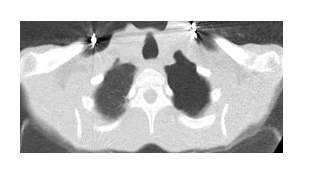
Figure 1: Pneumothorax view in left hemithorax on thorax CT.
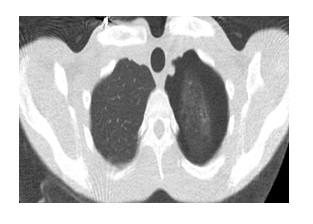
Figure 2: Lung parenchyma view in thorax CT.
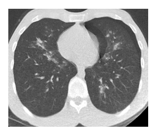
Figure 3: Bilateral infiltration view.
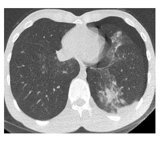
Figure 4: Ground glass in left lower lobe and pneumothorax in left hemithorax.
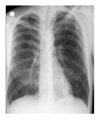
Figure 5: Posteroanterior chest x-ray after tube thoracostomy.
3. Discussion
The new coronavirus was identified and isolated by three groups of Chinese scientists. A consortium coordinated by W. Tan from the China Centers for Disease Control and Prevention (China CDC) achieved eight complete viral genome sequences by sequencing from RNA isolation and bronchoalveolar lavage fluid (BALF) in nine patients [11]. Although Covid-19 has a lower mortality rate than SARS-CoV and MERS-CoV, which are from the corona virus family, its spread rate is much higher than these viruses [6]. In 2019, the WHO-China Coronavirus disease joint mission report published the main signs and symptoms of Covid-19 as fever, dry cough, sputum complaint, shortness of breath, fatigue, muscle pain or arthralgia, headache and sore throat. The China Centers for Disease Control and Prevention (China CDC) identified 44 672 Covid-19 positive patients on February 14. 965 of them (2.2%) were under 20 and the mortality rate in this age group was 0.1%. 77.8% of the patients reported that they were between the ages of 30 and 69 [12]. Our patient was a 24-year-old young healthy man with no additional disease, previous operation and history of pneumothorax. In his complaints, he had high fever, cough, shortness of breath, and chest pain on the left.
Currently, the RT-PCR test is used as a standard in the definitive diagnosis of Covid-19 infection despite false negativity rates. Chest CT is also recommended by the Chinese National Health Commission for the clinical diagnosis of Covid-19. However, the final diagnosis of Covid-19 must be confirmed by positive RT-PCR or gene sequencing [13]. In the study conducted by Tao Ai et al., In those with RT-PCR positive CT; found the sensitivity to be 97% and to diagnose CT as 88% (888 patients) [14]. Due to its high sensitivity, chest CT is an important screening tool for suspected Covid-19 patients. In Covid-19 pneumonia, the finding seen in CT is GGO in the subpleural areas of the lower lobes [15]. These findings occur in the early stages of Covid-19 pneumonia, and this finding is caused by alveolar septal inflammation caused by infection [16]. Radiology imaging methods can be guiding in cases with RT-PCR negative but symptom. In PCR positive patients, imaging methods are needed again for the course of the disease. Increasing data and research over time will enable us to get to know this disease better.
In our patient's CT, there were bilateral irregular consolidation areas and ground-glass densities in the lower lobes. On the left, the ground glass was accompanied by pneumothorax. There was no underlying lung disease, smoking history, and structural abnormality in the body that would predispose our patient to pneumothorax formation. We think that in severe Covid-19 pneumonia, strong cough attacks that can cause widespread alveolar damage and a sudden increase in alveolar pressure may cause pneumothorax.
Sana S et al. They investigated imaging findings in 919 Covid-19 positive patients and detected ground-glass densities in 88% of cases. 87.5% of parenchymal attitudes were observed to be bilateral. none of them encountered pneumothorax [17]. In severe acute respiratory failure syndrome, sudden alveolar pressure increase may cause interstitial emphysema and air leak, leading to the development of mediastinal emphysema [18]. In this study, we present a 24-year-old young patient with Covid-19 pneumonia and pneumothorax. We discussed the patient and his radiological findings in the light of the literature. Pneumothorax may develop in Covid-19 pneumonia due to alveolar damage. This can cause increased mortality and morbidity. For this reason, pneumothorax should be kept in mind in the treatment and follow-up of Covid-19 infection.
Conflict of Interest
All authors declare that they have no conflict of interest.
Financial Support
No financial support was recieved for study.
Author Contributions
All authors contributed equally.
References
- Huang C, Wang Y, Li X, et al. Clinical features of patients infected with 2019 novel coronavirus in Wuhan, China. Lancet 395 (2020): 497-506.
- Li Q, Guan X, Wu P, et al. Early transmission dynamics in Wuhan, China, of novel coronavirus infected pneumonia. N Engl J Med 382 (2020): 1199-1207.
- Weiss SR, Leibowitz JL. Coronavirus pathogenesis. Adv Virus Res 81 (2011): 85-164.
- Eurosurveillance Editorial Team. Note from the editors: novel coronavirus (2019-nCoV). Euro Surveill 25 (2020).
- Cui J, Li F, Shi ZL. Origin and evolution of pathogenic coronaviruses. Nat Rev Microbiol 17 (2019): 181-192.
- WEi Zhao, Zheng Zhong, Xingzhi Xie, et al. Relation Between Chest CT Findings and Clinical Conditions of Coronavirus Disease (COVID-19) Pneumonia: A Multicenter Study. American Journal of Roentgenology 214 (2020): 1072-1077.
- World Health Organization Coronavirus disease 2019 (COVID-19) Situation Report-74 (2019).
- Wang C, Horby PW, Hayden FG, et al. A novel coronavirus outbreak of global health concern. Lancet 395 (2020): 470-473.
- Chest Radiographic and CT Findings of the 2019 Novel Coronavirus Disease (COVID-19): Analysis of Nine Patients Treated in Korea. Yoon SH et al. Korean J Radiol 21 (2020): 494-500.
- Zheng Ye, Yun Zhang, Yi Wang. Chest CT manifestations of new coronavirus disease 2019 (COVID-19): a pictorial review. European Radiology (2020).
- Harald Brüssow. The Novel Coronavirus-A Snapshot of Current Knowledge, Microbial Biotechnology 13 (2020): 607-612.
- Report of the WHO-China Joint Mission on Coronavirus Disease (COVID-19) (2019).
- Wenjie Yang, Fuhua Yan. Patients with RT-PCR-confirmed COVID-19 and Normal Chest CT. Radiology 295 (2020).
- Tao Ai, Zhenlu Yang, Hongyan Hou, et al. Correlation of Chest CT and RT-PCR Testing in Coronavirus Disease 2019 (COVID-19) in China: A Report of 1014 Cases. Radiology (2020).
- Lin X, Gong Z, Xiao Z, et al. Novel coronavirus pneumonia outbreak in 2019: computed tomographic findings in two cases. Korean J Radiol 21 (2020): 365-368.
- Xu Z, Shi L, Wang Y, et al. Pathological findings of COVID-19 associated with acute respiratory distress syndrome. Lancet Respir Med 8 (2020): 420-422.
- Sana Salehi, Aidin Abedi, Sudheer Balakrishnan. Coronavirus Disease 2019 (COVID-19): A Systematic Review of Imaging Findings in 919 Patients. American Journal of Roentgenology (2019): 1-7.
- Ooi GC, Khong PL, Müller NL, et al. Severe acute respiratory syndrome: temporal lung changes at thin-section CT in 30 patients. Radiology 230 (2004): 836-844.