Manabu Yamamoto MD1, Norifumi Iseda MD1, Hiroko Yano MD1, Yohei Tominaga MD1, Ayako Sugino MD2, Hiroyuki Kobayasi MD2
1Department of Surgery, Fukuoka Sanno Hospital, Momochi-hama 3-6-45, Fukuoka 814-0001, Japan
2Department of Gastroenterology, Fukuoka Sanno Hospital, Momochi-hama 3-6-45, Fukuoka 814-0001, Japan
*Corresponding Author: Dr. Manabu Yamamoto, Department of Surgery, Fukuoka Sanno Hospital, Momochi-hama 3-6-45, Fukuoka 814-0001, Japan
Received: 22 July 2019; Accepted: 05 August 2019; Published: 13 November 2019
Although tumor lysis syndrome is well described after chemotherapy for non-solid tumors, it is rarely seen or suspected in solid malignancies. Early recognition of this condition is paramount to reduce morbidity and mortality. We report on a case of tumor lysis syndrome induced by surgery in a patient with colon cancer with multiple liver metastases. We also review fifteen cases of diagnosed tumor lysis syndrome accompanying colorectal cancers. Though tumor lysis syndrome is rare in solid tumors, we should realize this possibility in patients with solid tumors of larger volumes, in particular, with enlarged liver metastases.
Colon cancer; Tumor lysis syndrome; Surgery; Liver metastases
Abbreviations:
TLS-Tumor Lysis Syndrome; CEA- Carcinoembryonic Antigen; DIC-Disseminated Intravascular Coagulation
1. Introduction
Tumor lysis syndrome (TLS) is a well described oncologic emergency. The syndrome is deemed spontaneous when it occurs before initiation of any cytotoxic or definitive treatment. Here, we report the rare case of a 70-year old man with colon cancer and extensive liver involvement manifesting with TLS following surgery. Previous investigators had demonstrated only fourteen cases with TLS associated in colorectal cancer [1-14]. Ten of the fourteen cases were induced by chemotherapy, three were spontaneous tumor lysis syndrome (STLS), and one was induced by radiofrequency ablation (RFA). This is therefore the first report of TLS induced by surgery in case of colon cancer with liver metastases.
2. Case Presentation
A 70-year old Japanese man, who had been in good health until two months prior to this admission, presented with worsening abdominal pain, accompanied by loss of appetite, fever and subjective weight loss. The patient also complained of postprandial abdominal pain in the right upper quadrant. Blood examination revealed anemia and elevated levels of the tumor marker, carcinoembryonic antigen (CEA). His past medical history detailed two previous abdominal operations, due to intestinal obstruction fifty years ago. His social history was negative for tobacco use, alcohol use and use of illicit drugs.
Physical examination revealed right upper quadrant tenderness on deep palpation with no signs of peritonitis, and bilateral pitting edema up to the knees. Cardiovascular, pulmonary, and neurological examinations were grossly normal. Laboratory findings at the time of presentation included: white blood cells: 9.35 × 10³/μL, hemoglobin: 7.7 g/dL, platelets: 353 × 103/μL, potassium: 4.5 mmol/L, serum creatinine: 0.77 mg/dL, albumin: 2.9 g/dL, total bilirubin: 0.7 mg/dL, alanine aminotransferase: 1037 U/L, aspartate aminotransferase: 40 U/L, alkaline phosphatase: 47 U/L and lactate dehydrogenase levels: 979 U/L. Elevated CEA was observed at 283.6 ng/UL. Colonoscopy revealed a mass in the transverse colon: biopsy of the mass confirmed a moderately- to well-differentiated invasive adenocarcinoma (Figure 1). Computed tomography scan of the abdomen and pelvis revealed hepatomegaly with multiple liver lesions alongside with mural thickening of the transverse colon, with ascites across the whole body (Figure 2). The patient underwent right hemicolectomy as the palliative surgery to prevent anemia or colon obstruction. On the first postoperative day, the patient experienced tumor lysis, with lactate dehydrogenase levels: 20072 U/L, uric acid: 8.7 mg/dL, serum potassium: 6.8 mEq/dL, serum calcium: 7.4 mg/dl, and serum phosphate: 6.3 mg/dL (Table 1). Kidneys ultrasound showed normal renal parenchyma with no evidence of obstructive uropathy. We diagnosed this as TLS. All available supportive measures, including continuous hemodiafiltration, were initiated from 1 to 7 postoperative days. Then, the patient also experienced liver failure and disseminated intravascular coagulation (DIC) from 3 to 11 postoperative days (Table 1). The patient subsequently recovered from TLS, and was administered the anticancer-drugs (FOLFOX) on postoperative day 21.
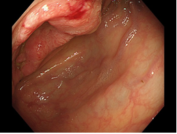
Figure 1: Colonoscopy showing a Bormann II type tumor at the transverse colon (arrow).
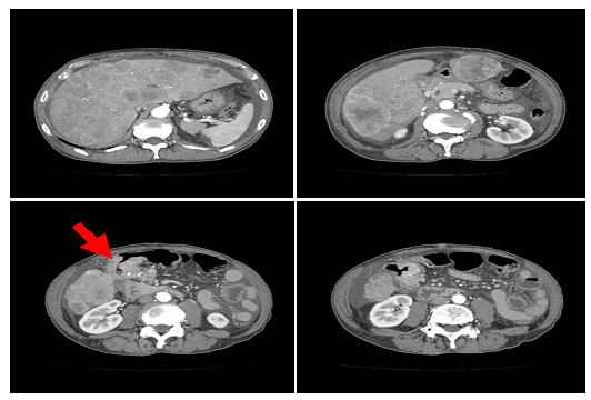
Figure 2: Computed tomography scan revealing multiple liver metastasis and ascites across the whole peritoneal cavity. The arrow represents the tumor at the transverse colon.
|
1POD |
7POD |
14POD |
21POD |
|
|
WBC (µl) |
10220 |
23760 |
17130 |
10320 |
|
Hemoglobin (g/dl) |
13.5 |
9.2 |
7 |
8 |
|
Platelets (x104/µl) |
33 |
12.2 |
21.7 |
29.1 |
|
BUN (mg/dl) |
18 |
15 |
9 |
6 |
|
Cre (mg/dl) |
1.35 |
0.88 |
0.62 |
0.56 |
|
K (mEq/l) |
6.8 |
3 |
4 |
4.3 |
|
Ca (mg/dl) |
7.4 |
7.4 |
7 |
7.9 |
|
P (mg/dl) |
6.3 |
2.4 |
3.1 |
3 |
|
T-Bil (mg/dl) |
2.3 |
8.6 |
3.2 |
2.3 |
|
GOT (U/l) |
1190 |
48 |
39 |
33 |
|
GPT (U/l) |
286 |
38 |
24 |
22 |
|
LDH (U/l) |
20072 |
591 |
643 |
530 |
|
Uric Acid (mg/dl) |
8.7 |
2.8 |
2.9 |
5.3 |
|
CK (U/l) |
1609 |
68 |
98 |
95 |
|
CRP (mg/dl) |
11.31 |
15.63 |
5.49 |
4.09 |
|
Continuous hemodiafiltration 1POD~7POD |
||||
|
DIC 3POD~11POD |
||||
POD: postoperative day; DIC: disseminated intravascular coagulation
Table 1: Laboratory trends in the patient presented.
3. Discussion
We present here the first case of TLS as a complication after surgery for metastatic colon adenocarcinoma. TLS is an oncological emergency precipitated by massive release into the circulation of intracellular tumor contents, manifesting in distinct laboratory abnormalities including hyperuricemia, hyperkalemia, hyperphosphatemia, and hypocalcemia. While TLS is most commonly observed in hematological cancers, it can also be seen in solid malignancies with large tumor burdens [15]. These include breast cancer, small cell lung cancer, germ cell tumor, melanoma, Merkel cell carcinoma, head and neck cancer, non-small cell lung cancer, ovarian cancer, vulva cancer, prostate cancer, hepatocellular carcinoma, colorectal cancer, gastric cancer, sarcoma, neuroblastoma, medulloblastoma, hepatoblastoma, gestational trophoblastic neoplasia, renal cell carcinoma, transitional cell carcinoma, and thymoma [6].
In most patients, TLS occurs as a result of chemotherapy, however, it may also be induced by other anti-neoplastic treatments, including radiotherapy, hormone therapy and immunotherapy, or may develop spontaneously before initiation of therapy [16, 17]. STLS is defined as the TLS occurring in the absence of any definitive treatment [18]. The incidence of STLS in solid tumor is rare. The first step for TLS diagnosis is based on the laboratory findings in an appropriate clinical context. Hyperuricemia, hyperkalemia, hyperphosphatemia, and hypocalcemia are the primary indicators of TLS [15, 16]. Clinical TLS is considered when traditional laboratory findings are accompanied by diarrhea, nausea, vomiting, lethargy, cardiac arrhythmias, seizures, or sudden death [17, 18]. Patient care when TLS has been established should include astute monitoring of uric acid, potassium, phosphorous, calcium, lactate dehydrogenase and serum creatinine levels, alongside cardiac monitoring and monitoring urine output. Allopurinol may be a useful prophylactic to lower uric acid production in patients at risk of TLS. However, allopurinol use may result in elevated xanthine levels by inhibiting xanthine oxidase in patients with TLS [19-21]. Xanthine may also precipitate in the renal tubules, contributing to obstructive uropathy in a similar manner as uric acid [20, 21]. As a result, rasburicase, which converts uric acid to soluble allantoin, is the recommended drug for hyperuricemia in TLS [15, 16, 18, 19]. The dialysis need to be initiated for hyperkalemia, hyperphosphatemia, or hypocalcemia [15]. The present case was not treated with rasburicase, due to a sufficient decrease in uric acid by the second postoperative day through continuous hemodiafiltration.
The first reported case of TLS in a nonhematological malignancy occurred in 1977 [22]. Approximately 120-140 reported cases of tumor lysis in solid tumors have since been reported, mostly after treatment, between 1977 and 2018 [6, 19]. Although there was different origins of primary tumors, extensive liver involvement has been noted in 82.8% of all cases. When the liver was not involved, patients inevitably had a large tumor burdens accompanied by necrosis. As noted by Gemici et al. [16], liver involvement seems to predispose patients to the TLS. This may be due to a high purine pool in the liver, which may be released during necrosis, or due to impaired uric acid metabolism if hepatic function is impaired by a high tumor burden [16]. TLS in solid tumors is unpredictable and carries a worse prognosis when compared to hematological malignancies [16]. Mortality rates are 20%–50% across all cases of TLS in solid tumors if undiagnosed or if diagnosed too late [19, 23].
Our literature review of TLS in patients with colon cancer revealed only three cases of STLS, ten of tumor lysis induced by a chemotherapy, one of tumor lysis induced by RFA, and the present case of tumor lysis induced by surgery [1-14] (Table 2). This case may represent a pre-process to STLS during surgery, though it is difficult to understand why TLS was induced by surgery. Of the fifteen cases, including the present case identified, most cases had multiple liver metastasis, with larger tumor volumes, and only five (33.3%) recovered from TLS. Early diagnosis and treatment are crucial if patients are to survive TLS.
|
No. |
Age |
Gender |
Tumor sites |
Treatment |
Onset of TLS |
Prognosis |
Ref |
|
1 |
27 |
M ? |
Liver, lung, pleura |
STLS |
Died |
[1] |
|
|
2 |
82 |
F |
Extensive liver involvement |
STLS |
Recovery |
[2] |
|
|
3 |
49 |
F |
Liver mass |
STLS |
Died |
[3] |
|
|
4 |
49 |
F |
Multiple liver |
Etoposide+cisplatin |
1 days |
- |
[4] |
|
5 |
83 |
F |
Peritoneal dissemination |
FOLFIRI+cetuximab |
5 days |
Died |
[5] |
|
6 |
59 |
M |
Numerous liver |
FOLFOX |
3 days |
Recovery |
[6] |
|
7 |
51 |
F |
Enlarger liver |
FOLFOX |
- |
Recovery |
[7] |
|
8 |
64 |
M |
Liver |
FOLFIRI+bevacizumab |
7 days |
Died |
[8] |
|
9 |
62 |
M |
Lung |
FOLFIRI+bevacizumab |
2 days |
Died |
[9] |
|
10 |
66 |
M |
Liver, lung |
FOLFIRI |
3 days |
Died |
[10] |
|
11 |
64 |
M |
Lung |
Cetuximab |
18 hr |
Recovery |
[11] |
|
12 |
38 |
F |
Liver |
Irinotecan |
6 days |
Died |
[12] |
|
13 |
42 |
F |
Multiple liver |
Irinotecan |
8 days |
Died |
[13] |
|
14 |
- |
- |
Liver |
RFA |
- |
- |
[14] |
|
Present case |
70 |
M |
Extensive liver involvement |
Surgery |
18 hr |
Recovery |
STLS, spontaneous tumor lysis syndrome; RFA, radiofrequency ablation;
FOLFIRI, 5-fluorouracil, leucovorin and irinotecan; FOLFOX: 5-fluorouracil, leucovorin and oxaliplatin.
Table 2: Fifteen cases induced tumor lysis syndrome in colorectal cancers.
In conclusion, we reported the first documented case of TLS induced by surgery in a colon cancer with multiple liver metastases. This patient recovered from TLS by early diagnosis and treatment, including continuous hemodiafiltration due to resolve hyperuricemia and hyperkalemia.
Conflict of Interest
The authors declared that they have no competing interests.
Consent for Publication
The informed consent for publication and presentation was obtained from the patient.
Funding
This work was no supported from any funding.
Authors’ Contribution
MY drafted the manuscript. NI, HY, YT, AS, and HK acquired data and revised the manuscript. All authors have read and approved the final manuscript.
Acknowledgments
We thank Dr. Masafumi Ohya for pathological diagnosis.
References
- Frestad D, Perner A, Pedersen UG. Acute onset and rapid progression of multiple organ failure in a young adult with undiagnosed disseminated colonic adenocarcinoma. BMJ Case Rep (2014) doi: 10.1136/bcr-2014-205002.
- Vaisban E, Braester A, Mosenzon O, et al. Spontaneous tumor lysis syndrome in solid tumors: really a rare condition? Am J Med Sci 325 (2003): 38-40.
- Sommerhalder D, Takalkar AM, Shackelford R, Peddi P. Spontaneous tumor lysis syndrome in colon cancer: a case report and literature review. Clin Case Rep 5 (2017): 2121-2126.
- El Mesbahi O, Beaudoin A, Louvet D, De Gramont A. Tumor lysis syndrome after chemotherapy for colon small cell carcinoma. Rev Med Interne 25 (2004): 768-769.
- Matsuyama S, Kuramoto T, Tanaka R, Hashiguchi K. Tumor lysis syndrome after FOLFIRI+cetuximab for ascending colon cancer. Nihon Shoukakibyo Gakkai Zasshi 112 (2015): 714-720.
- Kim HD, Ha KS, Woo IS, et al. Tumor lysis syndrome in a patient with metastatic colon cancer after treatment with 5-fluorouracil/leucovorin and oxaliplatin: case report and literature review. Cancer Res Treat 46 (2014): 204-207.
- Gouveia HS, Lopes SO, Faria AL. Management of tumor lysis syndrome during first-line palliative chemotherapy for high volume colorectal cancer. BMJ Case Rep (2018) Doi:10.1136/bcr-2017-223474.
- Krishnan G, D’Silva K, Al-Janadi A. Cetuximab-related tumor lysis syndrome in metastatic colon carcinoma. J Clin Oncol 26 (2008): 2406-2408.
- Hentrick M, Schiel X, Scheidt S, et al. Fatal tumor lysis syndrome after irinotecan/5-FU/folic acid/bevacizumab-containing therapy in a patient heavily pretreated for metastatic colon cancer. Acta Oncol 47 (2018): 155-156.
- Oztop I, Demarkan B, Yaren A, et al. Rapid tumor lysis syndrome in a patient with metastatic colon cancer as a complication of treatment with 5-fluorouracil/leucovorin and irinotecan. Tumori 90 (2004): 514-516.
- Kalmkerian GP, Darwish B, Varterasian L. Tumor lysis syndrome in small cell carcinoma and other solid tumors. Am J Med 103 (1997): 363-367.
- Nikolic-Tomasevic Z, Jelie S, Popv I. Irinotecan as second-line treatment in metastatic colorectal cancer: dilemmas regarding patient selection and toxicity prediction. J Chemother 12 (2000): 244-251.
- Boisseau M, Bugat R, Mahjoubi M. Rapid tumour lysis syndrome in a metastatic colorectal cancer increased by treatment (CPT11). Eur J Cancer 32A (1996): 737-738.
- Barry BD, Kell MR, Redmond HP. Tumor lysis syndrome following endoscopic radiofrequency interstitial thermal ablation of colorectal liver metastases. Surg Endosc 16 (2002): 1109.
- Coiffier B, Altman A, Pui CH, et al. Guidelines for the management of pediatric and adult tumor lysis syndrome: an evidence?based review. J Clin Oncol 26 (2008): 2767-2778.
- Gemici C. Tumour lysis syndrome in solid tumours. Clin Oncol (R Coll Radiol) 18 (2006): 773-780.
- Woo IS, Kim JS, Park MJ, et al. Spontaneous acute tumor lysis syndrome with advanced gastric cancer. J Korean Med Sci 16 (2001): 115-118.
- Nagaiah G, Truong Q, Monga M. Oncologic emergencies and paraneoplastic syndromes in Abraham J, editor, Gulley JL, editor, Allegra CJ, editor, eds. The Bethesda handbook of clinical oncology, 5th ed. Lippincott Williams & Wilkins, Philadelphia (2014).
- Vodopivec DM, Rubio JE, Fornoni A, Lenz O. An unusual presentation of tumor lysis syndrome in a patient with advanced gastric adenocarcinoma: case report and literature review. Case Rep Med (2012) Doi.10.1155/2012/468452.
- Cairo MS, Bishop M. Tumour lysis syndrome: new therapeutic strategies and classification. Br J Haematol 127 (2004): 3-11.
- LaRosa C, McMullen L, Bakdash S, et al. Acute renal failure from xanthine nephropathy during management of acute leukemia. Pediatr Nephrol 22 (2007): 132-135.
- Crittenden DR, Ackerman GL. Hyperuricemic acute renal failure in disseminated carcinoma. Arch Intern Med 137 (1977): 97-99.
- Coiffier B. Acute tumor lysis syndrome-a rare complication in the treatment of solid tumors. Oncol Res Treat 33 (2010): 498-499.