Joseph N Salama*, Sylvia Arce, Harold W Hunt, Diego A L Garcia
Department of Orthopaedic Surgery and Sports Medicine University of Florida, 345 Hull Road, Gainesville, FL, USA
*Corresponding Author: Dr. Joseph N Salama, Department of Orthopaedic Surgery and Sports Medicine University of Florida, 345 Hull Road, Gainesville, FL, USA
Received: 05 August 2026; Accepted: 10 August 2026; Published: 21 August 2026
Background: Anatomic variants of the median nerve and its accompanying vasculature, specifically a bifid median nerve (BMN) and persistent median artery (PMA) are well-recognized predisposing factors for carpal tunnel syndrome (CTS). Acute thrombosis of a PMA is a rare but important cause of sudden-onset wrist pain and median neuropathy, particularly in young, otherwise healthy individuals.
Case Presentation: We present the case of a 21-year-old left-hand–dominant collegiate baseball player who presented with acute volar wrist pain and median-distribution hyperesthesia following a forceful throwing motion. Electromyography (EMG) was normal. Computed tomography demonstrated no osseous abnormality. Magnetic resonance imaging (MRI) revealed a bifid median nerve with an adjacent persistent median artery demonstrating intraluminal signal abnormality and surrounding edema, suspicious for acute thrombosis.
Conclusion: This case underscores the importance of considering vascular anomalies such as PMA which is an uncommon yet clinically significant cause of acute median nerve symptoms in athletes. Recognition of this entity on MRI is critical to avoid diagnostic delay and guide appropriate conservative or surgical management to prevent long-term neurological deficit.
Median nerve; Bifid median nerve; Persistent median artery; Carpal tunnel syndrome; Thrombosis; Wrist MRI; Athlete; Baseball.
Median nerve articles; Bifid median nerve articles; Persistent median artery articles; Carpal tunnel syndrome articles; Thrombosis articles; Wrist MRI articles; Athlete articles; Baseball articles
Introduction
Carpal tunnel syndrome (CTS) is the most common compressive neuropathy of the upper extremity, typically the result of chronic mechanical compression, repetitive stress or tenosynovitis. Acute CTS is uncommon and most often associated with direct trauma, hemorrhage or other space-occupying lesions. Congenital anatomic variants of the median nerve and its vascular supply represent uncommon but important causes of acute median neuropathy. A bifid median nerve (BMN) is characterized by bifurcation of the median nerve into two discrete bundles proximally to or within the carpal tunnel. A persistent median artery (PMA) is an embryologic remnant that may course along or between these nerve bundles. The reported prevalence of BMN ranges from 2% to 26%, while PMA ranges between 0.6% and 30% [1,2].
While PMA is typically asymptomatic, it may become clinically significant when enlarged, aneurysmal or thrombosed resulting in acute median nerve compression [3,5,6,8,9–13]. This is particularly relevant in athletes who are exposed to repetitive loading, forceful wrist flexion and repetitive microtrauma.
Case Presentation
A 21-year-old left-hand–dominant collegiate baseball pitcher presented to the sports medicine clinic with acute onset of severe pain in the volar aspect of the wrist. The pain began abruptly during a high-velocity throw followed by forced wrist flexion. He described sharp, stabbing pain accompanied by hyperesthesia in the thumb, index, and middle fingers. There was no history of prior wrist injury, inflammatory arthropathy, coagulopathy, or anticoagulant use.
Physical Examination
Examination revealed mild swelling and tenderness over the volar wrist. Active wrist flexion exacerbated pain and paresthesia. Tinel and Phalen maneuvers reproduced symptoms. Sensation was mildly altered in the median nerve distribution, with preserved motor strength. Radial and ulnar pulses were palpable and symmetric and there were no signs of pallor or digital ischemia.
Electrodiagnostic Testing
Electromyography and nerve conduction studies of the left upper extremity were normal, with no evidence of median or ulnar neuropathy. Specifically, there was no electrodiagnostic evidence of ulnar nerve entrapment at the hook of the hamate, wrist, cubital tunnel, medial epicondyle or retrocondylar groove. The ulnar nerve demonstrated normal anatomic curvature adjacent to the medial triceps. Normal electrodiagnostic findings do not exclude acute compressive median neuropathy. Electromyography and nerve conduction studies may remain normal during the early stages of acute nerve compression because detectable abnormalities generally reflect axonal degeneration or demyelination, which require time to develop. Consequently, imaging plays a particularly important role when acute vascular compression is suspected despite normal electrodiagnostic testing [9,10].
Imaging
CT: Noncontrast CT of the left wrist demonstrated no acute fracture, malalignment, or other osseous abnormality to account for the patient's symptoms.
MRI: MRI of the left wrist demonstrated a Type II/III bifid median nerve within the carpal tunnel. Interposed between the two branches of the bifid median nerve was a persistent median artery (Figures 1 and 2). The vessel demonstrated loss of the expected vascular flow void, mild fusiform enlargement, intermediate-to-high T1 signal intensity, and heterogeneous T2 signal, compatible with acute intraluminal thrombus. Prominent T2-hyperintense perivascular edema extended into the adjacent carpal tunnel soft tissues with mild reactive perineural edema involving the adjacent bifid median nerve (Figure 3). The degree of surrounding inflammatory change was disproportionate to that expected for an uncomplicated persistent median artery and favored acute vascular pathology. The radial and ulnar arteries maintained normal flow voids without surrounding inflammatory change, supporting pathology localized to the persistent median artery (Figure 4). Mild volar bowing of the flexor retinaculum reflected minimal mass effect from the thrombosed artery and associated inflammatory change (Figure 5). Collectively, these imaging findings are most compatible with acute thrombosis of the persistent median artery. Although Doppler ultrasound was not obtained for confirmation, the MRI findings together with the patient's abrupt symptom onset were considered highly suggestive of acute thrombosis [10].
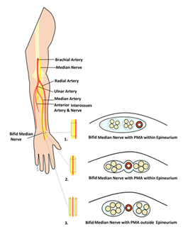
Figure 1: Illustration of the major anatomic variants of the bifid median nerve and persistent median artery within the carpal tunnel. The current case demonstrates a Type II/III configuration in which the persistent median artery courses interposed between the two branches of the bifid median nerve.
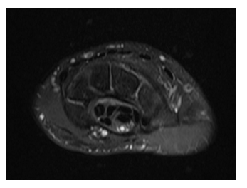
Figure 2: Axial proton-density fat-suppressed MR image demonstrating a Type II/III bifid median nerve (asterisks) with a centrally positioned persistent median artery (arrow), an uncommon congenital anatomic variant that predisposes to median nerve compression when thrombosed.
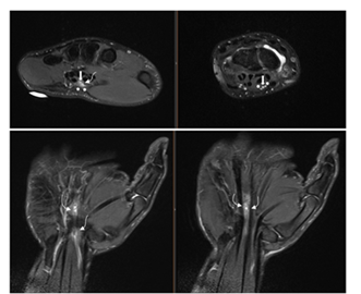
Figure 3: Axial and coronal T2-weighted fat-suppressed MR images demonstrate the thrombosed persistent median artery (arrow) with loss of the normal flow void, mild fusiform enlargement, and surrounding T2 hyperintense perivascular edema. Mild reactive signal alteration of the adjacent bifid median nerve (asterisks) is present, compatible with reactive neuritis.
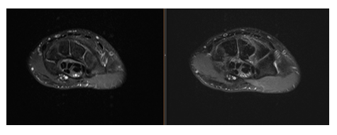
Figure 4: Axial T2-weighted fat-suppressed MR image demonstrates loss of the normal flow void within the persistent median artery (arrow), while the radial (RA) and ulnar (UA) arteries maintain normal flow voids, supporting focal thrombosis of the persistent median artery.
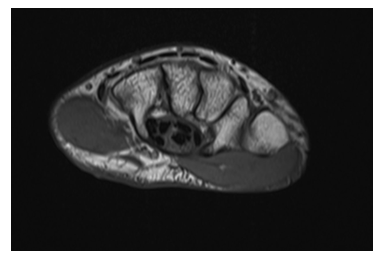
Figure 5: Axial non-fat-suppressed T1-weighted image demonstrates mild volar bowing of the flexor retinaculum (arrowheads) secondary to minimal mass effect from the thrombosed persistent median artery and adjacent inflammatory change. The bifid median nerve is indicated by asterisks.
Ultrasound
Color Doppler ultrasound was recommended for confirmation of arterial occlusion but was not obtained because the patient's symptoms improved with conservative management.
Management and Clinical Course
Orthopedic hand surgery consultation discussed both conservative and surgical options with the patient, including Guyon canal and carpal tunnel release. Given the acute nature of the injury and mild symptoms without significant neurological deficit, conservative management was recommended. The patient was treated with wrist bracing, rest and nonsteroidal anti-inflammatory medication. Surgical intervention was deferred if symptoms persisted beyond four weeks. The patient has since not had further follow up.
Discussion
This case highlights an uncommon cause of acute median nerve symptoms in a young athlete with suspected thrombosis of a persistent median artery in association with a bifid median nerve.
Pathophysiology
During embryogenesis, the median artery serves as the primary arterial supply to the hand before regressing as the radial and ulnar arteries mature. Persistence into adulthood can occur and in some cases the PMA accompanies a bifid median nerve as it courses within the carpal tunnel [2]. Acute thrombosis may occur due to repetitive microtrauma, mechanical compression, or abrupt wrist motion such as high-velocity throwing in our case, leading to rapid increases in tunnel pressure, ischemia, edema and nerve compression [3,5,6,8,9]. Recent surgical case reports have further demonstrated that PMA thrombosis can directly distort or compress the bifid median nerve, producing clinical CTS and necessitating operative decompression with persistent or progressively worsening symptoms [8].
Athletes participating in overhead throwing sports may represent a unique population at risk for symptomatic thrombosis of a persistent median artery. Repetitive high-velocity throwing subjects the volar wrist to substantial tensile, compressive, and torsional forces during the late cocking, acceleration, and follow-through phases of the throwing motion. Abrupt wrist hyperflexion and repetitive traction may produce transient compression of the persistent median artery within the confined carpal tunnel, resulting in endothelial injury, intimal disruption, and activation of the coagulation cascade. Recurrent microtrauma may further promote localized inflammation and edema, increasing intracarpal tunnel pressure and exacerbating compression of the adjacent median nerve. Although direct evidence remains limited because of the rarity of this condition, the biomechanical demands placed upon overhead athletes provide a plausible mechanism for the acute onset of symptoms observed in this patient and suggest that vascular etiologies should remain in the differential diagnosis of sudden volar wrist pain in competitive throwers.
Imaging Considerations
High-resolution ultrasound with color Doppler is considered the first-line modality for identifying PMA and BMN variants and evaluating arterial patency in real-time [4,13]. Absence of flow and intraluminal echogenicity suggests thrombosis. CT angiography or MRI can further delineate vascular anatomy and nerve morphology in addition to further evaluation of surrounding soft tissue to excluding alternative diagnoses such as other space-occupying processes. The combination of loss of the expected vascular flow void, abnormal intraluminal signal intensity, mild arterial enlargement, and disproportionate perivascular edema should prompt consideration of acute persistent median artery thrombosis rather than a normal vascular variant.
Management and Comparison with Literature
Management is largely dependent on symptom severity and neurologic findings. Mild cases may respond to conservative therapy. Progressive or severe cases where neurological deficits are evident, surgical decompression and possible excision of the thrombosed segment of the artery provide rapid relief and prevents nerve ischemia [5,7,8,10,12]. Early diagnosis is essential to prevent permanent nerve injury and facilitate timely return to sport.
A limited number of cases of symptomatic persistent median artery thrombosis have been reported, with most occurring in middle-aged adults presenting with acute carpal tunnel syndrome. Reports in competitive athletes remain exceptionally rare. Similar to previously published cases, MRI demonstrated loss of the normal arterial flow void with surrounding inflammatory change, while electrodiagnostic testing remained normal during the acute phase [8-13]. Unlike many reported patients who ultimately underwent surgical decompression because of persistent or progressive neurologic symptoms, our patient improved with conservative management, although long-term follow-up was unavailable.
Conclusion
Acute volar wrist pain in athletes should not be attributed solely to overuse or tendinous injury. Persistent median artery thrombosis should be considered in the differential diagnosis. Radiologist and clinicians should recognize the imaging manifestations of vascular anomalies within the carpal tunnel. Prompt recognition with early use of high-resolution imaging ensures timely diagnosis and appropriate management, potentially preventing long-term neurological sequelae and ensuring optimal functional recovery. Recognition of this uncommon vascular variant is particularly important for musculoskeletal radiologists interpreting MRI examinations performed for acute athletic wrist injuries.
Reference
- Walker FO, Cartwright MS. Sonographic representation of bifid median nerve and persistent median artery. J Diagn Med Sonogr 27 (2011): 89-94.
- Rodríguez-Niedenführ M, Vázquez T, Nearn L, et al. Median artery revisited. J Anat 195 (199): 57-63.
- Akgun AS, Ertan G, Ulus S. Acute carpal tunnel syndrome caused by thrombosed persistent median artery associated with bifid median nerve. BMJ Case Rep 2017 (2017): bcr2017221446.
- Cartwright MS, Hobson-Webb LD, Boon AJ, et al. Evidence-based guideline: neuromuscular ultrasound for the diagnosis of carpal tunnel syndrome. Muscle Nerve 46 (2012): 287-293.
- Bartels DW, Shin AY. Surgical excision of a thrombosed persistent median artery causing carpal tunnel–like symptoms. JBJS Case Connect 10 (2020): e20.00139.
- Lessard A, Lorange E, Lee J. Carpal tunnel syndrome secondary to acute thrombosis of persistent median artery. McGill J Med 17 (2019): 1-6.
- Vashistha A, Upadhyay A, Shanker V, et al. Persistent median artery and bifid median nerve: implications for carpal tunnel surgery. J Hand Surg Am 48 (2023): 45-52.
- Barr ML, Jain NS, Ghareeb PA, et al. Persistent median artery thrombosis causing a bifid median nerve and carpal tunnel syndrome: a case report. JBJS Case Connect 12 (2022): e22.00424.
- Abdouni YA, Brunelli JPF, Munia MAS. Acute carpal tunnel syndrome due to persistent median artery thrombosis: case report. Rev Bras Ortop (Sao Paulo) 58 (2023): 347-350.
- Miyashima Y, Gotani H, Okamoto K, et al. Median nerve neuropathy caused by persistent median artery thrombosis. Plast Reconstr Surg Glob Open 11 (2023): e4916.
- Qin J, Tan XX, Xue MQ, et al. Coexistence of anomalous muscle, persistent median artery, bifid median nerve causing carpal tunnel syndrome: a case report and literature review. Front Pediatr 11 (2023): 1043442.
- Hines EM, Roberts CJ, Frahm-Jensen G. Acute carpal tunnel syndrome from thrombosed persistent median artery in a patient with congenitally absent radial artery. Hand (N Y). 2024;32(4):896-899. doi:10.1177/17085381231164472.
- Yildizgoren MT, Ucar C. Carpal tunnel syndrome resulting from persistent median artery and bifid median nerve: the critical role of ultrasonography. Cureus 16 (2024): e54551.
Section 1 Genotyping Methods
Mouse Tail Genomic DNA Extraction
Genomic DNA was extracted from mouse tail biopsies using a proteinase K digestion–ethanol precipitation method. Approximately 0.3–0.5 cm of tail tissue was collected from mice older than 2 weeks of age (adjusted according to tail size to ensure comparable tissue volume) and transferred into labeled 1.5 mL microcentrifuge tubes.
Samples were incubated in 0.5 mL mouse tail lysis buffer (GenePool, Cat# GPQ19388) supplemented with 50 μL proteinase K and digested at 56 °C overnight with gentle agitation to ensure complete lysis. The lysates were centrifuged at 12,000 rpm for 10 min at room temperature.
The supernatant was transferred to a fresh tube, followed by the addition of 1 mL absolute ethanol. After inversion to precipitate DNA, samples were centrifuged at 13,000 rpm for 15 min at 4 °C, and the supernatant was discarded.
The DNA pellet was washed with 75% ethanol, centrifuged, briefly air-dried, and dissolved in 80–100 μL TE buffer (pH 8.0) or nuclease-free water.
PCR Amplification for Genotyping
PCR amplification was performed using a 2× Taq Master Mix (CWBIO, Cat# CW0682). Primer sequences, reaction components, and cycling conditions are summarized in Table S1.
PCR products were analyzed by agarose gel electrophoresis using a DNA ladder (DM2000, CWBIO) as a molecular weight reference.
Table S1: PCR Primers, Reaction System, and Cycling Conditions.
Primer Sequences
|
Primer |
Sequence (5’→3’) |
Type |
|
P1 |
ACTCCCCTAAGTCTGAACCA |
Forward |
|
P2 |
AACTGTGGGTCACTACCCTT |
Reverse |
|
P3 |
TGTTTGAGGGACGCTACAGA |
Reverse |
PCR Reaction Mixture
|
Component |
Volume (µL) |
|
ddH₂O |
7.8 |
|
2× Taq Master Mix |
10 |
|
Primer I |
0.4 |
|
Primer II |
0.4 |
|
Primer III |
0.4 |
|
Genomic DNA |
1 |
|
Total |
20 |
Cycling Conditions
|
Step |
Temp (°C) |
Time |
Note |
|
Initial denaturation |
98 |
2 min |
|
|
Denaturation |
98 |
20 s |
|
|
Annealing |
63 |
20 s |
|
|
Extension |
72 |
1 min |
35 cycles |
|
Final extension |
72 |
5 min |
|
|
Hold |
12 |
— |
Genotype Interpretation
- Wild type: 598 bp
- Heterozygote: 598 + 812 bp
- Homozygote: 812 bp
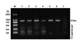
Figure S1: PCR Genotyping of Knockout Mice.
PCR products were generated using a three-primer strategy and analyzed by 1% agarose gel electrophoresis.
- Lane M: DNA marker
- Lanes 1–6: representative samples showing a single 812 bp band (homozygous knockout)
- Lane 7: negative control
The absence of the 598 bp band confirms successful disruption of the wild-type allele.
Section 2: MRI Methodological Details.
Table S2: MRI Acquisition Parameters for Quantitative T1 Mapping.
|
Parameter |
Value |
|
Scanner |
9.4 T Bruker BioSpec (Bruker, Ettlingen, Germany) |
|
RF coil |
26 mm mouse body volume coil |
|
Sequence |
Respiratory-gated VTR-FLASH |
|
T1 mapping strategy |
Variable repetition time (VTR) method |
|
Excitation flip angle |
15° |
|
Repetition times (TRs) |
Multiple TRs acquired under identical imaging geometry |
|
Echo time (TE) |
2.0 ms |
|
Field of view (FOV) |
30 × 30 mm² |
|
Matrix size |
256 × 256 |
|
Slice thickness |
0.5 mm |
|
In-plane spatial resolution |
0.15 × 0.15 mm² |
|
Respiratory gating |
Enabled throughout acquisition |
|
Contrast agent |
Gd-EOB-DTPA (Primovist®, Bayer Healthcare) |
|
Contrast dose |
0.025 mmol/kg |
|
Post-contrast acquisition time points |
10, 20, 30, and 40 min |
|
Acquisition time per T1 map |
Approximately 4–6 min |
Footnote
The acquisition duration varied slightly depending on respiratory gating efficiency and animal respiratory stability during imaging.
Section 3. Additional Quantitative Data
Table S3: Baseline and Post-Contrast Hepatic T1 Values.
|
Group |
Baseline T1 (ms) |
10 min |
20 min |
30 min |
40 min |
|
Control |
732.25±37.66 |
195.4±17.81 |
235.7±20.62 |
615.53±34.43 |
712.43 ± 36.62 |
|
KO |
752.45±33.02 |
676.25±38.1 |
696.98±31.33 |
726.27±44.35 |
741.7 ± 23.02 |
|
CCl₄ |
834.38±13.3 |
424.02±33.24 |
565.3±49.95 |
685.05±14.06 |
772.58 ± 17.82 |
|
MCD |
841.4±26.92 |
365.03±44.32 |
454.53±54.09 |
657.55±24.35 |
752.28 ± 20.52 |
|
AFLD |
865.43±16.63 |
493.52±69.65 |
598.65±50.79 |
668.1±38.88 |
767.7 ± 16.32 |
Values are presented as mean ± SD. Post-contrast T1 values were measured at multiple time points (10–40 min) following Gd-EOB-DTPA administration.
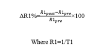
ΔR1 (%) was calculated as: