Michael Fry
Department of Biochemistry, Rappaport Faculty of Medicine, Technion – Israel Institute of Technology, POB 9649, Bat Galim Haifa, ISRAEL 31096
*Corresponding author: Michael Fry. Department of Biochemistry, Rappaport Faculty of Medicine, Technion – Israel Institute of Technology, POB 9649, Bat Galim Haifa, ISRAEL 31096
Received: 08 September 2026; Accepted: 15 September 2026; Published: 22 September 2026
Diphtheria, a potentially fatal human disease, is caused by the infective bacillus Corynebacterium diphtheriae which releases a cytotoxic diphtheria toxin (DT). DT damages various host tissues and organs, causing much of the pathology of the disease. Universal implementation of effective vaccination practically eradicated diphtheria by the 1940s. Thus, pre-vaccination era research that engaged in clinical and immunological aspects of diphtheria evolved after its eradication into curiosity-driven studies of the structure and mechanism of toxicity of DT. In a career that spanned almost 50 years, the American biologist Alwin Max Pappenheimer Jr., (1908-1995) has made significant contributions to the elucidation of the synthesis, structure, and mechanism of the toxic action of DT. This paper traces Pappenheimer’s pursuits of DT within the broader context of the advancement of DT research in the second half of the 20th century. Beginning with brief overview of the history and historiography of diphtheria, the paper next recounts Pappenheimer’s personal and professional biography and outlines his scientific accomplishments. Last, I argue that Pappenheimer’s scientific work is a representative case of a class of biomedical studies that while originally seeking solution to medical problems, ended up uncovering fundamental biological entities and principles.
Diphtheria, Corynebacterium diphtheriae, Diphtheria Toxin, Alwin Max Pappenheimer Jr., History of toxinology, Bacterial toxinology
Diphtheria articles, Corynebacterium diphtheriae articles, Diphtheria Toxin articles, Alwin Max Pappenheimer Jr., History of toxinology articles, Bacterial toxinology articles
Introduction
Diphtheria, a potentially deadly epidemic human disease is caused by virulent strains of the infective bacillus Corynebacterium diphtheriae. Normally confined to the upper respiratory tract, C. diphtheriae bacilli induce formation of a leathery pseudomembrane in the throat and release Diphtheria Toxin, (DT) that spreads and damages various uninfected organs such as the heart, lungs, kidneys, and adrenals. Although the disease was known under various other names since antiquity, it was only in 1826 that the French physician Pierre Bretonneau described the pathological characteristics of diphtheria and defined it as a distinct clinical entity: ‘une affection morbide sui generis’.
Diphtheria posed a grave threat to the health and life of predominantly children, until the introduction in the 1930-40s of highly efficacious universal vaccination that practically eradicated the disease. Studies in the pre-vaccination era focused on clinically relevant bacteriological, pathological, and immunological aspects of diphtheria. This phase of medically oriented research came to an end in the wake of the successful obliteration of the disease in the Western world (left arm of the timeline in Figure 1). Remarkably, however, just as diphtheria ceased to be a consequential medical problem, researchers launched vigorous long-term basic research programs that were divorced from clinical concerns. Begun in the 1930s and continuing today, (right arm of Figure 1) these efforts advanced the understanding at the cellular and molecular levels of the nature, structure, and mode of toxic action of diphtheria toxin, (DT). This paper chronicles the history of the experimental elucidation of the structure and activity of DT, focusing on major contributions to this endeavor by the American biologist Alwin Max Pappenheimer Jr., (1908-1995). Dedicating nearly 50 years to studies of DT, Pappenheimer had made major contributions to the elucidation of its synthesis, structure, and mechanism of toxic action. This paper traces Pappenheimer’s pursuits of DT in the broader context of the general advancement of DT research in the second half of the 20th century. The work opens with a brief overview of the history of diphtheria, its clinical features, DT, and anti-diphtheria vaccination. It next surveys historiographic studies of diphtheria, recounts the personal and professional biography of Pappenheimer, outlines his scientific accomplishments, and places his work in the context of trends of 20th century biomedical research.
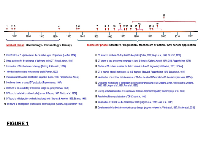
Figure 1: Timeline of research on C. diphtheriae and its toxin (DT). Research activity until the 1930s focused on clinical, bacteriological and immunological aspects of diphtheria. Introduction of effective universal vaccination in the 1930-1940s eradicated the disease and researchers turned to basic questions of DT structure and mechanism of toxicity.
Diphtheria: History, Clinical Features, Diphtheria Toxin, Immunotherapy, and Vaccination
Bacteriology and Clinical Presentation
Diphtheria is an acute, infective disease caused by exotoxin-producing club-shaped bacillus Corynebacterium diphtheriae. This bacterium, which infects and resides in the pharyngeal mucosa, was first isolated by the German Swiss physician Edwin Klebs [1]. Rigorously applying the criteria of Koch’s postulates, (see 3.1 below), the German bacteriologist Friedrich Loeffler established C. diphtheriae as the causative agent of diphtheria [2]. Prior to vaccination, transmission of the bacillus by direct contact, coughing, or sneezing, had spread diphtheria in Europe with an average annual mortality of 6-7 per 10,000 persons in the general population and up to 80% among affected children under 10 years of age. C. diphtheriae multiplies and ulcerates the mucosa of the upper respiratory tract, inducing formation of a white to black pseudomembrane of inflammatory cells, necrotic tissue, and bacteria. In severe cases, amassed pseudomembrane caused suffocation which was treated in some cases by life-saving tracheotomy. Although the C. diphtheriae bacilli usually remain in the pharyngeal mucosa, they release an exceptionally potent exotoxin, DT, which is spread to remote organs via the blood stream. Attacking heart, lungs, kidneys, and nerve cells, DT may evoke severe systemic pathology. Klebs was the first to propose that a ‘venom’ produced at the site of infection circulates in the blood stream and exerts ‘a severe alterating effect on the vascular wall’ [1]. Noting that the bacillus was present only in the pseudomembrane but not in deeper tissues, Loeffler explained the remote effect of C. diphtheriae by postulating, (without experimental proof), that it releases a toxin that damages uninfected distant tissues [2].
The Diphtheria Toxin
By demonstrating in 1888-90 that injected cell-free filtrates of C. diphtheriae cultures caused death in guinea pigs, Alexandre Yersin and Émile Roux of the Pasteur Institute demonstrated the existence in filtrates of C. diphtheriae of ‘venom’ or ‘toxin’ that was until then only a hypothetical entity. They also showed that rabbits and pigeons that were naturally more resistant to diphtheria than guinea pigs, were also more resistant to the injected filtrate. Furthermore, Yersin and Roux detected DT in urine that was collected shortly before the demise of sick children. Addressing in 1891 a Hygiene and Demography Congress in London, Roux proclaimed (English translation by [3]): “It is natural to conclude that microbes act through their chemical products, true poisons specific to each of them, and which determine the symptoms of the disease in man and in animals […] The infectious disease is therefore a poisoning: the source of the poison is the microbe settled in the tissues; it elaborates his toxin there at the expense of the living being that it is going to kill” . Roux also foresaw the use of filtrates of C. diphtheriae cultures for the induction of protective immunity. Distinguishing such procedure from vaccination with attenuated whole bacilli, he named it “chemical vaccination”:
“It is therefore not necessary for the refractory state to be acquired, that the microbes penetrate into the body, it suffices that the substances prepared in the artificial cultures be introduced into it. If, therefore, by swarming in the organism, microbes give immunity, it is undoubtedly because they produce the same chemicals that we find in in vitro cultures.”
Immunotherapy and Vaccination
Emil von Behring and Shibasaburō Kitasato discovered that exposure of naïve guinea pigs to serum from animals that were injected with immunizing sub-lethal amounts of tetanus toxin, rendered the exposed animals resistant to live tetanus bacilli [4]. Behring, (without Kitasato) next showed that antiserum (“antitoxin”) that guinea pigs produced against filtrates of C. diphtheriae cultures imparted naïve animals with passive immunity against DT [5]. Large-scale clinical trials in Berlin in 1894 [6], and in Copenhagen in 1896-97 [7], established that treatment of human patients with anti-diphtheria antisera, (serum therapy; serotherapy) significantly lowered the proportions of fatal cases. In a first attempt to induce permanent immunity, guinea pigs that were injected with a mixture of DT and its antitoxin were found to develop immunity against diphtheria [8]. Subsequently, similar toxin-antitoxin treatment successfully immunized susceptible children against diphtheria [9]. Gaston Ramon of the Pasteur Institute next provided the means for effective general vaccination. Treating DT with formaldehyde and heat, he produced nontoxic but immunogenic ‘toxoid’ that effectively induced production of anti-diphtheria antibodies in injected individuals [10]. Vaccination with the diphtheria toxoid, which was first launched in the 1930s, became universal in industrialized countries by the late 1940s. As a result, incidence and mortality in North America of ~200 and 15 cases per 100,000 population in 1920, were correspondingly lowered in the 1990s to ~0.001 and 0.0001[11].
The Molecular Mechanism of DT Toxicity
After vaccination had practically eradicated diphtheria, research into the disease lost its medical exigency. Yet Pappenheimer and other scientists launched in the late 1930s research programs aimed at elucidating the structure and mechanisms of the toxicity of DT and of related bacterial toxins. As detailed below, it was found that DT exerted its cytotoxic activity by inhibiting cellular protein synthesis through modification and inactivation of elongation factor 2, (EF-2) an essential component of the cellular protein synthesis machinery.
Historiography of Bacteriological and Immunological Facets of Diphtheria
In studying diphtheria, historians of biology and medicine assessed the discovery of its causative agent, C. diphtheriae, in the context of German bacteriology of the late 19th century whereas other historians studied various aspects of the development of passive immune treatment of the disease, (serum therapy) and of anti-diphtheria vaccination.
Loeffler and the Koch School of Bacteriology
Loeffler’s discovery that diphtheria was caused by C. diphtheriae must be viewed in a wider context of the Robert Koch school of medical bacteriology. Loeffler and Georg Gaffky, (who later identified Bacillus salmonella typhi as the causative agent of typhoid disease), were the first trainees to join in 1880 Koch’s newly established Berlin laboratory. This laboratory soon became the epicenter of German bacteriology that side-by-side with the laboratory of Koch’s adversary Louis Pasteur, had shaped medical microbiology in the late 19th century. Koch’s introduction of the technique of growing bacteria on solid media opened the way to his subsequent triumphant cloning and isolation of stable bacterial species [12, 13]. Employing this method, Koch and his associates identified and isolated between 1877 and 1906 twenty different bacterial pathogens [14] p. 290. Loeffler concocted in this context a solid medium, (‘Loeffler’s medium’) specifically suited for the growth and isolation of pure cultures of C. diphtheriae. Once obtaining the isolated bacteria, he meticulously followed the edicts of Koch’s postulates to unequivocally demonstrate that this bacterial species was the causative agent of diphtheria. Loeffler professed that his eight years in the Koch laboratory were his happiest after which he was appointed in 1888 professor of hygiene at the University of Greifswald [15]. There he conducted studies on the foot and mouth virus which pioneered virological research in Germany [15, 16].
Passive (Serotherapy) Treatment of Diphtheria and Active (Vaccination) Prevention of the Disease
Historians of diphtheria mostly focused on the development and application of passive (serotherapy) and active (vaccination) for the respective treatment and prevention of the disease. Historiographies were mainly concerned with scientific, social, political, and economic aspects of passive anti-diphtheria immunization in the late 19th century. Derek Linton comprehensively examined Von Behring’s life and scientific work, both successful, (serotherapy of tetanus and diphtheria) and failed, (development of anti-tuberculosis vaccine) [17]. Linking Behring’s introduction of serum therapy to his prior military-related studies of disinfectants, Jonathan Simon argued that he originally considered serotherapy to be a disinfection procedure and he only later came by more intelligible theories of immunity [18]. Historians paid considerable attention to the standardization and quality control of diphtheria antisera, delineating the major contribution of Paul Ehrlich to this endeavor, [19-21] and describing parallel efforts at the Pasteur Institute [22, 23]. Other studies charted the administrative regulation of the potency and quality of diphtheria antitoxin in late 19th century Germany and France [24-26, 20] and the conflict between philanthropy and business in the administration of serotherapy in fin-de-siècle France and Germany [24, 26, 25].
As to active immunization (vaccination), some authors investigated the life and scientific contributions of Gaston Ramon, the deviser of diphtheria toxoid [27, 28]. Others addressed the subsequent history of the development of diphtheria vaccination [29, 30]. Also, attention was paid to local histories of the production, administration, and failures of vaccination programs in countries such as the United States (Philadelphia, and New York) [31, 32], England [33], and Iran [34]. Unlike the well-researched histories of medically oriented studies of the bacteriology and immunology of diphtheria in the late 19th and very early 20th century, there are virtually no historiographies of the non-medical phase of diphtheria research that, starting in the 1930s, that was directed at the elucidation of the structure and mode of action of the diphtheria toxin, (DT). In this paper I survey the history of pivotal 20th century developments in DT research, focusing on the life and work of Alwin Max Pappenheimer Jr., a major contributor to the clarification of the structure of DT and its mechanism of toxicity.
Alwin Max Pappenheimer, Jr., Biographical Notes
The following concise biographical notes are based on Pappenheimer’s own short memoir [35] and on his portrayals by others: [36-40]; [41], 71-78. Born in 1908 in Cedarhurst, New York, Alwin Pappenheimer Jr., (Figure 2) was the eldest child of Alwin Max Pappenheimer Sr., a Columbia University professor of pathology. His younger siblings were Anne Pappenheimer Forbes, a Radcliff and Harvard educated endocrinologist and a clinical professor at the Harvard Medical School, and John Richard Pappenheimer, a Cambridge University educated physiologist and Harvard professor. Pappenheimer’s grandfather, who arrived in New York as a penniless Jewish immigrant from southern Germany later prospered in the tobacco business. His son Alwin Max Pappenheimer Sr. was educated in Harvard College and in the Columbia University College of Physicians and Surgeons, becoming a prominent professor of pathology in Columbia whose research interests encompassed bacteriology, serology, endocrinology, virology and vitamin deficiencies.
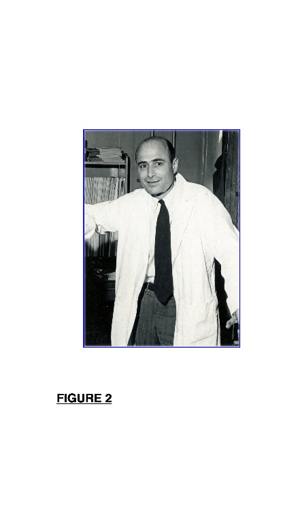
Figure 2: Alwin Max Pappenheimer Jr. in 1954. Image courtesy of The Lillian and Clarence de la Chapelle Medical Archives at New York University.
Alwin Max Pappenheimer Jr., (named ‘Pappenheimer’ in this paper) grew up in a highly cultured and musical household. After concluding his studies in the long since defunct Lincoln School of Teachers College in New York, Pappenheimer graduated in 1929 from Harvard College. Against expectations, he chose not to study medicine but rather to pursue PhD research in organic chemistry under the guidance of James B. Conant who later served as the President of Harvard University. Although his PhD thesis was in organic chemistry, Pappenheimer, who was attracted to biological questions, also worked on the oxidation potential of the hemoglobin-methemoglobin system. After obtaining a PhD degree in 1932, he was awarded a National Research Council Fellowship and worked in 1932-1934 in the National Institute for Medical Research in London under the neurophysiologist and 1936 Nobel laureate Sir Henry Dale. There he unsuccessfully attempted to isolate a hypothetical bacterial growth factor. Back in America, Pappenheimer spent the years 1935-1937 in Harvard, serving as research fellow and instructor in immunology. In parallel he also worked as a senior chemist in the Antitoxin and Vaccine Laboratory in Jamaica Plain, Massachusetts. The original mission of this Institute which was established in 1894 by the Massachusetts Board of Health, was the production of diphtheria antitoxin. It later undertook preparation of vaccines against smallpox, typhoid, tetanus, and pertussis. During World War II the Laboratory produced transfusable human albumin and immune globulins and in 1950 it was the first manufacturing site of Diphtheria-Tetanus-Pertussis (DTP) combination vaccine. Renamed MassBiologics, the Laboratory is operated since 1997 by the University of Massachusetts Chan Medical School. It was in this laboratory the Pappenheimer launched his life-long pursuit of DT by successfully isolating a highly purified form of DT and discovering that DT production in C. diphtheriae was regulated by iron (see section 5).
Despite his productive sojourn in the Antitoxin Laboratory, Pappenheimer’s missed a university environment and thus joined in 1939 the Department of Bacteriology at the University of Pennsylvania. Inopportunely, however, heavy teaching duties and limited research prospects left him unsatisfied in Pennsylvania. He thus willingly accepted in 1941 an offer from Colin MacLeod, the designated Chair of a new Department of Bacteriology in New York University (NYU), to join its faculty as an assistant professor. Other recruits to the department included the biochemist Efraim Racker, the bacteriophage expert Mark Adams, the epidemiologist Royal Christianson, and the bacterial toxinologist Alan Bernheimer. Just six months after joining NYU, the United States entered World War II and Pappenheimer put aside his scientific work to serve in the in the US Army Medical Corps in the Pacific theatre. Discharged in 1945, Pappenheimer returned to NYU and soon recruited his first graduate student, an acquaintance from the army, Melvin Cohn, who became in time a leading immunologist and cofounder of the Salk Institute for Biological Studies in La Jolla. Shortly thereafter his laboratory attracted additional mentees: Sherwood Lawrence, a later expert on lymphocyte function and head of the NYU Department of Infectious Diseases and Immunology, Lane Barksdale, a later professor of microbiology at NYU, Jonathan Uhr who became a leading expert on toxins (including DT) as potential cancer therapeutics, and Matthew Scharff, a later Distinguished Professor of immunology at the Albert Einstein College of Medicine. Pappenheimer’s productive research work at NYU established him as a leader in the DT area. During that period, he expanded his 1936-37 studies on the effect of iron on DT production, investigated relationship of a lysogenic b phage that carries the DT gene into its C. diphtheriae host, and examined the effect of DT on the metabolism of silkworm. Importantly, he urged Norman Strauss and Edelmira Hendee in his laboratory to conduct experiments that revealed that DT inhibited protein synthesis in cultured HeLa cells. This finding that was a first critical step in the consequent unraveling of the mechanism of DT toxicity (see Section 5 below).
After Colin MacLeod has left NYU, Pappenheimer replaced him in 1956 as chair of the NYU Department of Microbiology. Shortly thereafter, however, the Harvard biochemist John Edsall invited Pappenheimer to serve as professor of biology at Harvard and to chair its Board of Tutors in Biochemical Sciences, a tutorial program for students of cellular and molecular biology. In 1961 he was also appointed Master of Harvard’s Dunster House. Beginning in 1958 and lasting till the 1980s Pappenheimer conducted in Harvard experimental studies on the structure of DT and its toxic activity. There he also acted as an influential mentor to students and fellows, many of whom later became prominent scientists on their own. As detailed in the next section, Pappenheimer’s main achievements during the Harvard period included the discovery of the molecular mechanism of inhibition of protein synthesis by DT, studies on DT structure-function relationships, and investigations of mechanisms of DT entry into target cells and its subsequent intracellular processing. Elected in 1973 to the National Academy of Sciences USA, Pappenheimer also served as president of the American Association of Immunologists. He died in 1995 at the age of 86.
The Elucidation of the Structure and Toxic Action of DT
Once diphtheria ceased to be a threat to public health with the introduction of effective vaccination in the 1940s, research on this disease lost its medical impetus and became instead curiosity driven. Pappenheimer delved in the late 1930s into basic studies of the diphtheria toxin (DT). These early experiments presaged his next fifty years of quest after DT structure and mechanism of toxicity. Pappenheimer’s own research work and that of his mentees augured the emergence the presently expansive field of microbial toxinology. The historical case of Pappenheimer’s career exemplifies the basic and potential practical merits of biomedical research with no direct clinical exigency. This section describes Pappenheimer’s contributions to the study of DT in a broader context of a collective endeavor of many researchers to determine the structure of DT and explain the mechanism of its toxic action.
Pappenheimer Purified DT and Established its Proteinaceous Nature
Pappenheimer launched in 1936-37 his career-long pursuit of DT by isolating a highly purified form of toxically potent DT from filtrates of C. diphtheriae cultures. This was not a trivial undertaking as up to that time C. diphtheriae cells were cultivated in complex growth media that contained proteins and peptides which masked the minute amounts of toxin that the bacteria released into the medium. Pappenheimer overcame this problem by formulating a growth medium that contained free amino acids but no proteins or peptides [42, 43]. Growing C. diphtheriae in this medium he isolated from cell-free filtrate of the medium a ~70,000 Da potent proteinaceous toxin at a 95-98% purity [42, 44, 45]. Precise measurements later determined that the actual molecular weight of DT is 58,350 Da. Pappenheimer speculated already in the 1930s that a few or a single molecule of DT sufficed to kill one host cell [35]. Decades later an Osaka University research team confirmed this prediction by experimentally demonstrating that a single DT molecule induced death in one cell [46]. Parallel to Pappenheimer, the Harvard microbiologist Monroe Eaton devised a stepwise purification procedure that allowed isolation of minuscule amounts of highly purified DT from filtrates of protein-containing growth medium [47-49]. Eaton’s and Pappenheimer’s independent similar findings resolutely established that DT was a protein in its entirety. This was a turning point since Pappenheimer initially thought that the DT protein moiety served as mere carrier for a poisonous cofactor such as strychnine or curare [35]. However, when his analysis of acid hydrolysate of the highly purified DT revealed that it contained only amino acids, he realized that DT was wholly a protein. This raised a question how a proteinaceous DT exerted its toxicity, a riddle that Pappenheimer and others pursued in subsequent decades.
Studies on the Regulation of DT Production by Iron
As they were preparing a protein-deficient C. diphtheriae growth medium, Pappenheimer and his assistant Sylvia Johnson noticed that concentration of iron salt in the medium determined the levels of DT that the bacteria produced [50, 51]. Iron at low concentration was required for the synthesis and release of maximum amounts of DT but higher levels of the metal strongly inhibited toxin production. This discovery, which heralded the birth of a fertile area of research on regulation of DT biosynthesis by iron, earned Pappenheimer the 1941 Eli Lilly award. As Pappenheimer later attested, (cited in [39]) that this modest beginning had set the course for his career-long scientific endeavor: “As a graduate student and even as a postgraduate fellow, it worried me a great deal that I did not have an important problem in mind. However, with the discovery of the importance of iron in controlling the production of diphtheria toxin, this insecurity left me and I have never since had to worry about finding something to work on. One thing always leads to another”. Pappenheimer returned in the late 1940s to the question of the mechanism of iron control of DT production. Perhaps because of his past doctoral work on hemoglobin, he focused on cytochromes, which like hemoglobin, also contain an essential heme cofactor with an iron atom at its core, as potential keys to the regulation of DT synthesis by iron. Work in the next several years misled him to believe that DT toxicity was due to its interference with the synthesis or action of the cytochrome system in infected cells.
Pappenheimer and his NYU associate Edelmira Hendee measured release from C. diphtheriae of DT and of the heme precursor coproporphyrin III, measured iron content in the bacteria, and determined activities of their iron-containing enzymes cytochromes and catalase [52]. They found that DT and coproporphyrin III were secreted only during the terminal stages of bacterial growth after all the iron in the growth medium was exhausted. Also, reduction of the bacterial iron enzymes was positively correlated with the secretion of DT and coproporphyrin III. Based on these results, Pappenheimer speculated that DT represented the protein moiety of C. diphtheriae cytochrome and that infected cells died because this DT/cytochrome protein interfered with the synthesis or activity of their own cytochrome system. Subsequent experiments on the Platysamia cecropia silkworm appeared to support to this speculation. Collaborating with the Harvard zoologist Carrol Williams, Pappenheimer found that DT inhibited cytochrome synthesis in developing pupae [53, 54]. However, the cytochrome hypothesis of DT toxicity collapsed when later experiments in Pappenheimer’s own laboratory showed that DT killed host cells by inhibiting their protein synthesis and that the interference with the cytochrome system was a secondary outcome of this inhibition (see below).
Considerable parts of the true mechanism of the control of DT production by iron were ultimately elucidated by two of Pappenheimer’s former mentees, John Murphy of Boston University and Randall Holmes at the Uniformed Services University. Essentially, it was found that transcription of the DT encoding gene tox was controlled by an iron-activated repressor protein, DtxR. Once higher concentrations of the metal activated DtxR, it bound to a specific regulatory sequence in the tox gene to repress its transcription [55, 56] reviewed in [56]. In sum, Pappenheimer’s key contribution was his initial discovery that DT production was controlled by iron. However, his attempt to link this phenomenon to the mechanism of DT toxicity proved to be incorrect.
Studies of Immunological Aspects of DT
Much of Pappenheimer’s research work from the late 1930s to the 1950s was devoted to immunological facets of DT. Diverging from his later focus on cellular and biochemical features of DT, this direction of research was taken because DT toxoid vaccination had still left some questions unanswered. Also, the foci of interest of Pappenheimer’s MD mentees at the time, Sherwood Lawrence and Jonathan Uhr, were mainly immunological and medical. An early line of Pappenheimer’s immunological investigations dealt with the interaction of DT with its antitoxin [57-60]. Lawrence and Pappenheimer later attempted to minimize adverse allergic reactions to immunization among adults by modifying the preparation protocol and mode of injection of the diphtheria toxoid [61-63]. A third line of studies dealt with delayed inflammatory hypersensitivity reaction to diphtheria toxin, toxoid, and toxin-antitoxin complexes. Presaging today’s understanding that delayed hypersensitivity is mediated by monocytes/macrophages and not by circulating antibodies, Pappenheimer, Lawrence, and Uhr found that the delayed allergic reactions to injected diphtheria toxin or toxoid occurred in the absence of circulating anti DT antibodies [64-67].
Pappenheimer Used Phage Encoded DT to Study the Toxin Protein
DT research took an unexpected turn when it was discovered that the toxin was encoded by a gene of a C. diphtheriae specific bacteriophage and not by a bacterial gene. Victor Freeman, then at the University of Washington, fortuitously observed in 1951 that infection with a specific bacteriophage transformed avirulent strains of C. diphtheriae into DT producing bacteria [68, 69]. This initial observation was followed by a demonstration that corynephage b was solely responsible for the conversion of avirulent strain of C. diphtheriae into DT producing virulent bacteria, implying that DT was encoded by a structural gene in the prophage genome [70-73]. A later work in Pappenheimer’s laboratory strengthened this idea [74]. Finally, this notion was validated when the toxin encoding gene, tox, was mapped within the prophage genome [75]. Further, the amount of produced DT was directly proportional to the number of integrated prophage genomes in the host bacteria genome [76].
Pappenheimer was mostly interested in studying the normal and mutant protein products of the corynephage tox gene. He first investigated with Barksdale ultraviolet light (UV) induction of lysis of prophage-hosting bacteria [77]. More than a decade later he returned to corynephages by studying morphologies of B and b corynephage strains [78]. A concurrent study of DT protein products of normal and mutant corynephage tox genes left Pappenheimer uncertain whether the toxin encoding gene resided in the bacterial or the phage genomes [79]. However, subsequent studies of mutated tox gene that encoded a nontoxic but immunologically cross-reacting protein, (CRM), and purification and analysis of the CRM, strongly suggested that tox dwelt in the phage genome [74, 80]. This was validated by a demonstration that purified DNA from normal tox+ and mutant tox- phage strains directed the respective in vitro syntheses of DT and its related nontoxic CRM [81]. Pappenheimer later employed CRM proteins to probe structure-function relationships in the DT protein molecule (see ‘DT structure-function’ below).
Discovery of the Mechanism of DT Toxicity
Pappenheimer’s career-long pursuit of the diphtheria toxin reached its apex with his discovery of key elements of its mechanism of its toxic action. A first step toward uncovering of the molecular basis of DT toxicity was a seemingly modest observation that DT was lethal to cultured cells from toxin sensitive organisms [82, 83]. Norman Stauss and Edelmira Hendee of the Pappenheimer NYU laboratory next observed that DT arrested incorporation of the amino acid methionine into HeLa cell proteins, indicating that the toxin inhibited synthesis of cellular protein [84]. Furthermore, because repression of protein synthesis preceded the interruption of DNA and RNA syntheses and of energy metabolism, the blocking of protein synthesis appeared to be the primary cause of cell death [85-87]. Later in Harvard. Pappenheimer collaborated with his then doctoral student John Collier who later went on to independently study DT, first in the University of California, Los Angeles (UCLA) and later in Harvard. His contributions to this area were recognized by several prestigious awards. Collier and Pappenheimer reported that DT inhibited synthesis of proteins in a HeLa-derived cell-free system [88]. This in vitro system proved to be a vital platform for subsequent experimental discoveries of molecular details of DT toxicity. First, during his postdoctoral fellowship in Geneva, Collier used this system to find that DT inhibited cellular protein biosynthesis by inactivating elongation factor 2 (EF-2), an essential component of the protein synthesis apparatus that acts as a polypeptidyl-tRNA translocase that mediates a translocation step in protein synthesis [89]. In parallel, Pappenheimer and his doctoral student Ronald Goor showed that Nicotinamide Adenine Dinucleotide (NAD), (Figure 3A) was a required cofactor for DT inhibition of protein synthesis in the cell-free system [90-92].
Soon, experiments performed by Tasuku Honjo and associates in Kyoto University, and by Michael Gill in the Pappenheimer laboratory, linked the requirement for NAD to the mechanism of EF-2 inactivation by DT [93-95]. Part of the DT protein, (‘fragment A’, see next section) was found to act as an enzyme that hydrolyzed NAD, releasing its nicotinamide moiety and covalently bonding the remaining ADP-ribose part to EF-2 [96-98], (Figure 3A). ADP ribosylation inactivated EF-2 thus blocking protein synthesis. The contribution of Pappenheimer and his mentees to the elucidation of the molecular basis of the toxic action of DT heralded succession of discoveries on analogue and dissimilar toxic mechanisms of many other bacterial toxins [99-101].
Investigations of DT Structure-Function Relationships
In the 1970s Pappenheimer’s attention turned to the relationship between the structure of DT and its function and to related questions on the mode of entry of the toxin into cells and its subsequent intracellular processing. A distinct domain in DT was found to catalyze ADP ribosylation of EF-2. Two of Pappenheimer’s former mentees, John Collier and Michael Gill separately showed that DT was synthesized as a single intact polypeptide chain that contained two disulfide bridges and no free sulfhydryl groups [102, 103] (illustrated at the top of Figure 3B). Work in the laboratories of Collier, Gill, Pappenheimer, and others, [104, 105, 103, 106-110] revealed that in vitro nicking of DT by a protease such as trypsin followed by reductive breakage of its disulfide bridges, yielded two distinct polypeptide chains. One chain, (‘fragment A’) which corresponded to the amino terminal part of DT catalyzed ADP ribosylation of EF-2. The other chain, (‘fragment B’) represented the carboxy terminal portion of the toxin and was devoid of enzymatic activity (see Figure 3B).
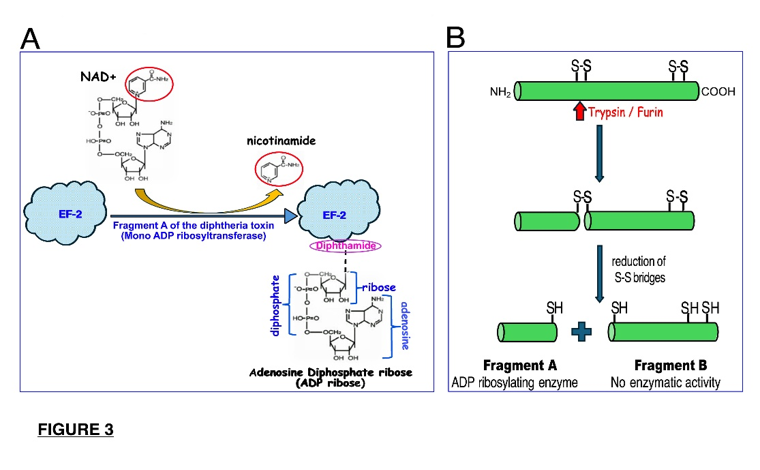
Figure 3: ADP ribosylation of EF-2 by fragment A of DT and intracellular processing of the toxin. A. Fragment A of DT acts as an NAD hydrolyzing mono ADP ribosyltransferase enzyme that releases nicotinamide and covalently binds the ADP ribose moiety to a specific modified histidine residue, (diphthamide) in the EF-2 molecule. As a result, EF-2 is inactivated and cellular protein synthesis is inhibited. B. Processing of the intact diphtheria toxin molecule. The full DT polypeptide is internally nicked by a protease such as trypsin (in vitro) or furin (in vivo), and inter-segment disulfide bridges are reduced. The DT molecule is subsequently split into two free polypeptide segments, an enzymatically active N-terminal fragment A and a C-terminal non-enzymatic fragment B.
Experiments showed that isolated fragment A was incapable of entering the cells and that the fragment B portion of DT mediated the penetration of the intact toxin molecule into the cell.
In a genetic line of inquiry, Tsuyoshi Uchida and Pappenheimer generated different nontoxic but serologically cross-reacting mutant CRM proteins of DT. Analysis of different mutant CRMs revealed that mutations in the fragment A region of DT nullified its ADP ribosylation enzymatic activity whereas mutations in the fragment B domain impeded entry of the toxin into cells [80, 111]. Also, by using normal DT and nontoxic CRM mutant proteins that he synthesized in E. coli-derived cell-free system, John Murphy, Pappenheimer’s postdoctoral fellow at the time, obtained first hints for the existence of a host-encoded iron binding repressor of toxin synthesis [81]. Later studies identified and characterized a C. diphtheriae encoded iron-binding protein, (DTxR) that repressed transcription of the DT tox gene in the presence of excess iron: [112-114].
Studies on the Entry of DT into Target Cells and its Subsequent Intracellular Processing
Taking a biochemical approach, Pappenheimer and his then postdoctoral fellow Patrice Boquet, (later at the Pasteur Institute) studied the interaction of DT with cell membranes. Based on their experimental results, they offered an early model of the interaction of DT with the cell membrane under which the DT fragment B region initially associates reversibly with a hydrophobic locale in the cell membrane [115, 116]. This early interaction is followed by a major irreversible conformational change in the toxin molecule that capacitates its insertion via the fragment B domain into the lipid bilayer of the cell membrane. Once DT reaches the inner surface of the membrane, a cellular protease nicks the link between its A and B domains, and the disulfide bridges are reductively broken. A freed fragment A then catalyzes in the cytoplasm ADP ribosylation of EF-2 whereas fragment B is degraded [116]. Pappenheimer modified this model when a third intermediate hydrophobic domain was identified as the first DT segment that penetrates the plasma membrane [117]. In 1982 Pappenheimer presented a refined final model of the entry of DT into target cells and its subsequent intracellular processing [118]. Details of the complex processes of DT entry into cells and its intracellular processing were elucidated after Pappenheimer retired from active research. DT was found to bind to a specific proHB-EGF cell membrane receptor and to be cleaved intracellularly by the protease furin, [119, 120]. Yet, although Pappenheimer could not be aware of these subsequent discoveries, the contours of his model remain valid today.
From Pappenheimer’s Basic Research to Deployment of DT in Cancer Therapy
Pappenheimer’s scientific oeuvre steadfastly adhered to investigations of foundational aspects of DT with no attention to possible practical applications. Other investigators, however, were alert to the potential of using DT and other toxins to selectively target and destroy cancer cells. The idea was to render a toxin selective for malignant cells by its conjugation to tumor-specific antibody, forming a so-called ‘immunotoxin’. As the antibody component of the immunotoxin binds specifically to the tumor cell, its toxin part becomes internalized and kills the cell. A first proof of this principle was provided when a DT conjugate with antibody against a surface antigen on mumps virus-infected cells killed mumps infected but not normal cultured cells [121]. This initial report was promptly followed by similar works [122, 123] that heralded emergence of a new field of immunotoxin-mediated anti-tumor therapy [124-128]. Multiple clinical trials of various immunotoxins have already yielded a few FDA approved anti-cancer immunotoxins [129-132]. Thus, although Pappenheimer himself adhered to basic research on DT, his work contributed to the use of DT and other toxins as cancer therapeutics.
Pappenheimer’s Science as a Representative Case of 20th Century Medical Studies that Yielded Discoveries of Fundamental Biological Entities and Principles
A widely held belief among biomedical researchers of the middle to late 20th century was that progress in medicine is best served by curiosity-driven basic investigations. The biochemist and 1959 Nobel laureate Arthur Kornberg, lucidly articulated this view [133]. Broaching representative historical cases, he argued that historically, curiosity motivated investigations proved to be generators of major practical advances in medicine. For instance, basic bacteriological investigations yielded penicillin, the fundamental discovery of X-rays furnished medicine with potent diagnostic tools and with radiation therapy, basic virological studies produced anti-viral vaccines, and techniques of genetic engineering yielded multifarious medicinal and pharmaceutical applications. Overall, Kornberg asserted [133]:
The pursuit of curiosity about the basic facts of nature has proven, with few exceptions throughout the history of medical science, to be the route by which the successful drugs and devices of modern medicine were discovered. Though it seemed unreasonable and impractical, counterintuitive even to scientists, to solve an urgent problem of disease by exploring apparently unrelated questions in biology, chemistry and physics, these basic studies proved time and again to be utterly practical and cost-effective.
A closer look at the history of biomedical science in the second half of the 20th century reveals, however, a parallel reverse direction of research. In some instances, studies that were originally aimed at solving specific medical problems, evolved to yield new foundational biological insights. Pappenheimer’s work followed such a pattern. His initial efforts to answer medically relevant immunological questions concerning diphtheria vaccination evolved into curiosity-driven experimentation that produced new information on the structure of DT and its mechanism of toxicity, opening a basic field of bacterial toxinology. In full circle, however, gained basic insights contributed to development of immunotoxin cancer therapy. Pappenheimer’s route of medical to basic research is not a solitary episode as two other representative cases illustrate.
The Identification of DNA as the Genetic Material
This landmark discovery represents an archetypical instance of medically motivated study that transformed into pursuit of a key basic question. The seed for this discovery was sown by the British clinical bacteriologist Frederick Griffith. Conducting epidemiological study of different serotypes of Streptococcus pneumoniae (pneumococcus) that were isolated from pneumonia patients, he unexpectedly discovered that mixing a killed and thus non-lethal virulent serotype with a live avirulent serotype resulted in transformation of the latter into virulent bacteria [134], narrated in [135], pp. 49-59. This observation was later taken up by Oswald Avery, a Rockefeller Institute bacteriologist whose whole career was dedicated to (unsuccessful) pursuit of effective vaccination against bacterial pneumonia. Based on Griffith’s observations, Avery launched in the 1930s a search for a “transforming principle” i.e., the constituent in the killed virulent bacteria that mediated the transformation of avirulent into virulent bacteria. Painstaking careful experiments culminated in the isolation from virulent pneumococci of purified transforming material that was chemically defined as DNA,[136]. For a history of this discovery and its mute or negative initial reception, see [135], pp. 46-49 and 62-105. Avery, Macleod, and McCarty, guardedly suggested, therefore, that DNA represented the hereditary material of cells. Thus, what began as medically oriented pursuit of anti-pneumonia vaccine ended up in the discovery that DNA was the genetic material. This watershed revelation rapidly engendered countless basic insights and practical applications that revolutionized the life sciences.
The Discovery of transfer RNA
Work of Paul Zamecnik and Mahlon Hoagland in the 1950s offers another example of medical research that evolved into cardinal basic discovery. Trained as a physician, Zamecnik launched in the late 1940s a medically motivated research program aimed at identifying putative differences in protein biosynthesis by normal and malignant cells [137]. To build experimental platform for these studies, Philip Siekevitz, then a postdoctoral fellow in the Zamecnik laboratory, constructed liver-derived cell-free protein synthesis system [138]. Realizing the potential power of this in vitro system, Zamecnik gave up his original medical objective and instead undertook with Hoagland to identify components of the cell-free protein synthesis system. Stepwise experimentation revealed that each amino acid was carried to the site of protein synthesis, (ribosomes) by specific molecules of transfer RNA (tRNA) that were charged with corresponding amino acids by specialized enzymes, aminoacyl tRNA synthetases [139, 140]. For historical and philosophical analyses of these discoveries see [141, 142]. This fundamental discovery explained how the nucleic acid ‘language’ of the genetic code is translated into the ‘language’ of amino acids and translated protein.
Statements
Ethical Approval Statement:
Human or animal studies: Not applicable
Patient consent: Not applicable
Informed consent: Not applicable
Clinical trials: Not applicable
Data availability: Not applicable
Conflict of Interest:
None declared
Permission for reused copyright material:
Not applicable
Funding disclosure:
No funds, grants, or other support was received
Author contribution:
The author alone gathered relevant scientific material, critically evaluated it, and wrote the paper.
References
- Klebs E. Ueber Diphtherie. Verh Kongr Innere Med 2 (1883): 139-154.
- Loeffler F. Untersuchungen uber die bedeutung der mikroorganismen fur die entstehung der Diphtherie beim menschen: Bei der traube und beim kalbe. Mitt K Gesundheitsamte 2 (1884): 421-499.
- Cavaillon J-M. From bacterial poisons to toxins: The early works of pasteurians. Toxins 14 (2022): 11.
- Behring E von, Kitasato S. Ueber das zustandekommen der diphtherie-immunität und der tetanus-immunität bei thieren. Dtsch Med Wochenschr 16 (1890): 1113-1114.
- Behring E. Untersuchungen ueber das Zustandekommen der Diphtherie-Immunität bei Thieren. Dtsch Med Wochenschr 16 (1890): 1145 - 1148.
- Hüntelmann AC. Diphtheria serum and serotherapy. Development, production and regulation in fin de siècle Germany. Dynamis 27 (2007): 107-131.
- Hróbjartsson A, Gøtzsche P, Gluud C. The controlled clinical trial turns 100 years: Fibiger's trial of serum treatment of diphtheria. BMJ (Clinical research ed) 317 (1998): 1243-1245.
- Smith T. The degree and duration of passive immunity to diphtheria toxin transmitted by immunized female guinea-pigs to their immediate offspring. J Med Res 16 (1907): 359-379.
- Park W H. Toxin-antitoxin immunization against diphtheria. J Am Med Assoc 79 (1922): 1584-1591.
- Ramon G. Sur le pouvoir floculant et sur les proprietes immunisantes d'une toxin diphterique rendu anatoxique (anatosine), C R Acad Sci 177 (1923): 1338-1340.
- Hardy IRB. Diphtheria. in: Bacterial Infections of Humans: Epidemiology and Control (Evans A S, Brachman P S. eds) (1998): 253-268.
- Smith T. Koch's views on the stability of species among bacteria. Ann Med Hist 4 (1932): 524-530.
- Weiss RA. Robert Koch: The grandfather of cloning? Cell 123 (2005): 539-542.
- Brock TD. Robert Koch: A Life in Medicine and Bacteriology. ASM Press, Washington DC (1999).
- Wittmann W. The legacy of Friedrich Loeffler - the Institute on the Isle of Riems. in: 100 Years of Virology: The Birth and Growth of a Discipline (Calisher C H, Horzinek M C. eds) (1999): 25-42.
- Schmiedebach H-P. The Prussian State and microbiological research - Friedrich Loeffler and his approach to the "invisible" virus. in: 100 Years of Virology: The Birth and Growth of a Discipline (Calisher, C H & Horzinek, M C, eds) (1999): 9-23.
- Linton DS. Emil von Behring: Infectious Disease, Immunology, Serum Therapy. American Philosophical Society Press, Philadelphia (2005).
- Simon J. Emil Behring’s medical culture: From disinfection to serotherapy, Med. Hist 51 (2007): 201-218.
- Silverstein AM. Paul Erlich’s Receptor Immunology: The Magnificant Obsession. Academic Press, San Diego (2002).
- Hüntelmann AC. The dynamics of Wertbestimmung. Sci Context 21 (2008): 229-252.
- Prüll C-R. Paul Ehrlich’s standardization of serum: Wertbestimmung and its meaning for twentieth-century biomedicine in: Evaluating and Standardizing Therapeutic Agents, 1890-1950 (Gradmann C, Simon J. eds) (2010): 13-30.
- Gachelin G. The designing of anti-diphtheria serotherapy at the Institut Pasteur (1888-1900): The role of a supranational network of microbiologists. Dynamis 27 (2007): 45-62.
- Simon J. Quality control and the politics of serum production in: Evaluating and Standardizing Therapeutic Agents, 1890-1950 (Gradmann C, Simon J. eds) (2010): 89-104.
- Hüntelmann AC. Two cultures of regulation. The production and state control of diphtheria serum at the end of the nineteenth century in France and Germany. Hygiea Int 6 (2007): 99-119.
- Gradmann C. Locating therapeutic vaccines in nineteenth-century history. Sci Context 21 (2008): 145-160.
- Hess V. The administrative stabilization of vaccines: Regulating the diphtheria antitoxin in France and Germany, 1894-1900. Sci Context 21 (2008): 201-227.
- Ebisawa I. The encounter of Gaston Ramon (1886-1963) with formalin: A biographical study of a great scientist. Kitasato Arch Exp Med 60 (1987): 55-70.
- Chippaux J-P. Gaston Ramon’s big four. Toxins 16 (2024): 33.
- Lévy MF. The fiftieth anniversary of diphtheria and tetanus immunization, Prev Med 4 (1975): 226-237.
- Malito E, Rappuoli R. History of diphtheria vaccine development. in: Corynebacterium diphtheriae and Related Toxigenic Species: Genomics, Pathogenicity and Applications (Burkovski A. ed) (2014): 225-238.
- Liebenau JM. Public health and the production and use of diphtheria antitoxin in Philadelphia. Bull Hist Med 61 (1987): 216-236.
- Hammonds EM. Childhood's Deadly Scourge: The Campaign to Control Diphtheria in New York City, 1880-1930. Johns Hopkins University Press, Baltimore (1999).
- Mortimer PP. The diphtheria vaccine debacle of 1940 that ushered in comprehensive childhood immunization in the United Kingdom, Epidemiol Infect 139 (2011): 487-493.
- Azizi MH, Bahadori M, Raees-Jalali G-A. A historical profile of diphtheria in Iran during the 19th and 20th centuries, Arch Iran Med 15(2012): 181-186.
- Pappenheimer MA Jr. The story of a toxic protein, 1888-1992. Protein Sci 2 (1993): 292-298.
- Cohn M. An introduction to “Le Pap”. Cell Immunol 66 (1982): 11-16.
- Thomas L. An appreciation of Alwin M. Pappenheimer Jr. Cell Immunology 66 (1982): 41-42.
- Lawrence HS. Dedication to Alwin M. Pappenheimer Jr., S.B. Ph.D. Cell Immunol 66 (1982): viii-1.
- Lawrence HS. Alwin Max Pappenheimer Jr in Biographical Memoirs, National Academies of Sciences, Engineering, and Medicine (1999): 265-280.
- Cohn M. The Institut Pasteur attic dwellers: Their origins, their paths to discovery. Res Microbiol 165 (2014): 318-324.
- Bourgeois S. Genesis of The Salk Institute: The Epic of its Founders. University of California Press, Berkeley. (2013).
- Pappenheimer MA Jr. Studies in diphtheria toxin production. II: Production of potent diphtheria toxin on a simple amino-acid medium. Br J Exp Pathol 17 (1936): 342-344.
- Pappenheimer MA Jr, Mueller J H, Cohen S. Production of potent diphtherial toxin on a medium of chemically denned composition. Proc Soc Exp Biol Med 36 (1937): 795-796.
- Pappenheimer MA Jr. Diphtheria toxin I. Isolation and characterization of a toxic protein from corynebacterium diphtheriae filtrates. J Biol Chem 120 (1937): 543-553.
- Pappenheimer MA Jr, Lundgren PH, Williams JW. Studies on the molecular weight of diphtheria toxin, antitoxin, and their reaction products. J Exp Med 71 (1940): 247-262.
- Yamaizumi M, Mekada E, Uchida T, et al. One molecule of diphtheria toxin fragment A introduced into a cell can kill the cell. Cell 15 (1978): 245-250.
- Eaton DM. The purification and concentration of diphtheria toxin. I. Evaluation of previous methods; description of anew procedure. J Bact 31 (1936): 347-366.
- Eaton DM. The purification and concentration of diphtheria toxin: II. Observations on the nature of the toxin. J Bact 31 (1936): 367-383.
- Eaton DM. The purification and concentration of diphtheria toxin: III. Separation of toxin from bacterial protein. J Bact 34 (1937): 139-151.
- Pappenheimer MA Jr, Johnson JS. Studies in diphtheria toxin production. I: The effect of iron and copper. Br J Exp Pathol 17 (1936): 335-341.
- Pappenheimer MA Jr, Johnson JS. Studies in diphtheria toxin production. III: A simple gelatin hydrolysate medium and some properties of the toxin produced thereon. Br J Exp Pathol 18 (1937): 239-244.
- Pappenheimer MA Jr, Hendee DE. Diphtheria toxin: IV. The iron enzymes of corynebacterium diphtheriae and their possible relation to diphtheria toxin. J Biol Chem 171 (1947): 701-713.
- Pappenheimer MA Jr, Williams MC. The effects of diphtheria toxin on the cecropia silkworm. J Gen Physiol 35 (1952): 727-740.
- Pappenheimer MA Jr, Williams MC. Cytochrome b5 and the dihydrocoenzyme i-oxidase system in the cecropia silkworm. J Biol Chem 209 (1954): 915-929.
- Tao X, Schiering N, Zeng H-Y, et al, and the regulation of diphtheria toxin expression. Mol Microbiol 14 (1994): 191-197.
- Parveen S, William BR, Murphy RJ. Corynebacterium diphtheriae: Diphtheria Toxin, the tox operon, and its regulation by Fe2+ activation of apo-DtxR. Microbiol Spect 7 (2019).
- Pappenheimer M A Jr, Robinson SEA. Quantitative study of the Ramon diphtheria flocculation reaction. J Immunol 32 (1937): 291-300.
- Pappenheimer MA Jr, Studies on diphtheria toxin and its reaction with antitoxin, J Bact 43 (1942): 273-289.
- Cohn M, Pappenheimer MA Jr. A quantitative study of the diphtheria toxinantitoxin reaction in the sera of various species including man. J Immunol 63 (1949): 291-312.
- Pappenheimer MA Jr, Yoneda MA. Reinvestigation of the diphtheria toxin-antitoxin flocculation reaction using 355-methionine-labelled toxin. Br J Exp Pathol 38 (1957): 194-206.
- Lawrence HS, Pappenheimer MA Jr. Immunization of adults with diphtheria toxoid; Immunological properties of formalinized diphtherial protein fractions from culture filtrates. Am J Hyg 47 (1948): 226-232.
- Pappenheimer MA Jr, Lawrence HS. Immunization of adults with diphtheria toxoid; an analysis of the pseudoreactions to the Schick test. Am J Hyg 47 (1948): 233-240.
- Pappenheimer MA Jr, Lawrence HS. Immunization of adults with diphtheria toxoid; highly purified toxoid as an immunizing agent. Am J Hyg 47 (1948): 241-246.
- Lawrence HS, Pappenheimer MA Jr. Transfer of delayed hypersensitivity to diphtheria toxin in man. J Exp Med 104 (1956): 321-336.
- Uhr WJ, Pappenheimer, MA Jr, Yoneda M. Delayed hypersensitivity. Induction of hypersensitivty to diphtheria toxin in guinea pigs by infection with corynebacterium diphtheriae. J Exp Med 105 (1957): 1-9.
- Uhr WJ, Salvin SB, Pappenheimer AM Jr. Delayed hypersensitivity. II. Induction of hypersensitivity in guinea pigs by means of antigen-antibody complexes. J Exp Med 105 (1957): 11-24.
- Uhr WJ, Pappenheimer MA Jr. Delayed hypersensitivity: III. Specific desensitization of guinea pigs sensitized to protein antigens. J Exp Med 108 (1958): 891-904.
- Freeman VJ. Studies on the virulence of bacteriophage-infected strains of corynebacterium diphtheriae. J Bact 61 (1951): 675-688.
- Freeman VJ, Morse UI. Further observations on the change to virulence of bacteriophage-infected avirulent strains of corynebacterium diphtheriae. J Bacteriology 63 (1952): 407-414.
- Groman NB, Lockart RZ. A study of the application of standard phage techniques to the host-phage system of Corynebacterium diphtheriae. J Bact 66 (1953): 178-183.
- Groman NB. Evidence for the induced nature of the change from nontoxigenicity to toxigenicity in corynebacterium diphtheriae as a result of exposure to specific bacteriophage. J Bact 66 (1953): 184-191.
- Groman NB. The relation of bacteriophage to the change of Corynebacterium diphtheriae from avirulence to virulence. Science 117 (1953): 297-299.
- Groman NB. Evidence for the active role of bacteriophage in the conversion of nontoxigenic Corynebacterium diphtheriae to toxin production. J Bact 69 (1955): 9-15.
- Uchida T, Gill MD, Pappenheimer MA Jr. Mutation in the structural gene for diphtheria toxin carried by temperate phage β. Nat New Biol 233 (1971): 8-11.
- Laird W, Groman NB. Prophage map of converting corynebacteriophage beta. J Virol 19 (1976): 208-219.
- Rappuoli R, Michel LJ, Murphy RJ. Integration of corynebacteriophages beta tox+, omega tox+, and gamma tox- into two attachment sites on the Corynebacterium diphtheriae chromosome. J Bact 153 (1983): 1202-1210.
- Barksdale LW, Pappenheimer MA Jr. Phage-host relationships in nontoxigenic and toxigenic diphtheria bacilli. J Bact 67 (1954): 220-232.
- Mathews MM, Miller AP, Pappenheimer MA Jr. Morphological observations on some diphtherial phages. Virology 29 (1966): 402-409.
- Miller PA, Pappenheimer MA Jr, Doolittle WF. Phage-host relationships in certain strains of Corynebacterium diphtheriae. Virology 29 (1966): 410-425.
- Uchida T, Pappenheimer MA Jr, Greany R. Diphtheria toxin and related proteins: I Isolation and properties of mutant proteins serologically related to diphtheria toxin. J Biol Chem 248 (1973): 3838-3844.
- Murphy JR, Pappenheimer MA Jr, de Borms S T. Synthesis of diphtheria tox-gene products in Escherichia coli extracts, Proc. Natl Acad Sci U S A 71 (1974): 11-15.
- Lennox ES, Kaplan AS. Action of diphtheria toxin on cells cultivated in vitro. Proc Soc Exp Biol Med 95 (1957): 700-702.
- Sousa PC, Evans DG. The action of diphtheria toxin on tissue cultures and its neutralization by antitoxin. Br J Exp Pathol 38 (1957): 644-649.
- Strauss N, Hendee ED. The effect of diphtheria toxin on the metabolism of HeLa cells. J Exp Med 109 (1959): 145-163.
- Strauss N. The effect of diphtheria toxin on the metabolism of hela cells: II. Effect on nucleic acid metabolism. J Exp Med 112 (1960): 351-359.
- Kato I, Pappenheimer MA Jr. An early effect of diphtheria toxin on the metabolism of mammalian cells growing in culture. J Exp Med 112 (1960) 329-349.
- Collier JR, Pappenheimer MA Jr. Studies on the mode of action of diphtheria toxin. I. Phosphorylated intermediates in normal and intoxicated hela cells. J Exp Med 120 (1964): 1007-1018.
- Collier JR, Pappenheimer MA Jr. Studies on the mode of action of diphtheria toxin. II. Effect of toxin on amino acid incorporation in cell-free systems. J Exp Med 120 (1964): 1019-1039.
- Collier JR. Effect of diphtheria toxin on protein synthesis: Inactivation of one of the transfer factors. J Mol Biol 25 (1967): 83-98.
- Goor RS, Pappenheimer AM Jr, Ames E. Studies on the mode of action of diphtheria toxin: V. Inhibition of peptide bond formation by toxin and NAD in cell-free systems and its reversal by nicotinamide. J Exp Med 126 (1967): 923-939.
- Goor RS, Pappenheimer MA Jr. Studies on the mode of action of diphtheria toxin. IV. Specificity of the cofactor (NAD) requirement for toxin action in cell-free systems. J Exp Med 126 (1967): 913-921.
- Goor RS, Pappenheimer MA Jr. Studies on the mode of action of diphtheria toxin: III. Site of toxin action in cell-free extracts. J Exp Med 126 (1967): 899-912.
- Honjo T, Nishizuka Y, Hayaishi O, et al. Diphtheria toxin-dependent adenosine diphosphate ribosylation of aminoacyl transferase ii and inhibition of protein synthesis. J Biol Chem 243 (1968): 3553-3555.
- Honjo T, Nishizuka Y, Hayaishi O. Adenosine diphosphoribosylation of aminoacyl transferase II by diphtheria toxin. Cold Spring Harb Symp Quant Biol 34 (1969): 603-608.
- Honjo T, Hayaishi O. Enzymatic ADP-ribosylation of proteins and regulation of cellular activity. in: Current Topics in Cellular Regulation (Horecker B L, Stadtman E R. eds) (1973): 87-127.
- Van Ness BG, Howard JB, Bodley JW. ADP-ribosylation of elongation factor 2 by diphtheria toxin. NMR spectra and proposed structures of ribosyl-diphthamide and its hydrolysis products. J Biol Chem 255 (1980), 10710-10716.
- Van Ness BG, Howard JB, Bodley JW. ADP-ribosylation of elongation factor 2 by diphtheria toxin. Isolation and properties of the novel ribosyl-amino acid and its hydrolysis products. J Biol Chem 255 (1980): 10717-10720.
- Van Ness BG, Barrowclough B, Bodley JW. Recognition of elongation factor 2 by diphtheria toxin is not solely defined by the presence of diphthamide. FEBS Lett 120 (1980): 4-6.
- Holbourn KP, Shone CC, Acharya KRA. Family of killer toxins. FEBS J 273 (2006): 4579-4593.
- Masignani V, Pizza M, Rappuoli R. Molecular, functional, and evolutionary aspects of ADP-ribosylating toxins. in: The Comprehensive Sourcebook of Bacterial Protein Toxins. (Alouf J E, Popoff M R. eds) (2006): 213-244.
- Popoff MR. Overview of bacterial protein toxins from pathogenic bacteria: Mode of action and insights into evolution Toxins 16 (2024): 4.
- Collier JR, Kandel J. Structure and activity of diphtheria toxin. J Biol Chem 246 (1971): 1496-1503.
- Gill MD, Dinius LL. Observations on the structure of diphtheria toxin. J Biol Chem 246 (1971): 1485-1491.
- Collier JR, Cole HA. Diphtheria toxin subunit active in vitro. Science. 164(1969): 1179-1181.
- Drazin R, Kandel J, Collier JR. Structure and activity of diphtheria toxin: II. Attack by trypsin at a specific site within the intact toxin molecule. J Biol Chem 246 (1971): 1504-1510.
- Gill MD, Pappenheimer MA Jr. Structure-activity relationships in diphtheria toxin. J Biol Chem 246 (1971): 1492-1495.
- Michel A, Zanen J, Monier C, et al. Partial characterization of diphtheria toxin and its subunits. Biochim Biophys Acta 257 (1972): 249-256.
- DeLange RJ, Williams LC, Collier JR. The amino acid sequence of fragment A, an enzymically active fragment of diphtheria toxin. I. The tryptic peptides from the maleylated protein. J Biol Chem 254 (1979): 5827-5831.
- DeLange RJ, Williams LC, Drazin RE, et al. The amino acid sequence of fragment A, an enzymically active fragment of diphtheria toxin. III. The chymotryptic peptides, the peptides derived by cleavage at tryptophan residues, and the complete sequence of the protein. J Biol Chem 254 (1979): 5838-5842.
- Drazin RE, Collier JR, Williams LC, et al. The amino acid sequence of fragment A, an enzymically active fragment of diphtheria toxin. II. The cyanogen bromide peptides. J Biol Chem 254 (1979): 5832-5837.
- Uchida T, Pappenheimer AM Jr, Harper AA. Diphtheria toxin and related proteins: II. Kinetic studies on intoxication of hela cells by diphtheria toxin and related proteins. J Biol Chem 248 (1973): 3845-3850.
- Boyd J, Oza NM, Murphy RJ. Molecular cloning and DNA sequence analysis of a diphtheria tox iron-dependent regulatory element (dtxR) from Corynebacterium diphtheriae. Proc Natl Acad Sci USA 87 (1990): 5968-5972.
- Schmitt PM, Holmes KR. Iron-dependent regulation of diphtheria toxin and siderophore expression by the cloned Corynebacterium diphtheriae repressor gene dtxR in C. diphtheriae C7 strains. Infect Immun 59 (1991): 1899-1904.
- Sun L, vanderSpek J, Murphy RJ. Isolation and characterization of iron-independent positive dominant mutants of the diphtheria toxin repressor DtxR. Proc Natl Acad Sci USA 95 (1998): 14985-14990.
- Boquet P, Silverman MS, Pappenheimer MA Jr, Vernon WB. Binding of triton X-100 to diphtheria toxin, crossreacting material 45, and their fragments, Proc Natl Acad Sci USA 73 (1976): 4449-4453.
- Boquet P, Pappenheimer MA Jr. Interaction of diphtheria toxin with mammalian cell membranes. J Biol Chem 251 (1976): 5770-5778.
- Pappenheimer MA Jr. Diphtheria toxin. Ann Rev Biochem 46 (1977): 69-94.
- Pappenheimer MA Jr. Mechanisms of cell injury by certain protein toxins. Paper presented at the Neuronal-glial Cell Interrelationships, Berlin, Heidelberg (1982).
- Tsuneoka M, Nakayama K, Hatsuzawa K, et al. Evidence for involvement of furin in cleavage and activation of diphtheria toxin. J Biol Chem 268 (1993): 26461-26465.
- Chiron MF, Fryling CM, FitzGerald DJ. Cleavage of pseudomonas exotoxin and diphtheria toxin by a furin-like enzyme prepared from beef liver. J Biol Chem 269 (1994): 18167-18176.
- Moolten FL, Cooperband SR. Selective destruction of target cells by diphtheria toxin conjugated to antibody directed against antigens on the cells. Science 169 (1970): 68-70.
- Thorpe PE, Ross WCJ, Cumber AJ, et al. Toxicity of diphtheria toxin for lymphoblastoid cells is increased by conjugation to antilymphocytic globulin. Nature 271 (1978): 752-755.
- Casellas P, Brown JP, Gros O, et al. Human melanoma cells can be killed in vitro by an immunotoxin specific for melanoma-associated antigen p97. Int J Cancer 30 (1982): 437-443.
- Collier JR, Kaplan DA. Immunotoxins. Sci Am 251 (1984): 56-64.
- Vitetta ES, Fulton RJ, May RD, et al. Redesigning nature's poisons to create anti-tumor reagents. Science 238 (1987): 1098-1104.
- Pastan I, Hassan R, FitzGerald DJ, et al. Immunotoxin therapy of cancer. Nat Rev Cancer 6 (2006): 559-565.
- Mei X, Chen J, Wang J, et al. Immunotoxins: Targeted toxin delivery for cancer therapy. Pharm Fronts 1(2019): e33-e45.
- Shafiee F, Aucoin MG, Jahanian-Najafabadi A. Targeted diphtheria toxin-based therapy. Front Microbiol 10 (2019): 2340.
- Frankel AE. Clinical Applications of Immunotoxins. Springer, Berlin (1998).
- Meng L, Liu Z-S, Liu X-L, et al. Clinical targeting recombinant immunotoxins for cancer therapy. Onco Targets Ther 10 (2017): 3645-3665.
- Hagerty BL, Pegna GJ, Xu J, et al. Mesothelin-targeted recombinant immunotoxins for solid tumors. Biomolecules 10 (2020): 973.
- Kreitman RJ, Pastan I. Development of recombinant immunotoxins for hairy cell leukemia. Biomolecules 10 (2020): 1140.
- Kornberg A. Basic research, the lifeline of medicine, Nobel Prize Outreach.
- Griffith F. The significance of Pneumococcal types. J Hyg 27 (1928): 113-159.
- Fry M. Landmark Experiments in Molecular Biology. Elsevier/Academic Press, Amsterdam (2016).
- Avery TO, Macleod MC, McCarty M. Studies on the chemical nature of the substance inducing transformation of pneumococcal types: Induction of transformation by a desoxyribonucleic acid fraction isolated from pneumococcus type III. J Exp Med 79 (1944): 137-158.
- Zamecnik PC, Frantz ID, Loftfield RB, et al. Incorporation in vitro of radioactive carbon from carboxyl-labeled dl-alanine and glycine into proteins of normal and malignant rat livers. J Biol Chem 175 (1948): 299-314.
- Siekevitz P. Uptake of radioactive alanine in vitro into the proteins of rat liver fractions J Biol Chem 195 (1952): 549-565.
- Zamecnik PC. Historical and current aspects of the problem of protein synthesis. Harvey Lectures. 1958-1959 (1960): 250-281.
- Zamecnik PC. Historical aspects of protein synthesis. Ann NY Acad Sci 325 (1979): 269-302.
- Rheinberger H-J. Toward a History of Epistemic Things: Synthesizing Proteins in the Test Tube. Stanford University Press, Stanford (1997).
- Fry M. Crick's adaptor hypothesis and the discovery of transfer RNA: Experiment surpassing theoretical prediction, PTPBio 14 (2022), 11.