Mariangela Rutigliani1#, Davide Lijoi2#, Andrea DeCensi3, Laura Paleari4*
1Pathology Unit, Galliera Hospital, Genoa, Italy
2Obstetrics and Gynecology Unit, Galliera Hospital, Genoa, Italy
3Medical Oncology Unit, Galliera Hospital, Genoa, Italy
4A.Li.Sa., Liguria Region Health Authority, Genoa, Italy
*Corresponding Author: Dr. Laura Paleari, A.Li.Sa., Liguria Region Health Authority, Genoa, Italy
#These authors contribute equally
Received: 03 February 2021; Accepted: 26 February 2021; Published: 12 March 2021
Ovarian hemangiomas are very rare benign tumors of the female genital tract. Most cases are small lesions that are discovered by chance. Although they are often an incidental finding at surgery, these lesions may sporadically be associated with systemic manifestations. Here, we describe a 63 year-old woman with a right ovary benign hemangioma who presented with a clinically suspicious left ovarian torsion.
Ovarian hemangiomas; Hamartomatous malformation; Ovarian torsion; Cavernous hemangioma
1. Introduction
Ovarian hemangiomas are very rare benign tumors of the female genital tract, usually found incidentally and with an unknown and controversial etiology [1]. Hemangiomas can be of different types: i) the cavernous type is composed of a non-lobular, proliferation of numerous dilated vessels with flattened endothelium; ii) the capillary type usually lobulated made up of numerous small capillaries often radiating from larger, more central vessels set; iii) the anatomizing type consists of a non-lobular vascular proliferation of capillary sized vessels which are usually intermixed with a larger vessel [2]. Hemangiomas, cavernous type, capillary type and anastomosing type, are found only occasionally in the ovary, with less than 100 cases reported in the literature [3, 4]. The origin of ovarian hemangiomas, as well as hemangioma in general, is a matter of debate and it is considered either a hamartomatous malformation or a true neoplasm. Most cases are small lesions but some lesions could be large with abdominal enlargement due of the presence of an ovarian mass [1]. However, most ovarian hemangiomas are of the cavernous type and consist of multiple, dilated, blood-filled vascular channels lined by a single layer of endothelium. Occasionally, OHs are associated with hemangioma of the genital tract or other sites. Here, we report a case of ovarian cavernous hemangioma found accidentally for clinical suspicion of contralateral ovarian torsion.
2. Case Presentation
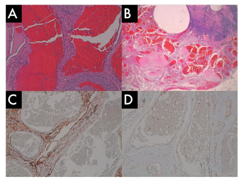
Figure 1: (A) The tumor is composed of numerous blood vessels, some of which contain red blood cells in the dilated lumens. (Hematoxylin & Eosin; magnification 20X); (B) The mass is well defined but not encapsulated with numerous, dilated vascular spaces adjacent to remnant ovarian tissue on the left side, vascular spaces were lined by flattened endothelial cells without cytologic atypia (H&E; magnification 4X); (C-D) IHC: CD34 and CD31 slides showing positive vascular endothelial cells (C: CD34, magnification 20X; D: CD31, magnification 20X).
3. Discussion
Ovarian hemangioma is a benign and very rare neo-formation often small and asymptomatic usually unilateral lesion, although bilateral ovarian hemangioma have been reported [5, 6]. When this kind of neo-formation is larger, it could appear with abdominal pain derived from the torsion of the ovary. The main risk factor for ovarian torsion is not the ovarian hemangioma itself but the dimension of the ovarian mass (diameter of > 5 cm). In fact, a large ovarian mass increases the possibility that the ovary could rotate on the axis reducing venous outflow and arterial inflow [7]. Some authors described an association between ovarian hemangioma, massive ascites and elevated CA-125 marker [8] or non-ovarian tumors (cervical carcinoma, endometrial or tubal carcinoma) [9]. Ovarian hemangioma have been considered hematoma-like malformations or neoplasm by itself, generally located in the medulla and hilar regions with a controversial etiology [1, 10]. Pregnancy, hormonal stimulation and infections have been considered potential important factors for its growth [11]. In the case reported, the patient presented to the hospital with acute abdominal pain and the hemangioma was discovered incidentally during histopathological examination. Similar to other cases in literature, abdominal pain was due to ovarian torsion. The precise site of the hemangioma could not be localized because the large tumor (about 8 cm) and consequent ovarian torsion leaded to ovarian architecture destruction. Contralateral ovary appeared normal and this was confirmed by microscopical examination. We did not find ascites, and serum tumor markers were normal.
In conclusion, ovarian hemangioma may manifest itself as an ovarian mass with a sudden increase. Clinicians should consider this rare event in their diagnostic work-up.
Conflict of Interest Statement
The authors have no personal, financial, or institutional interest concerning the authorship and/or publication of this manuscript.
References
- Kim SS, Han SE, Lee NK, et al. Ovarian Cavernous Hemangioma Presenting as a Large Growing Mass in a Postmenopausal Woman: A Case Report and Review of the Literature. Journal of Menopausal Medicine 21 (2015): 155-159.
- Dundr P, Nemejcová K, Laco J, et al. Anastomosing Hemangioma of the Ovary: A Clinicopathological Study of Six Cases with Stromal Luteinization. Pathol Oncol Res 23 (2017): 717-722.
- Ziari K, Alizadeh K. Iran Ovarian Hemangioma: a Rare Case Report and Review of the Literature. J Pathol 11 (2016): 61-65.
- Lawhead RA, Copeland LJ, Edwards CL. Bilateral ovarian hemangiomas associated with diffuse abdominopelvic hemangiomatosis.. Obstet Gynecol 65 (1985): 597-599.
- Uppal S, Heller DS, Majmudar B. Ovarian hemangioma--report of three cases and review of the literature. Arch Gynecol Obstet 270 (2004): 1-5.
- Talerman A. Hemangiomas of the ovary and the uterine cervix. Obstetrics & Gynecology 30 (1967): 108-113.
- Varras M, Tsikini A, Polyzos D, et al. Uterine adnexal torsion: pathologic and gray-scale ultrasonographic findings. Clin Exp Obstet Gynecol 31 (2004): 34-38.
- Gehrig PA, Fowler WC, Jr, Lininger RA. Ovarian capillary hemangioma presenting as an adnexal mass with massive ascites and elevated CA-125. Gynecol Oncol 76 (2000): 130-132.
- Miliaras D, Papaemmanouil S, Blatzas G. Ovarian capillary hemangioma and stromal luteinization: a case study with hormonal receptor evaluation. Eur J Gynaecol Oncol 22 (2001): 369-371.
- DiOrio J, Lowe LC. Hemangioma of the ovary in pregnancy: a case report. J Reprod Med 24 (1980): 232-234.
- Katayoun Z, Kamyab A. Ovarian Hemangioma: a Rare Case Report and Review of the Literature. Iran J Pathol 11 (2016): 61-65.