Chandra Bhushan Tiwary*
Assistant Professor, Department of Zoology, Munshi Singh PG College, Motihari (Bihar), India
Corresponding Author: Chandra Bhushan Tiwary, Assistant Professor, Department of Zoology, Munshi Singh PG College, Motihari (Bihar), India.
Received: 11 May 2026; Accepted: 18 May 2026; Published: 11 August 2026
DOI: 10.26502/fjwhd.2644-288400153
ShareAndrogens are essential sex steroid hormones for both sexes. Women synthesize a greater quantity of androgens compared to estrogens which presence in peripheral system is related with menstrual cycle and gradually in order of decreasing serum concentrations. Androgen precursors are derived in the adrenal cortex and ovaries may converted into estrogens but testosterone formation held in peripheral system. Testosterone also produced in steroidogenic organs through a series of enzymatic reactions. The classic action of androgens on target or¬gans is mediated through the androgen receptor, which regulates nuclear receptor gene transcription. However, the androgen receptor complex may also interact directly with membrane proteins or signaling molecules to exert more rapid effects. The androgen is secreted by ovaries, and DHT is the main peripheral product of testosterone in postmenopausal women. These hormones influence the ovaries, endometrium, vagina, and vulva to maintain organized reproductive system and provide regulation and protection to various body parts and functions. However, the androgen effects can become destructive when present in excess than normal content. This review presents the current concepts of androgen synthesis and physiology.
Androgens; Dehydroepiandrosterone sulfate (DHEAS); Dehydroepiandrosterone (DHEA); Androstenedione; Testosterone; Dihydrotestosterone; Menopause; Ttestosterone therapy
Androgens articles; Dehydroepiandrosterone sulfate (DHEAS) articles; Dehydroepiandrosterone (DHEA) articles; Androstenedione articles; Testosterone articles; Dihydrotestosterone articles; Menopause articles; Ttestosterone therapy articles
Introduction
Synthesis and physiology, and correlates these effects to respective women congenital Androgens are steroid hormones that are essential for human sexual development and reproduction, but they also modulate other organs, including bone, muscle, adipose tissue, skin, hair, the brain and the cardiovascular system, thereby affecting growth, body shape, and human behavior.Androgens are produced in the adult female ovaries and in the adrenal glands, from where they are secreted into circulation to exert their biological effects on target organs. In addition, secreted androgens serve as substrates for peripheral organs for intermediate steroid metabolism and its action. Androgens also serve as precursors for estrogen biosynthesis in the ovaries and peripheral tissues. Ultimately, the androgens biosynthesized in the adrenals and gonads are metabolized in the liver and then excreted in urine [1].The main androgens based upon their presence in serum also consist of pro-androgens as dehydro-epiandrosterone sulphate (DHEAS), dehydro-epiandrosterone (DHEA) and androstenedione (A) which provide effects only after conversion to testosterone (T), whereas testosterone and dihydrotestosterone (DHT) directly acts upon body metabolism, and Androgen biosynthesis performs in both the adrenal gland and ovary and is modified by mitochondrial enzymes that facilitate side-chain separation through 17-hydroxylation with 17-20 bond breakage in cholestrol required for 17-C hormones such as DHEA and androsterone [2].
The testosterone content are 10 to 15-fold lower in a 30 year female than in a same-aged male [3,4] and is further reduced due to slow-steroid formation in post-menopausal women. The hypothalamus-pituitary-gonadal (HPG) axis, luteinizing hormone (LH) and follicular-stimulating hormone (FSH) control over androgen synthesis in the adult ovary, and consist of balanced positive and negative feedback loops. The adrenocorticotropic hormone (ACTH) still co-regulates androgen production by the adrenal gland in adult female [5]. Testosterone in the circulation present in free form or bound to albumin and the inactive one bound to sex hormone-binding globulin (SHBG). The SHBG protein produced by the liver and showed a high level association with sex steroids [6]. The SHBG exhibits primarily as a transport protein, however, it also serves as a metabolic marker in the evaluation of polycystic ovary syndrome (PCOS) due to its direct association with excess insulin, diabetes, and metabolic syndrome [7]. Low SHBG levels in postmenopausal women have been associated with adverse lipid profiles, visceral fat deposit, and an elevated risk of diabetes [8]. In addition, low SHBG levels cause fatty liver disease, especially when induced by dietary fructose. Androgens are activated when binds to androgen receptor (AR), and the complex reaches the nucleus to work as a transcription factor. This review aims to provide an update on current advancements in androgenand metabolic disturbances with close insights upon therapeutic approaches of androgens in women.
Androgen biosynthetic Pathways
The cholesterol is prime metabolite to biosynthesize primary androgen within the ovaries of females, and the brain can synthesize DHEA, whilst peripheral tissues, such as the skin and adipose tissues, also participate in androgen production through specific alterations of steroid intermediates into different androgenic molecules [9, 10, 11]. The partial synthesis of androgen is also confined to three distinct regions of adrenal cortex, where 11-keto-androgens (KTs) are produced mainly in the middle zona fasciculata and inner zona reticularis layers [12]. However, metabolic inter-conversion in adipose tissue, the brain, and several other peripheral sites underscore the dynamic and tissue-specific regulation of steroid hormone activity in human physiology.
The classic pathway of androgen biosynthesis in the adrenal cortex and ovaries in women is initiated by steroidogenic acute regulatory (StAR) protein, which transports cholesterol into the mitochondria [13]. The next step is held within the inner mitochondrial membrane by cytochrome P450 enzyme that mediates gradually catalyzes 20α-hydroxylation, 22-hydroxylation, and side-chain cleavage of cholesterol to generate pregnenolone as rate-limiting step. This mitochondrial enzyme is encoded by the CYP11A1 gene located on the chromosome and its catalytic activity involves the NADPH-mediated electron transport system. In the adrenal cortex, both 17α-hydroxylase and 17, 20-lyase P450 are expressed, which enables DHEA production and ultimately changes into testosterone [14]. However, 17-hydroxyprogesterone also produces testosterone to a lesser extent, and, DHEA is also converted into androstenedione by the 3β-hydroxysteroid dehydrogenase enzyme [15].
The alternative biosynthesis pathway is governed by the combined activity of several key enzymes on progesterone and 17-hydroxyprogesterone as substrates, in which 5α-reductase type-1 (SRD5A1) is expressed in the human fetal ovary. This enzymatic step is important for conveying steroid intermediates toward alternative mode of androgen biosynthesis.
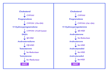
Figure 1:Classic and alternative pathways of Androgen biosynthesis.
There 17α-hydroxylase activity of the critical P450 enzyme has been reported upon dihydroprogesterone and allopregnanolone substrates, whereas 17, 20-lyase venture has been also cited on 17-hydroxyallopregnanolone as the most accurate precursor [16]. These reactions highlight the versatile role of P450c17 in both androgen biosynthetic routes. In addition, accessory enzymes such as AKR1C2/4, 17β-hydroxysteroid dehydrogenase type 5 (17βHSD5 or AKR1C3), and 17β-hydroxysteroid dehydrogenase type 6 (17βHSD6), which are also known as retinol dehydrogenase (RoDH), are key enzymes that promote the gradual conversion of required intermediates for testosterone-independent dihydrotestosterone synthesis as a mandatory precursor [17]. The alternative route of androgen synthesis can activate congenital adrenal hyperplasia (CAH), or polycystic ovary syndrome (PCOS), inducing excess androgens and virilization in female newborns [18, 19]. The relative role of P450, rather than 5α-reductase type 1, is especially important in determines whether progesterone and 17-hydroxyprogesterone are directed toward the classic or alternative biosynthetic pathways.
Cell reception of Androgen
Androgen mainly works after binding with androgen receptor that exposes the appearance of several genes by target cells and its signal modulation. The AR is 3-ketosteroid receptors closely related to the progesterone, glucocorticoid and aldosterone receptors [20]. The diverse action of androgen in XX-female is associated with its location at X-chromosome.
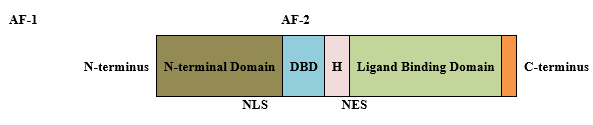
Figure 2:Domains of the Androgen Receptor (AR).
The AR protein is composed by three primary functional domains as the N-terminal transcription regulator domain, the DNA-binding domain (DBD) and the ligand-binding domain (LBD). The most variable region is the N-terminal, whereas the DBD present as the most highly conserved region that contains two zinc fingers to recognize specific DNA sequences. The DBD is joined with LBD by a hinge region, where LBD facilitate the interaction between heat shock and chaperone proteins with the AR beside interact to N-terminal to stabilize bound androgens [21].
There AF-1 along the N-terminal is essential to maximize AR performance, whereas the ligand-subsidized AF-2 provides co-regulator binding site in the nucleus and N/C interactions [22-24]. The nucleus localization signal (NLS) imports receptor into the nucleus, whilst NES exporting the AR to the cytoplasm upon ligand withdrawal. Androgens diffuse across the plasma membrane and binds to cytoplasmic AR linked to heat-shock protein (HSPs) and molecular chaperons, resulting in a confirmation change, dissociation of chaperone proteins and exposure of the NLS [25]. The androgen/AR-complex enters into the nucleus where dimerize and binds to androgen response elements (AREs) within classical target genes at promoter region to modulate gene transcription [26]. The action of co-regulators (specific proteins) modulates transcriptional activity of the androgen linked to AR through chromatin remodeling and histone modifications, and also recruits transcriptional co-activators such as SRC-1 and CBP/p300 regulating gene expression [27].
Androgen effect on Metabolism
Androgens modulating the differentiation of adipose stem cells into mature adipocytes as evidenced by differential distribution of adipose tissues in male and female body. The androgen affects on early aspects of human SC abdominal adipogenesis could alter the numbers of adipocytes in this adipose depot and thereby affect its capacity to safely store fat in women with androgen excess. In females, testosterone also reduces lipolysis by preventing adipose-sensitive lipases, where visceral obesity is linked to insulin resistance resulting in excess insulin as similar to insulin-like growth factor- 1 [28]. Testosterone in physiological concentrations causes a depot-specific reduction of catecholamine-stimulated lipolysis in subcutaneous fat cells, probably due to reduced protein expression of beta (2)-adrenoceptors and hormone-sensitive lipase. This could be an important pathogenic factor underlying regional differences in lipolysis and development of insulin resistance and hyperandrogenic polycystic ovary syndrome [29].
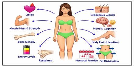
Figure 3:Androgens impact on menstrual and menopausal women.
These situations gradually raise androgen levels in ovary and lower the SHBG synthesis by the liver. Then elevated androgen contents and sustains a cycle of polycysticovary syndrome (PCOS) and metabolic syndrome. Also, similar event may occur in post-menopause females when testosterone extents rise due to declined estrogen and SHBG [30].
Neuro-protective and Cognition-Regulating Effects
The brain similar to other body organs is influenced by the declined ovarian harmones, extremely in menopausal women because estrogen and testosterone both present anti-inflammatory and neuron-protective features within the brain and subscribe regulation of cognition and mood and temper. The considerable cognitive changes exhibits in some menopause women and particularly younger women who have passed through surgical removal of ovary experiences major hormonal imbalance that impact their quality of life, especially younger women who have face surgical removal of ovary [31]. Androgen receptors (ARs) are well dispersed all over the central nervous system and are involved in functions like as sexual desire, temperature regulation, sleep, visual-spatial potential, and language. Moreover, testosterone works to eliminate oxidative stress, prevent abnormal formation of beta proteins, and collaborate as its critical role against degenerative neurological conditions such as Alzheimer's disease [32].
The testosterone dose in postmenopausal women evidenced experimental progress in verbal learning and cognition as postmenopausal women who applied transdermic 300 µg/day of testosterone gel for 26 weeks exhibited significant improvements in verbal learning and memory functions, however, minor effects on overall well-being were observed [33]. The cognitive benefits of testosterone in postmenopausal women appear to be independent of its conversion to estradiol. This study revealed no major changes in cognitive function among patients [34].
Androgen mediated Bone health
The bone metabolism has major impact of androgen and estrogen receptors as osteoclast and osteoblast advances by them, where activated AR induces osteoclast growth and blocks osteoclast venture [35, 36]. Similarly, active estrogen receptor also quashes osteoclast proliferation and promote apoptosis to declined bone re-absorption. The past research reported more estrogen associated mechanism in females as supported by low amount of free testosterone in their late reproductive phase rapidly decrease bone density [37]. Another research denoted that older women with lower free intrinsic testosterone exhibited reduced bone densities in the lumbar and hip regions [38]. The observational study on the women’s health revealed that internal biosynthetic testosterone are linked with low risk of hip fractures, even not dependent on estradiol and sex hormone-binding globulin (SHBG) contents. The past researches have focused on internal testosterone concentration relation with bone health, however, available investigations about the exogenous testosterone on bone mineral density has inconsistent and contradictory results [39, 40]. As a result, androgen-based therapies are not presently considered a key outlook add to bone density.
Impact on Skin and Hair
While the adrenal glands and ovaries are the primary sources of androgen production, the skin also functions as a site for dihydrotestosterone (DHT) synthesis with participation of androgen receptors (ARs) that are located in sebocytes, dermal papilla cells, root sheaths of hair follicle, sweat glands, endothelial mural cells, and both epidermal and follicular keratinocytes [41]. The ARs induce cellular growth and sebum production within sebaceous glands after activation, however, in persons with a genetic disposition contributes to the anagen phase of hair follicles lastly resulting in hair loss. The androgen activity in other sites of body enables vellus hair transition into terminal hairs [42]. Although ARs are involved in the pathogenesis of blemishes, but only in low secretion condition in acute acne exhibited high androgen level as clinical features. The hirsutism is mainly related to increased androgen activity, with most affected women demonstrating elevated androgen levels [43].
Androgen upon muscular activity
It is widely accepted that androgens play a key role in buildup muscle mass and upgrade output in both males and females. Many sportsperson, globally utilize testosterone derivatives to increase their athletic performance, and, also athletes with high androgen levels even due to polycystic ovary syndrome (PCOS) have better muscle mass providing competitive advantage [44, 45]. The androgen products may be beneficial in women after menopause to increase muscle density for athletic performance and also can be used in the treatment of neuromuscular disturbances [46-48].
Androgens and Reproductive Health
Ovarian Function
Androgens in the ovary are vital for normal follicle growth, serving as the essential chemical building blocks for estrogen production and improving how follicles respond to growth signals. The mRNA and proteins of Androgen receptor (AR) in the females are expressed by overall hypothalamic-pituitary-gonadal (HPG) axis, which including the brain, ovarian stroma, ovarian follicles, and corpora lutea [49]. The most stages of ovarian follicular growth presents differential spatial and temporal patterns in both fetal and adult stages, which suggesting unique participation of AR-facilitated activities during follicular development [50, 51]. The androgen receptors present in the overall cells of pre-antral follicles, whereas gradually diminishes in the outer mural granulosa cells in antral stage, whilst remaining pronounced in the cumulus cells [52, 53]. The androgens have been identified in primate ovarian granulosa cells of developing follicles, suggesting that, in addition to de novo synthesis, phosphorylation of AR may regulate receptor levels and function [54]. Furthermore, the evolutionary conservation of ovarian AR expression across human supports a universal role for AR-mediated androgen actions in ovarian function [55].
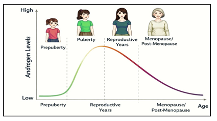
Figure 4:Androgen content in stages of women life.
The distinct role of androgens in ovarian function is contingent upon AR knockout animal models, which explained the energizing early follicle growth and their gradual use in inter-follicular communication to carry on follicle health and later on induce egg development [56].The recent use of pro-androgen or aromatase inhibitor advised to hyperandrogenic females in fertilization clinics, however, positive effects of androgen on follicular development are really only possible within an perfect concentration limit [57]. The concentration doze above optimum level may unexpectedly problems start to exhibit clinical symptoms comparable to polycystic ovary syndrome (PCOS), which evidenced by PCOS growth by response of altered androgen receptor signaling pathway [58].
Endometrial Effect
Androgens can provoke both controlling and restorative physiology in the endometrium and also exhibits pathological processes through alteration in AR-based pathways and estrogen aromatization [59]. The expression of androgen receptors significantly changes during the menstrual cycle, as enhancement in epithelial cells during the growth period and declined in the secretory phase, and, it is considered that ARs facilitate repair and durability in endometrial tissue [60]. In later reproductive phase of women, stimulation of endometrial stromal fibroblasts by DHEA is responsible to decidualization, which provides positive effect on fertility. [61]. The DHEA has also been evidenced to increase endometrial receptivity by antioxidant effects on the endometrial stroma in animal models. [62, 63].
The recent research does not strongly evidences that testosterone treatment can initiate endometrial cancer, however, long-period testosterone therapy; atrophic changes are more observed than prolonged progesterone users. In another study, higher pre-diagnostic total and free testosterone level were associated with endometrial malignancy, whereas circulating androgens as androstenedione and DHEAS were showed any risk of cancer. [64].
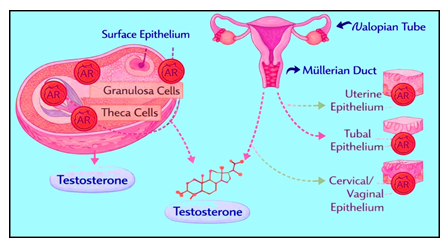
Figure 5:Androgen receptor sites in reproductive organs (@Bio Render).
Vulvo-Vaginal Effects
The estrogen use increases outer epithelial cells in vulvo-vaginal tissues and reduces vaginal pH are well established, and this process underpins in the genitourinary problems of menopause (GSM) females. The recent researches in both human and animal tissues have evidenced ARs in the all vaginal layers as vulva, labium and clitoris. [65,66].
The working of androgen receptors can be illustrated by rise and fall regulatory mechanisms as evidenced by about 1000 receptor mutations mostly identified in the females with androgen insensitive females or trans-genders. The androgen doze suggested improvements on vulvo-vaginal tissues, as an outcome by increase of vaginal weight in experimented mice. [67] However, despite these findings, androgens have not been incorporated into routine clinical practice for the treatment of vulvo-vaginal problems. Moreover, similar to estrogen, there remains no clear concept about the benefits and risks associated with either systemic or local androgen therapy. Furthermore, there is no definitive consensus regarding the benefits and risks associated with the systemic or local application of androgens, similar to estrogens [68].
Androgen and Women health
Genitourinary Syndrome of Menopause
Up to 70% of women who have gone through menopause experience GSM, a condition previously referred to as vulvo-vaginal atrophy. It encompasses urinary, genital, andsexual dysfunctions resulting from declining sex hormone levels [69].The clinical presentation typically encompasses dyspareunia, vaginal dryness, irritation, dysuria, increased urinary frequency and urgency, recurrent urinary tract infections, and a shift towards alkalinity in vaginal pH.However, research on menopausal women utilizing androgen products has identified sexual disturbances resulted from decreased sex hormone levels, whereas hormonal formulations, especially vaginal estrogen and dehydroepiandrosterone (DHEA), are considered as the most effective treatments for genitourinary syndrome of menopause (GSM) [70,71]. The Food and Drug Administration has approved intra-vaginal 6.5mg DHEA for GSM treatment, which has revealed refinements in cell maturation, vaginal pH, dyspareunia, and sexual function without disturbing the endometrium [72].
Hypoactive Sexual Desire Disorder
The variation in women’s sexual behavior is fashioned by diverse factors of biological and psychological components. The twist of androgens with their biosynthesis and reception makes it strenuous to efficient measuring in laboratory conditions. Nevertheless, it is widely recognized that androgens are vital in managing women's sexual function. Testosterone and its progenitors exercise a key regulation on sexual function, desire, and arousal, evidenced by past research as a linkage between testosterone levels in blood and sexual function in both young and old women populations [73]. Even though hypoactive sexual desire disorder (HSDD) remains a subject of debate in certain clinical and academic contexts, testosterone therapy in specific communities must emerging as key point in perspective studies. The exact method is short-term trans-dermal therapy in low concentration as recommended by clinical guidelines as a six-month trial of such testosterone for women diagnosed with HSDD [74]. In clinical practice, careful dosing is essential; typically, women should receive no more than one-tenth of the standard male dose in order to avoid supra-physiological exposure because testosterone products has adverse effect on women’s health. Topical preparations are generally applied to areas such as the inner thigh, buttocks, abdomen, or vulvar region, while application to the breasts and arms is discouraged. Oral testosterone is not recommended due to first-pass hepatic metabolism and its associated systemic effects. Similarly, intramuscular injections and dermal pellet therapies are suspected because of their potential to produce long-term exposure and high hormone levels [75]. Although short-term use appears to be both safe and effective, there remains a clear need for further long-term studies to establish detailed safety and efficacy profiles. Testosterone treatment is applicable with initial content, improved content after 3-6 weeks and total content analysis after 2 years are needful to control overdose for safety objectives. The treatment must be continued only with efficient sexual function, less distress and normal weight and lagged only after 6 months under non-effective condition. The side-effects are always mild when treatment level remains within normal limits [76, 77].
Androgens and Breast Cancer
Breast cancer is the most frequent tumor affecting women worldwide. In breast cancer tissues may hold either positive or negative androgen receptors (ARs), irrespective of the position of estrogen receptors (ERs) [78]. The women with positive ARs have display boosted treatment impact and extended survival periods, however, role of androgen hormones are unclear and past researches revealed contradictory results but currently selective AR modulators are applied for the diagnosis of specific breast cancer subtypes [79]. The testosterone is not suitable for women with a history of breast cancer due to the risk of its conversion to estrogen [80].
Emerging Research and Future Directions
The future research within the reproductive systems in women is needful about exploration of androgens role in ovarian reserve, receptor polymorphisms, and reproductive processes from conception to sexual behavior. The studies related to cancer especially concerning the endometrium and breast, also about their impact on local pain perception. More researches are essential about effects of excessive androgen and reproductive metabolic syndromes. The study about effects of androgen after post-gender will be probably relevant to both transgender and non-transgender individuals
Conclusion
The androgens in women are synthesized by the adrenal glands, ovaries and peripheral system from cholesterol, where intravenous testosterone doze elevates serum levels, responds LH surge and the sensitivity of gonadotropin-releasing hormone. However, it remains uncertain whether reducing androgens content in serum would result in higher gonadotropins in circulation as cited in post-menopause period when ovaries mainly secreting androgens in lesser function of pituitary gland. Androgens are significant about women's reproductive and overall health as affecting ovarian cycle, endometrial repair, bone health, nerve protection and metabolic functions. The disturbances in androgen contents can lead to hyperandrogenic condition, as observed in PCOS, cause metabolic and reproductive problems, while deficiency is linked to conditions such as HSDD and GSM. Their systemic effects are modulated by interactions with estrogen and SHBG, affecting cardiovascular, cognitive, and musculoskeletal health. A significant challenge in effective androgen therapy is the lack of reliable biomarkers to assess tissue-level androgen activity. Current treatments aim to maintain total testosterone within normal physiological ranges, but there is insufficient evidence to justify initiating therapy based solely on low levels. HSDD is the only recognized indication for androgen replacement, underscoring the urgent need for standardized measures and long-term safety data.
References
- Olson P, Ristau BT. Review of adrenal androgen synthesis, hypersecretion and blockade. Urol Clin North Am 52 (2025):217-227.
- Endoh A, Kristiansen SB, Casson PR, et al. The zona reticularis is the site of biosynthesis of dehydroepiandrosterone and dehydroepiandrosterone sulfate in the adult human adrenal cortex resulting from its low expression of 3-beta-hydroxysteroid J Clin Endocrinol Metab. 81(1996): 3558-3565.
- Santoro N, Braunstein GD, Butts CL, Compounded bioidentical hormones in endocrinology practice: an endocrine society scientific statement. J Clin Endocrinol Metab 101(2016):1318-1343.
- Rothman MS, Carlson NE, Xu M, et al. Re-examination of testosterone, dihydrotestosterone, estradiol and estrone levels across the menstrual cycle and in postmenopausal women measured by liquid chromatography-tandem mass spectrometry. Steroids. 76 (2011): 177-182.
- Miller WL, Fluck CE, Breault DT. Chapter 14-The adrenal cortex and its disorders. In: Sperling MA, Editor. Sperling pediatric Endocrinology (5th edition). Philadelphia: Elsevier, (2021): 425.
- Peter A, Kantartzis K, Machann J. Relationships of circulating sex hormone-binding globulin with metabolic traits in humans. Diabetes 59 (2010): 3167-3173.
- Montalcini T, Migliaccio V, Ferro Y, et al. Androgens for postmenopausal women’s health? Endocrine 42 (2012): 514-
- Ding EL, Song Y, Manson JE.Sex hormone-binding globulin and risk of type-2 diabetes in women and men. N Engl J Med 361 (2009): 1152-1163.
- Zwain IH, Yen SSC. Dehydroepiandrosterone: Biosynthesis and metabolism in the brain. Endocrinology140 (1999): 880-887.
- Blouin K, Veilleux A, LuuThe V, et al.Androgen metabolism in adipose tissue: Recent advances. Mol.Cell.Endocrinol301 (2009): 97-103.
- Karbowska J, Kochan Z. Effect of DHEA on endocrine functions of adipose tissue, the involvement of PPARγ. BiochemPharmacol 70 (2005): 249-257.
- Miller WL, Anchus RJ. The molecular biolog, biochemistry and physiology of human steroidogenesis and its disorders. Endocr Rev 32 (2011): 81-151.
- Turcu AF, Auchus RJ. Clinical significance of 11-oxygenated androgens. Curr Opin Endocrinol Diabetes Obes24 (2017): 252-259.
- Black SM, Harikrishna JA, Szklarz GD, et al. The mitochondrial environment is required for activity of the cholesterol side-chain cleavage enzyme, cytochrome P450cc. Proc Natl Acad Sci USA 91 (1994) 7247-7261.
- Miller WL. Mini review: regulation of steroidogenesis by electron transfer. Endocrinology 146 (2005): 2544-2550.
- Miller WL. Disorders of androgen synthesis: From cholesterol to dehydroepiandrosterone. Med Princ Pract 14S11 (2005): 58-68.
- Gupta MK, Guryev OL, Anchus RJ. 5α-reduced C21 steroids are substrates for human cytochrome P450c17. Arch BiochemBiophy 418 (2003): 151-160.
- Krone N, Reisch N, Idkowiak J, et al.Genotype-phenotype analysis in congenital adrenal hyperplasia due to P450 oxidoreductase deficiency. J Clin Endocrinol Metab 97 (2012): E257-267.
- Rana K, Davey RA, Zajac JD. Human androgen deficiency: insights gained from androgen receptor knockout mouse models. Asian J Androl 16 (2014): 169-1
- Tan MH, Li J, Xu HE, et al. Androgen receptor: structure, role in prostate cancer and drug discovery. Acta Pharmacol Sin 36 (2015): 3-23.
- MacLean HE, Warne GL, Zajac JD. Localization of functional domains in the androgen receptor. J Steroid Biochem Mol Biol 62 (1997): 233-242.
- Heinlein CA, Chang C. Androgen receptor (AR) coregulators: an overview. Endocr Rev 23 (2002): 175-
- Callewaert L, Van Tilborgh N, Claessens F. Interplay between two hormone-independent activation domains in the androgen receptor. Cancer Res 66 (2006): 543-553.
- Slagsvold T, Kraus I, Bentzen T, et al. Mutational analysis of the androgen receptor AF-2 (activation function 2) core domain reveals functional and mechanistic differences of conserved residues compared with other nuclear receptors. Mol Endocrinol 14 (2000): 1603-1617.
- Wilson EM. Analysis of inter-domain interactions of the androgen receptor. Methods Mol Biol 776 (2011): 113-129.
- Eder IE, Culig Z, Putz T, et al. Molecular biology of the androgen receptor: from molecular understanding to the clinic. EurUrol 40 (2001): 241-251.
- Bevan C, Parker M. The role of co-activators in steroid hormone action. Exp Cell Res 253 (1999): 349-356.
- Dicker A, Rydén M, Näslund E, Muehlen IE, Wirén M, Lafontan M, Arner P. Effect of testosterone on lipolysis in human pre-adipocytes from different fat depots. Diabetologia 47 (2004) :420-428.
- Iwasa T, Noguchi H, Tanano R, et al. Yamanaka E, Takeda A, Tamura K, Aoki H, Sugimoto T, Sasada H, Kaji T. Age-dependent changes in the effects of androgens on female metabolic and body weight regulation systems in humans and laboratory animals. Int J Mol Sci 24 (2023): 16567.
- Mielke MM, Kapoor E, Geske JR, et al. Fields JA, Brasseur NK, Monrow MM, Winham SJ, Faubion LL, Castillo AM, Hofrenning E, Bailey KR, Rocca WA, Kantarki K. Long-term effects of premenopausal bilateral oophorectomy with or without hysterectomy on physical aging and chronic medical conditions. Menopause 30 (2023): 1090-1097.
- Rosario ER, Chang L, Head EH, et al. Stanczyk FZ, Pike CJ. Brain levels of sex steroid hormones in men and women during normal aging and in Alzheimer’s disease. Neuro biol Aging 32 (2011): 604-613.
- Pike CJ, Carroll JC, Rosario ER, et al.. Protective actions of sex-steroid hormones in Alzheimer’s disease. Front Neuroendocrinol 30 (2009): 239-258.
- Aleman A, Bronk E, Kessels RPC, et al. A single administration of testosterone improves visuo-spatial ability in young women. Psycho neuroendocrinology 29 (2004): 612-617.
- Huang G, Wharton W, Travison TG, Effects of testosterone administration on cognitive function in hysterectomized women with low testosterone levels: a dose-response randomized trial. J Endocrinol Invest 38 (2015): 455-461.
- Zhang H, Ma K, Li RM, et al. Association between testosterone levels and bone mineral density in females aged 40-60 years from NHANES 2011-2016. Sci Rep 12 (2022): 16426.
- Lee JS, La Croix AZ, Wu L, Associations of serum sex hormone-binding globulin and sex hormone concentrations with hip fracture risk in postmenopausal women. J Clin Endocrinol Metab 93 (2008):1796-1803.
- Zhang H, Ma K, Li RM, et al. Association between testosterone levels and bone mineral density in females aged 40-60 years from NHANES 2011-2016. Sci Rep 12 (2022): 16426.
- Vanderschueren D, Vandenput L, Boonen S, et al. Androgens and bone. Endocr Rev 25 (2004): 389-
- Slemenda C, Longcope C, Peacock M, et al. Hui S, Johnston CC. Sex steroids, bone mass, and bone loss. A prospective study of pre-, peri-, and post menopausal women. J Clin Invest 97(1996): 14-21.
- Chen JF, Lin PW, Tsai YR, et al. Androgens and androgen receptor actions on bone health and disease: from androgen deficiency to androgen therapy. Cells 8 (2019): 1318.
- Lai JJ, Chang P, Lai KP, et al.The role of androgen and androgen receptor in skin related disorders. Arch Dermatol Res 304 (2012): 499-510.
- Ceruti JM, Leirós GJ, Balañá ME. Androgens and androgen-receptor action in skin and hair follicles. Mol Cell Endocrinol 465 (2018): 122-
- Bienenfeld A, Azarchi S, Lo Sicco K et al. Androgens in women: androgen-mediated skin disease and patient evaluation. J Am Acad Dermatol 80 (2019): 1497-1506.
- Bermon S. Androgens and athletic performance of elite female athletes. Curr Opin Endocrinol Diabetes Obes 24 (2017): 246-251.
- Bermon S, Garnier PY, Hirschberg AL. Serum androgen levels in elite female athletes. J Clin Endocrinol Metab 99 (2014): 4328-
- Sheffield-Moore M, Paddon-Jones D, Casperson SL. Androgen therapy induces muscle protein anabolism in older women. J Clin Endocrinol Metab 91 (2006): 3844-3849.
- Huang G, Basaria S, Travison TG, Testosterone dose-response relationships in hysterectomized women with or without oophorectomy: effects on sexual function, body composition, muscle performance and physical function in a randomized trial. Menopause 21(2014):612-623.
- Rutkove SB, Parker RA, Nardin RA, et al. A pilot randomized trial of oxandrolone in inclusion body myositis. Neurology 58 (2002): 1081-1087.
- Walters KA, Handelsman DJ. Role of androgens in the ovary. Mol Cell Endocrinol 465 (2018): 36-47.
- Wilson CM, McPhaul MJ. 1996 A and B forms of the androgen receptor are expressed in a variety of human tissues. Molecular and Cellular Endocrinology 120 (1996):51-57.
- Suzuki T, Sasano H, Kimura N, et al. Immunohistochemical distribution of progesterone, androgen and oestrogen receptors in the human ovary during the menstrual cycle: relationship to expression of steroidogenic enzymes. Human Reproduction 9 (1994):1589-1595.
- Szoltys M, Slomczynska M. Changes in distribution of androgen receptor during maturation of rat ovarian follicles. Experimental and Clinical Endocrinology and Diabetes 108 (2000):228-234.
- Lenie S, Smitz J 2009 Functional AR signaling is evident in an in vitro mouse follicle culture bioassay that encompasses most stages of folliculogenesis. Biology of Reproduction 80(2009): 685-695.
- McEwan IJ, Mcguinness D, Hay CW, et al. Identification of androgen receptor phosphorylation inthe primate ovary in vivo. Reproduction 140 (2010): 93-104.
- Nielsen ME, Rasmussen IA, Kristensen SG, et al. In human granulosa cells from small antral follicles, androgen receptor mRNA and androgen levels in follicular fluid correlate with FSH receptor mRNA. Molecular Human Reproduction17(2011):63-70.
- Smith T, Batur P. Prescribing testosterone and DHEA: the role of androgens in women. Cleve Clin J Med 88 (2021): 35-43.
- Alexander SPH, Cidlowski JA, Kelly E, et al. Concise guide to pharmacology. 2019/20: Nuclear hormone receptors. Br J Pharmacol 176 (2019): S229-S246.
- Bianchi VE, Bresciani E, Meanti R, et al. Role of androgens in women’s health and wellbeing. Pharmacol Res 171 (2021): 105758.
- Simitsidellis I, Saunders PTK, Gibson DA. Androgens and endometrium: new insights and new targets. Mol Cell Endocrinol 465 (2018): 48-60.
- Evans SF, Hull ML, Hutchinson MR, et al. Androgens, endometriosis and pain. Frontiers in Reproductive Health 3 (2021): 792920.
- Wongwananuruk T, Sato T, Kajihara T, et al. Endometrial androgen signaling and decidualization regulate trophoblast expansion and invasion in co-culture: a time-lapse study. Placenta 47 (2016): 56-62.
- Qin A, Qin J, Jin Y. DHEA improves the antioxidant capacity of endometrial stromal cells and improves endometrium receptivity via androgen receptor. Eur J Obstet GynecolReprod Biol 198 (2016): 120-126.
- Young SL. Androgens and endometrium: new lessons from the corpus luteum via the adrenal cortex? Fertil Steril 109 (2018): 623-
- Allen NE, Key TJ, Dossus L, Endogenous sex hormones and endometrial cancer risk in women in the European Prospective Investigation into Cancer and Nutrition (EPIC). EndocrRelat Cancer 15 (2008): 485-497.
- Palacios S. The expression of androgen receptors in the structures of vulvo-vaginal tissue. Menopause 27 (2020): 1336-1342.
- Labrie F, Martel C, Pelletier G. Is vulvovaginal atrophy due to a lack of both estrogens and androgens? Menopause 24 (2017): 452-461.
- Traish AM, Kim SW, Stankovic M, et al. Testosterone increases blood flow and expression of androgen and estrogen receptors in the rat vagina. J Sex Med 4 (2007): 609-619.
- British Menopause Society: Tool for Clinicians.
- Traish AM, Vignozzi L, Simon JA, et al. Role of androgens in female genitourinary tissue structure and function: implications in the genitourinary syndrome of menopause. Sex Med Rev 6 (2018): 558-571.
- Scheffers CS, Armstrong S, Cantineau AEP, et al. Dehydroepiandrosterone for women in the peri- or postmenopausal phase. Cochrane Database of Systematic Reviews 2015 (1): CD011066.
- Cipriani S, Maseroli E, Ravelli SA, et al. The vagina as source and target of androgens: implications for treatment of GSM/VVA, including DHEA. Climacteric 26 (2023): 309-315.
- Wierman ME, Arlt W, Basson R, et al. Androgen therapy in women: a reappraisal: an Endocrine Society clinical practice guideline. J Clin Endocrinol Metab 99 (2014): 3489-510.
- Davis SR, McCloud P, Strauss BJ, et al. Burger H. Testosterone enhances estradiol’s effects on postmenopausal bone density and sexuality. Maturitas 21 (1995): 227-236.
- Traish AM, Kim N, Min K, et al. Role of androgens in female genital sexual arousal: receptor expression, structure, and Fertil Steril 77Sl4 (2002): S11-S18.
- Davis SR, Worsley R, Miller KK, et al. Androgens and female sexual function and dysfunction--findings from the fourth International Consultation of Sexual Medicine. J Sex Med 13 (2016): 168-178.
- Maseroli E, Santangelo A, Lara-Fontes B. The non-aromatizable androgen dihydrotestosterone (DHT) facilitates sexual behavior in ovariectomized female rats primed with estradiol. Psychoneuroendocrinology115 (2020):104606.
- Elraiyah T, Sonbol MB, Wang Z. The Benefits and harms of systemic testosterone therapy in postmenopausal women with normal adrenal function: a systematic review and meta-analysis. J Clin Endocrinol Metab 99(2014): 3543-3550.
- Kim Y, Jae E, Yoon M. Influence of androgen receptor expression on the survival outcomes in breast cancer: a meta-analysis. J Breast Cancer 18(2):134-142.
- Narayanan R, Dalton JT. Androgen receptor: a complex therapeutic target for breast cancer. Cancers (Basel) 8 (2016):108.
- Solomon ZJ, Mirabal JR,Mazur DJ, et al.Selective androgen receptor modulators: current knowledge and clinical applications. Sex Med Rev 7 (2019): 84-94.