Jan Hysing*,1, Jacob Thalamus1, Charlotte Gibbs4, Øystein Lunde Holla2, Keson Jaioun3, Geir Hoff3, Kristina H Haugaa5,6
1Department of Internal Medicine, Telemark Hospital Trust, Skien, Norway
2Department of Medical Genetics, Telemark Hospital Trust, Skien, Norway
3Department of Research and Innovation, Telemark Hospital Trust, Skien, Norway
4Department of Internal Medicine, Østfold Hospital Trust, Moss, Norway
5Faculty of Medicine, University of Oslo, Norway
6ProCardio center for research-based innovation, Department of Cardiology, Oslo University Hospital, Rikshospitalet, Oslo, Norway
*Corresponding author: Jan Hysing. Department of Internal Medicine, Telemark Hospital Trust, Skien, Norway.
Received: 23 July 2026; Accepted: 29 July 2026; Published: 17 August 2026
Objectives: To assess whether beta-blocker therapy is associated with improved prognosis in a defined cohort of patients with acquired long QT on ECG (aLQT).
Methods: We included patients with prolonged QT at Telemark Hospital, Norway, 2004-2014 with at least one ECG record of QTc > 500 ms. Genetic testing was performed to exclude congenital long QT syndrome (cLQTS). Statistical analyses were performed relating survival to clinical diagnosis, biochemical parameters and pharmacological data from the hospital medical records. Stepwise Cox regression was performed to identify predictors for survival.
Primary aim: To assess short (30 days) and long term (12 months) survival rate for patients with aLQT recruited from a hospital-wide ECG database stratified by beta blocker medication.
Results: We included ECGs from 1531 patients. Based on genetic testing, 33 patients with cLQTS were excluded, rendering 1498 individuals with acquired long QT (aLQT) for analysis. Mean age was 70.9 years, 59% were female. 47% were on beta blocking treatment at the time of ECG documenting prolonged QT. Patients on beta-blocker were older, and had more frequently heart failure, stroke, diabetes and coronary heart disease than patients who were not on beta-blockers. Short term survival rate (30 days) for patients on beta-blocker medication was 96%, compared to 81% without beta-blocker (p<0.001). Similarly, 12-month survival rate was 80% for patients on beta-blocker compared to 68% without beta-blocker (p<0.001).
Conclusions: In this observational study, beta-blocker treatment was associated with a 10-15% better survival in patients with aLQT, both short-term (30-day) and long-term (12-month) follow up.
QT prolongation, beta-blocker treatment
QT prolongation articles, beta-blocker treatment articles
Introduction
Acquired long QT (aLQT) is a relatively common ECG-finding in hospitalized patients. Depending on definition used and population examined, prevalence rates range from 2% to 18 % (1, 2). From Telemark Hospital Trust, Norway, serving a population of 170.000, we have previously reported that 2.4% of patients were found to have QTc ≥ 500 ms (3). Our group and others have shown that aLQT may be a powerful predictor of poor short- and long-term survival (2, 3). Treatment has been focused on removal or mitigation of causative factors while prescription of beta blocking medication has not been standard of care for these patients. This contrasts with congenital LQTS (cLQTS), where treatment with beta-blockers is a well-established treatment. Beta blockers have shown favorable outcome effects in a variety of cardiac conditions as illustrated by studies on myocardial infarction (4). The Pro-QTc risk score described by Haugaa et al. (2) was calculated in all patients with LQT. QT-prolonging conditions and factors were summarized in a pro-QTc score. This score included female sex, QT-affecting clinical diagnoses and conditions, QT-prolonging electrolyte disturbances, and QT-prolonging medication(s) presented in the Arizona CredibleMeds QT drug list (5). Pro-QTc risk score has been shown to be associated with reduced survival. In the present study, we hypothesized that beta-blockers have a graded, favorable effect on survival, depending on the Pro-QTc risk score.
Methods
Study population
Telemark Hospital Trust is a secondary care hospital serving a catchment area of approximately 170,000 individuals. The population is predominantly Caucasian, and Telemark Hospital Trust is the sole provider of hospital care in the region. The institutional ECG MUSE database comprised 225,117 ECGs from 63,286 patients.
ECG analyses and QT measurements
Since January 2004, Telemark Hospital Trust has used digital 12-lead ECG analyzed by the Marquette 12SL ECG analysis program. All digital ECGs, from both inpatients and outpatients, were stored in a central database (GE MUSE database). The ECG database was searched with the following criteria: QTc (Bazett’s formula) > 500ms, QRS width < 120ms, age > 15 years, heart rate (b.p.m.) > 30 and < 100 (due to the limitations of Bazett’s formula), no acute ST-elevation infarction, no atrial fibrillation or atrial flutter. An experienced cardiologist (JT) manually reviewed all ECGs with QTc > 500ms. The cardiologist was blinded to clinical data and outcome but not to the automated ECG measurements. The QT interval was measured manually in the lead showing the longest QT interval as the mean of three consecutive beats. We determined the end of the T wave by the tangent method, and U waves were not included if distinct from T waves (2,13). The average heart rate over the whole recording was used. For patients who had more than one ECG meeting the specified criteria, the first ECG with QTc > 500ms was chosen for inclusion (here called index ECG). The QTc interval was calculated according to the Bazett’s formula. The manually measured QT interval was defined as being in agreement with the automatically measured QT interval if it was within ±10ms. If the automatically measured QTc value differed by more than 10ms from the manually measured QTc, the manually measured QTc was used (3). All ECG recordings were manually reviewed, and 324 were excluded due to electromechanical artefacts and measurement deviations of more than 10ms from manual measurements.
Genetic analysis
DNA Sequencing: the 17 genes in which mutations are known to cause monogenic LQTS were sequenced: AKAP9 (NM_005751.4), ANK2 (NM_1148.4), CACNA1C (NM_000719.6), CALM1 (NM_006888.4), CALM2 (NM_001743.4), CALM3 (NM_ 005184.2), CAV3 (NM_033337.2), KCNE1 (NM_000219.5), KCNE2 (NM_172201.1), KCNH2 (NM_000238.3), KCNJ2 (NM_000891.2), KCNJ5 (NM_000890.3), KCNQ1 (NM_ 000218.2), SCN4B (NM_174934.3), SCN5A (NM_198056.2), SNTA1 (NM_003098.2), and TRDN (NM_006073.3) (6). The patients were not referred to testing in a clinical setting but were included in a research project based solely on prolonged QTc. Patients with a positive genetic testing for one of these LQTS genes were excluded from the cohort.
Medication data
Information on patients’ medication was extracted from the patients’ electronic medical records. Drugs potentially influencing QT according to the CredibleMeds list were recorded March 2021 and categorized into drugs with known, possible or conditional influence (7). Also, treatment with beta-blockers was recorded if the patient had taken the medication within 7 days prior to the index ECG. Of note, we did not register sotalol as a beta-blocker, but as a drug with known risk for Torsade des Pointes (TdP).
Calculation of the pro-QT score
We collected information on the use of drugs, electrolyte disturbances and clinical conditions associated with QTc prolongation obtained from the hospital electronic medical records. The Pro QT score described by Haugaa et al. (2) was calculated in all patients. QT-prolonging conditions and factors were summarized in a pro-QTc score. This score included female sex, QT-affecting clinical diagnoses and conditions, QT-prolonging electrolyte disturbances, and QT-prolonging medication(s) present on the Arizona CredibleMeds QT drug list (Table 1). To the purpose of this study, each QT-prolonging data point, drug, or medical condition was considered equipotent and was designated 1 point. High-risk patients were defined as those with a Pro-QTc score>1, and low-risk patients as those with a Pro-QTc score≤1. We compared short‑ and long‑term survival between low‑ and high‑risk patients and evaluated the effect of beta-blocker therapy on survival within these risk categories.
Outcome
Information of causes of death was obtained by patient identity linkage with the National Causes of Death register at the Norwegian Public Health Institute. It is compulsory by law to report all deaths in Norway to this register.
Statistical analysis
All statistical analyses were performed using SPSS version 25.0, IBM, Armonk, NY, USA. Continuous data were described with mean ± SD, the differences between groups were performed using unpaired Student’s t-test. We described categorical data as proportions and used the χ2 test for analysis. We used Kaplan-Meier analysis to create survival curves, all-cause mortality as outcome, and differences in survival between groups were compared using the log-rank test. Cox regression was performed to identify significant predictors, and a backward stepwise approach was used for multivariable analysis to obtain a parsimonious model by systematically eliminating variables that did not contribute significantly to the model. The variance inflation factor was analyzed to exclude multicollinearity. We explored that the predictors satisfied the proportional hazard assumption. A two-sided p value < 0.05 was considered statistically significant.
Results
Population
From the Telemark MUSE database, comprising 225,117 ECGs from 63,286 patients (2004–2014), we initially identified 2,709 ECGs from 1,855 unique patients fulfilling the predefined search criteria. After manual review and exclusion of ECGs with artefacts or significant measurement discrepancies, 1531 patients withQTc≥500 msremained (3). Additionally, 33 were excluded according to exclusion criteria of positive genetic testing for LQTS (6), leaving a cohort of 1498 patients with aLQT for analysis in the current study. In this cohort of 1498 patients, the mean age was 70.9 years, 59.1% were women, and 707 (47.2%) were receiving beta-blocker medication at the time of the index ECG. Clinical characteristics of the total cohort, stratified by beta-blocker treatment, are summarized in Table 1.
Table 1: Demographics of patients using and not using betablocker medication. Drugs risk graded as “known”, “possible”, “conditional” and “drugs to be avoided by cLQTS” according to the Arizona CredibleMeds QT drug list (5).
|
Total (n=1498) |
No beta blocker (n=791) |
Beta blocker (n=707) |
p-value |
|
|
Age (years) (mean + SD)* |
70.9±15.0 |
69.6±16.1 |
72.3±13.5 |
<0.01 |
|
Heart rate (bpm) (mean + SD)* |
79.0±12.9 |
80.9±12.8 |
76.9±12.7 |
<0.001 |
|
Female gender |
885 (59.1%) |
464 (58.7%) |
421 (59.5%) |
0.77 |
|
Heart failure* |
325 (21.7%) |
84 (10.6%) |
241 (34.1%) |
<0.001 |
|
Sepsis |
368 (24.6%) |
207 (26.2%) |
161 (22.8%) |
0.13 |
|
Survived cardiac arrest |
25 (1.7%) |
18 (2.3%) |
7 (1.0%) |
0.05 |
|
Diabetes mellitus |
256 (17.1%) |
120 (15.2%) |
136 (19.2%) |
0.04 |
|
Acute coronary syndome* |
293 (19.6%) |
48 (6.1%) |
245 (34.7%) |
<0.001 |
|
Stroke* |
75 (5.0%) |
56 (7.1%) |
19 (2.7%) |
<0.001 |
|
A.fib cardioversion* |
75 (5.0%) |
18 (2.3%) |
57 (8.1%) |
<0.001 |
|
Hypokalemia* |
439 (29.3%) |
257 (32.5%) |
182 (25.7%) |
<0.01 |
|
QTc (ms) |
523±29 |
524±31 |
523±27 |
0.05 |
|
Drugs with known risk |
430 (28.7%) |
226 (28.6%) |
204 (28.9%) |
0.9 |
|
Drugs with possible risk |
166 (11.1%) |
97 12.3%) |
69 (9.8%) |
0.12 |
|
Drugs with conditional risk* |
652 (43.5%) |
305 (38.6%) |
347 (49.1%) |
<0.001 |
|
Drugs to be avoided by cLQTS |
187 (12.5%) |
105 (13.3%) |
82 (11.6%) |
0.33 |
Overall, the cohort consisted of hospitalized, elderly patients with serious acute and chronic comorbidities. Patients treated with beta-blockers were older and had a higher prevalence of heart failure, diabetes mellitus, coronary syndromes, and prior stroke compared with those not treated with beta-blockers, whereas the proportion of women was similar in both groups. Table 1 lists the distribution of major diagnoses and risk factors, together with statistical comparisons between the groups.
Outcome mortality, short and long term
Table 2 shows the results of the stepwise Cox regression analysis for 30‑day mortality among the 1,498 patients with aLQT. As expected, recent cardiac arrest was strongly associated with increased mortality, with a hazard ratio (HR) of 5.68. Recent stroke was likewise associated with a markedly increased mortality risk (HR 3.34). Other significant risk factors for 30‑day mortality included left ventricular ejection fraction<40%(HR 3.14) and sepsis (HR 2.23). In contrast, any medication with beta-blocker during 7 days prior to the index ECG was associated with a highly statistically significant reduction in mortality risk. The HR for death among beta-blocker users was 0.12, suggesting a substantial protective effect. Consistent with this, beta-blocker treatment was associated with improved short‑term (30 days) and long‑term (12 months) survival (Figs 1 and 2). The 30‑day survival rate was 96% in patients receiving beta-blocker compared with 81% among patients who were not on beta-blocker medication (p<0.001). The 12‑month survival rate was 80% in the beta-blocker group compared to 68% in the non–beta-blocker group (p<0.001). Mean survival time was 126 days in patients on beta-blockers and 60 days in patients not on beta-blockers.
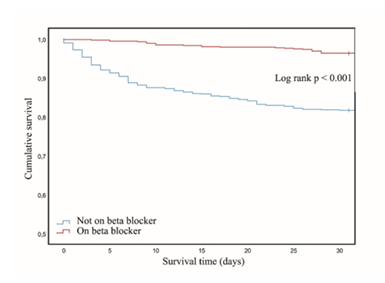
Figure 1: Kaplan-Meier plot showing 30-day survival of patients with aLQT according to their use of beta-blocker or no beta-blocker
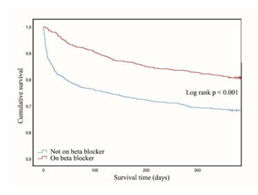
Figure 2: Kaplan-Meier plot showing 12-month survival of patients with aLQT according to their use of beta-blocker or no beta-blocker.
Risk assessment
Of the 1498 patients with aLQT, 171 (11.4%) died within 30 days after the index ECG. Of the deceased patients, 70 (41%) were diagnosed with cardiovascular diseases, 60 (35%) were diagnosed with infectious diseases, and 26 (15%) with malignancies. The lowest heartrate was 38 bpm and less than 10 % had heart rate < 60 bpm. The patients on beta-blocker were older; they were more likely to have heart failure and acute coronary syndrome (Table 1). They were also less likely to suffer from hypokalemia and stroke. As previously reported, eight factors (* in Table 1) significantly influenced patients’ survival (3) . Any use of beta-blocker within the last seven days before index ECG, however, was shown to improve survival in the stepwise Cox regression analyses, tables 2 and 3 (HR 0.12) (95% CI 0,08-0.19, p<0.001). This favorable impact was found both for short-term (30-day) and long-term (12-month) survival, as seen in figures 1 and 2. Subgroup analyzes on heart rate relation to this favorable effect revealing similar effects on all four heart rate quartiles (figures 3 and 4). The ProQT score showed an independent value for mortality prediction in multivariable analysis (Table 4).
Table 2: Cox regression analyses of 30-day mortality for 1498 patients with acquired LQT according to premorbid use of beta-blocker medication
|
Univariate analyses |
Multivariable analyses |
Multivariable analyses (Backward stepwise approach) |
|||||||
|
HR |
95% CI |
p-value |
HR |
95% CI |
p-value |
HR |
95% CI |
p-value |
|
|
Beta-blocker use N=718 |
0.19 |
0.13-0.29 |
<0.001 |
0.12 |
0.08-0.19 |
<0.001 |
0.12 |
0.08-0.19 |
<0.001 |
|
<24h after cardiac arrest N=25 |
6.9 |
3.91-12.15 |
<0.001 |
5.86 |
3.07-11.17 |
<0.001 |
5.68 |
3.01-10.44 |
<0.001 |
|
<7 days after stroke N=75 |
3.31 |
2.13-5.14 |
<0.001 |
3.32 |
2.08-5.30 |
<0.001 |
3.34 |
2.11-5.31 |
<0.001 |
|
Ejection fraction < 40% N=325 |
1.96 |
1.43-2.69 |
<0.001 |
2.88 |
1.97-4.20 |
<0.001 |
3.14 |
2.20-4.49 |
<0.001 |
|
Sepsis N=368 |
2.9 |
2.15-3.91 |
<0.001 |
2.1 |
1.53-2.88 |
<0.001 |
2.23 |
1.63-3.05 |
<0.001 |
|
Drugs with conditional risk N=652 |
1.37 |
1.01-1.84 |
0.04 |
1.61 |
1.17-2.21 |
<0.01 |
1.6 |
1.17-2.18 |
<0.01 |
|
Male gender N=613 |
1.4 |
1.04-1.90 |
0.03 |
1.44 |
1.04-1.98 |
0.03 |
1.39 |
1.01-1.91 |
0.05 |
|
Age (years) |
1.05 |
1.03-1.06 |
<0.001 |
1.05 |
1.03-1.06 |
<0.001 |
1.05 |
1.03-1.06 |
<0.001 |
|
Hypokalemia N=439 |
1.18 |
0.86-1.62 |
0.32 |
1.38 |
0.99-1.92 |
0.06 |
|||
|
Heart rate (bpm) |
1.01 |
1.00-1.02 |
0.26 |
1.01 |
0.99-1.02 |
0.29 |
|||
|
Diabetes (5)mellitus N=256 |
1.08 |
0.73-1.59 |
0.71 |
1.07 |
0.72-1.60 |
0.72 |
|||
|
<7 day after ACS N=293 |
0.99 |
0.68-1.45 |
0.96 |
1.36 |
0.88-2.11 |
0.16 |
|||
|
Syncope N=59 |
0.28 |
0.07-1.11 |
0.07 |
0.45 |
0.11-1.85 |
0.27 |
|||
|
<7 days after cardioversion N=75 |
0.11 |
0.02-0.75 |
0.03 |
0.17 |
0.02-1.21 |
0.08 |
|||
|
Drugs with known risk for TdP N=430 |
1.28 |
0.93-1.76 |
0.13 |
1.19 |
0.86-1.66 |
0.3 |
|||
|
Drugs with possible risk for TdP N=166 |
1.13 |
0.71-1.78 |
0.61 |
1.04 |
0.65-1.66 |
0.87 |
|||
|
Drugs to be avoided in cLQTS N=187 |
1.1 |
0.71-1.71 |
0.67 |
0.95 |
0.60-1.51 |
0.84 |
|||
Table 3: Cox regression analyses of 30-day mortality for 1498 patients with acquired LQTS. ProQT score included
|
Covariate |
Univariate analyses |
Multivariable analyses |
||||
|
HR |
95% CI |
p-value |
HR |
95% CI |
p-value |
|
|
Beta-blocker use, N=718 |
0.17 |
0.11-0.26 |
<0.001 |
0.15 |
0.10-0.22 |
<0.001 |
|
ProQT score |
1.26 |
1.17-1.36 |
<0.001 |
1.3 |
1.20-1.40 |
<0.001 |
|
Age |
1.06 |
1.04-1.07 |
<0.001 |
|||
|
Male gender |
2.08 |
1.53-2.84 |
<0.001 |
|||
Table 4: 30-day mortality of patients with aLQT according to Pro-QTc score and premorbid beta-blocker treatment. For all risk groups, beta-blocker treatment was associated with better survival.
|
Pro-QTc Score |
n |
Dead |
On beta blockers |
||||||
|
n |
% |
n |
% |
Dead n |
Dead % |
HR (95%CI)* |
p-value |
||
|
Pro-QTc Score 0-2 |
555 |
39 |
7 |
217 |
39.1 |
3 |
1.4 |
0.13 (0.04-0.42) |
<0.001 |
|
Pro-QTc Score 3 |
346 |
41 |
11.9 |
173 |
50 |
4 |
2.3 |
0.09 (0.03-0.25) |
<0.001 |
|
Pro-QTc score 4 |
280 |
43 |
15.4 |
134 |
47.9 |
5 |
3.7 |
0.12 (0.05-0.31) |
<0.001 |
|
Pro-QTc score ≥5 |
317 |
48 |
15.1 |
194 |
61.2 |
16 |
8.3 |
0.21 (0.11-0.39) |
<0.001 |
|
Total |
1498 |
171 |
11.4 |
718 |
47.9 |
28 |
3.9 |
0.18 (0.12-0.27) |
<0.001 |
*Hazard ratio (HR) with 95% Confidence interval (CI) for 30 day mortality adjusted for age, sex and heartrate
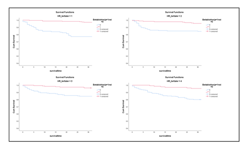
Figure 3: 30-day survival according to heartrate quartile.
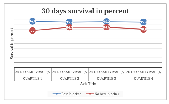
Figure 4: 30 days survival (%) on beta-blocker according to heartrate quartiles (Log Rank p< 0.001 for all quartiles)
Discussion
This retrospective single center observational study showed reduced mortality risk at 30 days and 120 days follow-up for patients with aLQT and beta-blocker medication. In our cohort heart failure, sepsis and coronary syndromes were common, affecting altogether 66% of the cohort. Other results might be found in a selected cohort with e.g. a high fraction of diabetic patients. However, aLQT prolongation was the only selection criterion in our study, and our cohort thus reflects all hospitalized patients in this hospital with acquired QT >500ms. Our study showed a markedly better short- and long-term survival for patients with aLQT when treated with beta-blockers. One explanation could be that that patients on beta-blocker treatment had a better prognosis due to the underlying condition. However, the proQTc score in patients on beta-blockers was not different from those without beta-blockers. Intriguingly, the beta-blocker group of patients were older than those not treated with beta-blockers, suggesting worse long-term prognosis by age itself.
Mechanisms of beta-blockers and impact on outcome
After Propanolol, as the first beta-blocker, was marketed for treatment of hypertension in 1964, it was soon discovered that beta-blockers also had cardio-protective properties (4). Information on which beta-blocker each patient used was not available in our database. However, during the years 2004 through 2014, sales figures have documented that metoprolol had 61% of the beta-blocker market in standard dosages. Atenolol had 17%, and carvedilol had 8%. Beta-blockers now constitute the standard guideline recommended care for patients with cLQTS. However, the literature is sparce and we have not succeeded in finding documentation of an effect of beta-blockers specifically for aLQTS. As QT prolongation reflects a reduced repolarization reserve of the cardiomyocyte membrane, independent of the triggering cause, our observation of a favorable beta-blocker effect in aLQT may not be surprising. Beside the heart, β1 adrenergic receptors are found in the kidneys, on platelets and on adipocytes. Stimulation of these receptors causes renin secretion, platelet activation and lipolysis. Both the cardiac and the extra-cardiac effects of beta-blockade must be considered of importance.
The data in our study only account for total deaths, and we do not have any data distinguishing between sudden death, cardiovascular death, or cancer, but we have data on actual diagnosis at the time of the first ECG recording of prolonged QT, and we found that 21.7 % of the cohort suffered from heart failure, 19.6 % had acute coronary syndrome, 24.6% had sepsis, 29.0% had hypokalemia, and 17.1 % had diabetes. Searching support in published data, we looked for beta blocker effects related to these diagnoses: Some studies elucidate why beta-blockers might be favorable in aLQT. For patients with heart failure and reduced ejection fraction, guidelines recommend beta-blockers (7). As for patients with acute coronary syndrome (ACS), the evidence is not so clear, but it seems that beta-blockers have a favorable effect - at least in ACS patients with EF< 50% (8, 9). Also, for arrhythmic disorders, such as catecholaminergic polymorphic ventricular tachycardia, guidelines recommend treatment with beta-blockers (10).
In our cohort, a large proportion had septicemia, and our findings support previous data indicating a protective role for beta-blockers in this setting (11, 12).
Observational studies suggest that treatment with beta-blockers may prevent sudden cardiac death in patients on hemodialysis. So, for three of the diagnoses in our cohort (heart failure, acute coronary syndrome and sepsis) accounting for 66% of the patients, favorable effects have been reported. For patients with diabetes, however, (17% of our patients) beta-blockers might increase the all-cause mortality (13). For patients with aLQT, we found a markedly better short-term survival in our cohort of patients on beta-blockers and the finding was consistent through major subgroups, through risk groups and through heart rate quartiles. The patients in this study are mostly hospitalized patients with severe acute illness, and one could speculate if the premorbid beta-blocker treatment blunts the beta-adrenergic effect in patients with severe illness leading to a favorable outcome.
Our cohort had relatively high heart rates with a mean heart rate of 79 bpm. This may suggest that the patients were suffering from beta-adrenergic stress and the slightly lower heartrate in the group with premorbid beta-blocker treatment may have been a favorable factor mediating part of the apparent protection against death seen in this group (14). The reduced heart rate among beta-blocker users cannot alone explain our findings as the reduction in mortality is evenly distributed over the range of heart rates in the cohort. However, beta-blocker mediated protection against arrhythmias or myocardial injury as e.g. Takotsubo cardiomyopathy could well play a major role. A high proportion of patients were treated with drugs known to prolong the QTc interval. Beta-blockers are known to shorten the QTc interval, and one could speculate that beta-blocker treatment might mitigate the negative effect of these drugs.
Clinical implications
Data from our study suggests not stopping beta-blocker treatment in patients with aLQT. Whether beta-blocker treatment should be started in aLQT needs further studies.
Limitation and Conclusion
In this cohort of 1498 patients with acquired LQTc, we have observed a favorable effect of beta-blocker treatment on overall mortality. This is a hypothesis generating, single center study presenting patients with a panel of diseases not necessarily generalizable to other populations. Hopefully, our study may encourage others with access to similar clinical databases like MUSE to analyze reproducibility of our results in their patient cohorts. The treatment of aLQT patients should be different from the cLQTS patients. After work-up of the patients in our database we found that the majority of the aLQT patients were treated with drugs liable to prolong the QT interval according to the CredibleMeds list. Furthermore, many of the patients had electrolyte disturbances. Once the cLQTS cases have been identified and taken care of, the risk factors for developing aLQT are the first to be addressed. So, removing unfavorable drugs, and correcting electrolytes have the highest priority (3, 10). However, if an aLQT patient already is treated with beta-blocker, our data suggests that the beta-blocker treatment should be continued.
Author Contributions
Conceptualization, Jan Hysing, and Jacob Thalamus; methodology: Jan Hysing, Jacob Thalamus Øystein Lunde Holla, andKeson Jaioun; software, Jacob Thalamus, Jan Hysing and Charlotte Gibbs; validation: Jacob Thalamus, Charlotte Gibbs and Keson Jaioun; formal analysis: Jacob Thalamus and Keson Jaioun; data curation, Charlotte Gibbs and Jacob Thalamus; writing—original draft preparation Jacob Thalamus; writing—review and editing Jan Hysing, Geir Hoff and Kristina Haugaa; visualization, Jacob Thalamus; supervision, Kristina Haugaa.; project administration, Jan Hysing; funding acquisition, The project was funded by a grant from Telemark Hospital Trust. All authors have read and agreed to the published version of the manuscript.
Funding:The project was funded by a grant from Telemark Hospital Trust.
Institutional Review Board Statement:The study was conductedin accordance withthe Declaration ofHelsinki, andapproved by the Norwegian Regional Committee for Medical and Health Research Ethics, protocol code 2013/1090 October 18. 2013
Informed Consent Statement:Informed consent was obtained from all subjects involved in the study.
Data Availability Statement:There are restrictions by law in Norway which make data sharing particularly difficult outside Norway.
Conflicts of Interest:The authors declare no conflict ofinterest.
References
- Tisdale JE, Wroblewski HA, Overholser BR, et al. Prevalence of QT interval prolongation in patients admitted to cardiac care units and frequency of subsequent administration of QT interval-prolonging drugs: a prospective, observational study in a large urban academic medical center in the US. Drug safety 35 (2012): 459–70.
- Haugaa KH, Bos JM, Tarrell RF, et al. Institution-wide QT alert system identifies patients with a high risk of mortality. Mayo Clinic proceedings 88 (2013): 315–25.
- Gibbs C, Thalamus J, Heldal K, et al. Predictors of mortality in high-risk patients with QT prolongation in a community hospital. Europace 20 (2018): f99–f107.
- Yusuf S, Peto R, Lewis J, et al. Beta blockade during and after myocardial infarction: an overview of the randomized trials. Prog Cardiovasc Dis 27 (1985): 335–71.
- RL W, KA R. www. Crediblemeds.org, QTdrugs list: Azcert, Inc. 1822 Innovation Park Dr., Oro Valley, AZ 85755 (2021).
- Gibbs C, Thalamus J, Tveten K, et al. Genetic and Phenotypic Characterization of Community Hospital Patients With QT Prolongation. J Am Heart Assoc 7 (2018): e009706.
- McDonagh TA MM, Adamo M, Gardner RS, et al. ESC Scientific Document Group. 2021 ESC Guidelines for the diagnosis and treatment of acute and chronic heart failure. Eur Heart J Sep 42 (2021): 3599–726.
- Silvain J, Cayla G, Ferrari E, et al. Beta-Blocker Interruption or Continuation after Myocardial Infarction. The New England journal of medicine 391 (2024): 1277–86.
- Munkhaugen J, Kristensen AMD, Halvorsen S, et al. Beta-Blockers after Myocardial Infarction in Patients without Heart Failure. The New England journal of medicine 393 (2025): 1901–11.
- Zeppenfeld K T-HJ, de Riva M, Winkel BG, et al. ESC Scientific Document Group. 2022 ESC Guidelines for the management of patients with ventricular arrhythmias and the prevention of sudden cardiac death. Eur Heart J 43 (2022): 3997–4126.
- Guz D, Buchritz S, Guz A, et al. Beta-Blockers, Tachycardia and Survival Following Sepsis- An Observational Cohort Study. Clin Infect Dis (2021).
- Morelli A, Ertmer C, Westphal M, et al. Effect of heart rate control with esmolol on hemodynamic and clinical outcomes in patients with septic shock: a randomized clinical trial. Jama 310 (2013): 1683–91.
- Tsujimoto T, Kajio H, Shapiro MF, et al. Risk of All-Cause Mortality in Diabetic Patients Taking β-Blockers. Mayo Clinic proceedings 93 (2018): 409–18.
- Jervell A, Lange-Nielsen F. Congenital deaf-mutism, functional heart disease with prolongation of the Q-T interval and sudden death. American heart journal 54 (1957): 59–68.