Huynh Dinh Chien*
College of Health Sciences, VinUniversity, Hanoi, Vietnam
*Corresponding author: Huynh Dinh Chien, College of Health Sciences, VinUniversity, Hanoi, Vietnam.
Received: 20 August 2026; Accepted: 24 August 2026; Published: 30 September 2026
DOI: 10.26502/jbb.2642-91280227
ShareStem and progenitor cells have traditionally been understood as agents of tissue formation primarily through their capacity to proliferate and differentiate into the specialized cell types of a mature organ. This review argues for a complementary, and in many contexts more mechanistically accurate, view: that the secretome — the coordinated array of soluble factors, enzymes, and extracellular vesicles a cell releases into its surroundings — functions as an active, instructive signal that directs organ formation, rather than as an incidental byproduct of cellular activity. We first outline the molecular composition and regulation of the stem cell secretome, before examining the mechanistic evidence linking secreted signals to key organogenic processes: morphogen gradient formation, reciprocal epithelial-mesenchymal induction, vesicle-mediated signal delivery, and extracellular matrix remodeling. We then survey organ-specific evidence for secretome-driven development across the heart, kidney, nervous system, liver, and pancreas, and show that organoid engineering functions largely by empirically reconstituting these same endogenous secretome combinations in vitro. Building on this mechanistic foundation, we examine translational applications — cell-free therapeutics, secretome-functionalized biomaterials, and secretome profiling as a candidate potency biomarker for cell therapy manufacturing — and explicitly connect the latter to the batch-to-batch variability that has undermined clinical trials of mesenchymal stromal cell therapies. Finally, we discuss persistent challenges, including secretome heterogeneity, unresolved causation, technical detection limits, and the gap between organoid and in vivo fidelity, and outline future directions — spatiotemporal secretome atlases, computational prediction, and synthetic secretome design — aimed at resolving them. We conclude that the next phase of progress in regenerative medicine depends less on identifying new stem cell sources than on learning to read, predict, and ultimately design the secretome signals cells use to build organs.
Stem cell secretome; Secretomics; Organogenesis; Extracellular vesicles; Organoids; Paracrine signaling; Regenerative medicine; Cell-free therapy
Stem cell secretome articles; Secretomics articles; Organogenesis articles; Extracellular vesicles articles; Organoids articles; Paracrine signaling articles; Regenerative medicine articles; Cell-free therapy articles
Introduction
From cell-centric to secretome-centric: a paradigm shift
The therapeutic and developmental logic of stem cell biology has historically been cell-centric: a stem or progenitor cell's value was understood to lie in its capacity to self-renew and to differentiate into the specific mature cell types required to build or repair a tissue [1]. This framing shaped both basic developmental biology and the early clinical translation of cell therapy, in which efficacy was generally attributed to donor cell engraftment and direct replacement of damaged tissue. Over the past two decades, however, this assumption has been substantially revised, particularly in the mesenchymal stromal cell (MSC) literature, where accumulating evidence indicates that transplanted cells frequently exert their effect while achieving only transient engraftment, with therapeutic activity instead attributable to the factors they secrete into the surrounding tissue [2,3]. This shift has run in parallel with a much older thread in developmental biology, dating to the classical discovery of embryonic induction by the Spemann-Mangold organizer, which established that one tissue can direct the fate of an adjacent tissue through diffusible signals rather than direct cellular contribution [4,5]. What is new is not the concept of secreted, inductive signaling itself, but the systematic, omics-scale toolkit now available to characterize it comprehensively — giving rise to secretomics as a discipline in its own right (Figure 1).
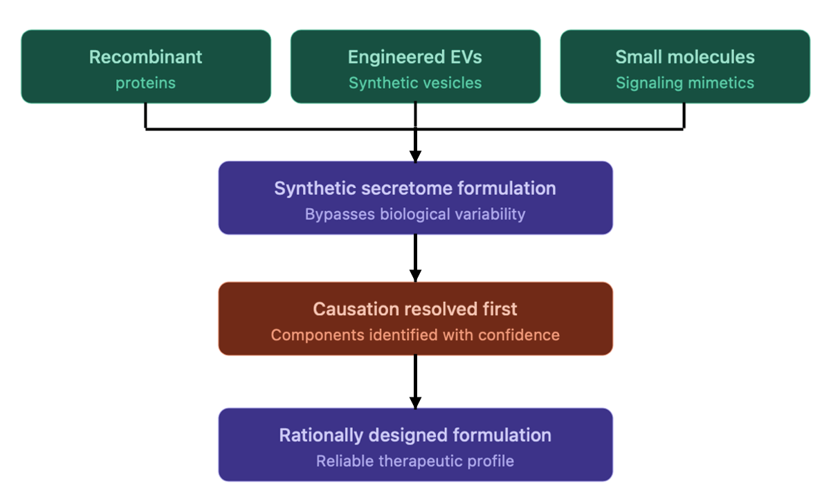
Figure 1: Pathway from cell-free therapy to synthetic secretome design.
Recombinant proteins, engineered extracellular vesicles (EVs), and small-molecule mimetics converge to form a synthetic, defined secretome formulation, which bypasses the donor-, tissue-, and culture-dependent variability inherent to cell-derived secretomes. However, translating this formulation into a rationally designed, reliable therapeutic product depends on first resolving the causation problem — identifying, with confidence, which specific components are responsible for a given therapeutic or morphogenetic effect. This dependency illustrates why the mechanistic and translational threads of this review remain fundamentally interdependent.
Defining the secretome
The secretome is most usefully defined as the complete set of molecules a cell, tissue, or organism actively releases into its extracellular environment, encompassing both freely soluble factors — growth factors, cytokines, chemokines, and morphogens — and vesicular cargo carried within exosomes and microvesicles, which can additionally include non-coding RNA species such as microRNAs [6,7]. This definition deliberately excludes the intracellular proteome and the surfaceome (stably membrane-anchored proteins that are not released), distinguishing secretomics from adjacent -omics disciplines that characterize what a cell contains or displays rather than what it exports to influence its surroundings. A cell's secretome is not a fixed property but a dynamic output that varies with differentiation state, microenvironmental context, and culture or physiological condition, a property that is central to both its developmental flexibility and its translational unpredictability.
Why the secretome qualifies as an instructive signal, not merely a byproduct
Characterizing the secretome as instructive, rather than incidental, rests on three converging lines of evidence: first, secreted morphogens form concentration gradients that are read out by recipient cells in a threshold-dependent manner to generate discrete positional and fate decisions, a relationship established through decades of gain- and loss-of-function embryology [8]; second, loss-of-function studies targeting specific secreted ligands or their cognate receptors yield predictable, often organ-specific developmental phenotypes, providing causal evidence that goes beyond mere correlation [9,10]; and third, reconstitution experiments — most directly, organoid systems — demonstrate that defined secretome combinations are frequently sufficient to drive self-organization of progenitor cells into organized, organ-like architecture in the absence of any other instructive template [11,12]. Together, these lines of evidence support treating the secretome as a primary causal driver of organ formation, on comparable mechanistic footing to genetic and transcriptional regulatory programs, rather than as a downstream consequence of cell identity alone.
Aims and scope of this review
This review is organized to move from mechanism to application to open problem. Following the Introduction (Section 1), Section 2 surveys the molecular composition, secretion mechanisms, and regulation of the stem and progenitor cell secretome, along with the analytical toolkit used to characterize it. Section 3 examines the specific mechanisms through which secreted signals direct organogenesis, and Section 4 grounds these mechanisms in organ-specific case studies spanning the heart, kidney, nervous system, liver, and pancreas, extending into the relationship between secretome biology and organoid engineering. Section 5 turns to translational applications, including cell-free therapeutics and secretome-based potency biomarkers, before Section 6 critically examines the heterogeneity, causal, technical, and fidelity limitations that currently constrain the field, and Section 7 outlines future directions aimed at resolving them. Section 8 concludes the review. We focus throughout on postnatal and embryonic organogenesis and regenerative applications; secretome biology in oncogenic or pathological contexts, while an active and related area of investigation, falls outside the primary scope of this review and is addressed only incidentally where directly relevant.
Foundational Biology of the Stem Cell Secretome
Molecular composition: soluble factors and vesicular cargo
The secretome comprises two broad, mechanistically distinct classes of output. Soluble factors include classical growth factors (e.g., FGF, VEGF, PDGF family members), cytokines and chemokines, and morphogens (Wnt, BMP, Shh ligands), each acting through specific cell-surface receptors on recipient cells [13,14]. Vesicular cargo is carried within extracellular vesicles (EVs), a heterogeneous category comprising exosomes — small vesicles of roughly 30–150 nm in diameter, originating from the endosomal system via multivesicular body formation and release — and microvesicles, typically larger and generated by direct outward budding of the plasma membrane [15,16]. EV cargo extends beyond protein to include messenger RNA fragments, microRNAs, and in some cases DNA fragments, giving EVs a capacity for combinatorial, multi-modal signaling that soluble factors alone do not possess [17,18] (Figure 2).
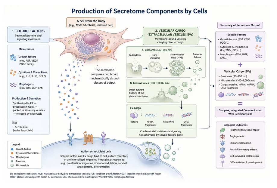
Figure 2: Production of secretome components.
Cells release bioactive molecules into their surroundings through two broad, mechanistically distinct pathways. (1) Soluble factors—growth factors, cytokines, chemokines, and morphogens—are synthesized in the endoplasmic reticulum, processed in the Golgi apparatus, and secreted via the classical secretory pathway to act on surface receptors of recipient cells. (2) Vesicular cargo—extracellular vesicles (EVs)—includes exosomes (30-150 nm), formed inside multivesicular bodies (MVBs) of the endosomal system and released by exocytosis, and microvesicles (100-1,000+ nm), which bud directly from the plasma membrane. EVs transport diverse cargo such as proteins, mRNA fragments, microRNAs, and DNA fragments, enabling combinatorial, multi-modal communication with recipient cells that extends beyond the capacity of soluble factors alone.
Secretion mechanisms: conventional, unconventional, and vesicular pathways
Proteins bearing an N-terminal signal peptide are secreted via the conventional endoplasmic reticulum-to-Golgi pathway, the route used by most classical growth factors and cytokines [19,20]. A substantial subset of biologically important secreted factors, however — including fibroblast growth factor 2 (FGF2) and interleukin-1β — lack a signal peptide and instead exit the cell via unconventional secretion routes, including direct plasma membrane translocation, lysosomal secretion, and membrane-blebbing mechanisms, which remain comparatively less well characterized than the conventional pathway [21,22]. EV biogenesis constitutes a third, mechanistically separate route: exosome formation proceeds through both ESCRT-dependent and ESCRT-independent pathways during multivesicular body maturation, while microvesicle shedding occurs through direct, largely ESCRT-independent budding at the plasma membrane, with cargo selection into each vesicle type governed by distinct, only partially understood sorting mechanisms [23,24].
Dynamic regulation of secretome output
Secretome composition is not a static cellular property but a dynamic output shaped by differentiation state, mechanical microenvironment, and metabolic condition. Substrate stiffness and other mechanical cues are transduced through pathways such as YAP/TAZ signaling to influence which factors a cell secretes, linking the mechanotransductive matrix remodeling to secretome regulation itself in a bidirectional loop [25]. Oxygen tension is similarly influential: hypoxic conditions stabilize hypoxia-inducible factor 1-alpha (HIF-1α), driving upregulation of angiogenic factors such as VEGF and shifting the broader secretome toward a pro-angiogenic profile, a relationship directly relevant to the hypoxic preconditioning strategies [26,27]. Inflammatory or cytokine priming of cultured cells produces further, often substantial shifts in secreted output, and donor age and senescence status introduce yet another axis of variation, foreshadowing the heterogeneity challenges.
The secretomics analytical toolkit
Characterizing the secretome experimentally draws on several complementary technology platforms, each with distinct strengths and limitations. Liquid chromatography-tandem mass spectrometry (LC-MS/MS)-based proteomics, used in either label-free or isotope-labeled (e.g., SILAC, tandem mass tag) quantitative formats, offers broad, relatively unbiased coverage of secreted proteins but can lack sensitivity for low-abundance factors present at picogram-per-milliliter concentrations [28,29]. Multiplex bead-based immunoassays (e.g., Luminex-type platforms) offer higher sensitivity and throughput for a predefined panel of cytokines and growth factors, at the cost of being limited to previously selected analytes rather than providing unbiased discovery [30]. EV-specific characterization relies on a further set of tools, including nanoparticle tracking analysis and single-vesicle flow cytometry for particle counting and sizing, and RNA sequencing of EV cargo for transcriptomic characterization, though standardization of EV isolation and single-vesicle-resolution analysis remains an active methodological challenge, directly underlying the technical detection limitations [31-33].
The Secretome as an Instructive Signal Directing Organogenesis (Figure 3)
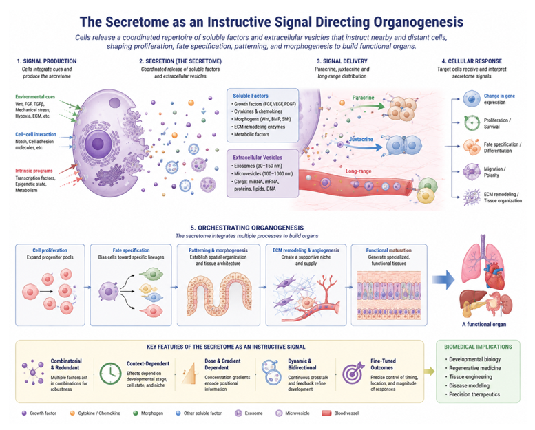
Figure 3: The Secretome as an Instructive Signal Directing Organogenesis.
Cells integrate environmental, cell–cell, and intrinsic cues to release a coordinated repertoire of soluble factors and extracellular vesicles, which act through paracrine, juxtacrine, and long-range mechanisms to regulate gene expression, proliferation, cell-fate specification, migration, extracellular matrix remodeling, and tissue organization. Through dynamic, context-dependent, combinatorial, and dose-dependent signaling, the secretome coordinates successive stages of organogenesis—from progenitor expansion and lineage specification to patterning, vascularization, and functional maturation—ultimately guiding the formation of functional organs and providing a conceptual framework for regenerative medicine and tissue engineering.
Morphogen gradients and positional patterning
A central, long-standing principle in developmental biology is that spatial patterning of a developing organ field is established not by cell-intrinsic programs alone, but by extracellular concentration gradients of diffusible signaling molecules — morphogens — secreted from discrete organizing centers [34,35]. Members of the Wnt, bone morphogenetic protein (BMP), Sonic hedgehog (Shh), and fibroblast growth factor (FGF) families are the principal morphogen classes implicated across nearly all organ systems, and stem and progenitor cell populations frequently constitute the local source of these gradients within a developing organ field [36-38]. Cells interpret their position within the gradient through concentration-dependent activation thresholds, translating a continuous extracellular signal into discrete transcriptional and fate decisions — the classical “French flag” model of positional information [39-41]. Notch-Delta signaling operates through a related but mechanistically distinct logic: because Notch ligands are membrane-tethered rather than secreted, this pathway acts juxtacrine rather than paracrine, and is more typically implicated in fine-grained fate decisions between adjacent cells (lateral inhibition) than in long-range patterning, a distinction worth preserving conceptually even though Notch is frequently discussed alongside secreted morphogens in the organogenesis literature [42,43].
Epithelial-mesenchymal paracrine crosstalk in branching morphogenesis
Branching organogenesis — the iterative process by which tubular organs such as the lung, kidney, and salivary and mammary glands generate their characteristic arborized architecture — depends on reciprocal paracrine signaling between adjacent epithelial and mesenchymal compartments, rather than on autonomous behavior of either tissue alone [44]. In the developing lung, mesenchymally secreted FGF10 acts on FGFR2b-expressing epithelium to direct the site and outgrowth of new branch tips, while epithelially derived Sonic hedgehog feeds back to restrain and spatially restrict FGF10 expression in the surrounding mesenchyme, generating a self-limiting, iterative signaling loop that produces stereotyped branch points [45,46]. An analogous reciprocal circuit operates in kidney development, in which mesenchymally secreted glial cell line-derived neurotrophic factor (GDNF) signals through the RET receptor on the ureteric bud epithelium to drive its outgrowth and branching, while the ureteric epithelium reciprocally secretes signals — including Wnt9b — that induce the surrounding metanephric mesenchyme to undergo mesenchymal-to-epithelial transition and form nephrons [47-49]. These examples illustrate a general principle: the secretome of one progenitor compartment functions as the developmental instruction set for its neighboring compartment, and organ-specific architecture emerges from the geometry and iteration of these paracrine circuits rather than from a pre-specified blueprint within either cell population alone.
Extracellular vesicles as selective, long-range signal carriers
Beyond soluble morphogens, extracellular vesicles (EVs) — exosomes and microvesicles — constitute a second, mechanistically distinct secretome-mediated signaling channel during organogenesis. EVs package selected protein and RNA cargo, including microRNAs capable of post-transcriptionally silencing specific target transcripts in recipient cells, allowing a secreting progenitor population to exert combinatorial, multi-target effects on neighboring or distant tissue through a single vesicular unit [18,50,51]. EV packaging has also been proposed to extend the effective signaling range of otherwise poorly diffusible or labile morphogens — for example, lipid-modified Wnt ligands, which are membrane-associated and diffuse poorly on their own, have been reported to be carried within exosomes, potentially enabling longer-range Wnt signaling than free ligand diffusion would otherwise permit [52,53]. This has led to the proposal that EVs function as discrete “signalosomes,” delivering pre-assembled combinations of morphogens, RNAs, and enzymes as a coordinated packet rather than as independently diffusing single factors, adding a layer of combinatorial and spatial precision to secretome-mediated patterning that soluble factors alone may not achieve [54,55].
Extracellular matrix remodeling as a secretome-mediated morphogenetic mechanism
A substantial component of the organogenic secretome is enzymatic rather than purely signaling in nature. Matrix metalloproteinases (MMPs) and their endogenous inhibitors, the tissue inhibitors of metalloproteinases (TIMPs), are secreted by progenitor and stromal populations to locally degrade and remodel the basement membrane and surrounding extracellular matrix (ECM), a process required for epithelial bud outgrowth and branch point formation during organogenesis [56-58]. This remodeling activity has a dual function: it physically permits tissue movement and reshaping, and it also liberates matrix-sequestered growth factors — including FGF and VEGF family members that are stored bound to heparan sulfate proteoglycans within the ECM — converting MMP-mediated matrix degradation into an indirect mechanism of growth factor release and signal activation [59-61]. The mechanical properties of the remodeled matrix further feed back onto cell behavior through mechanotransduction pathways, meaning that the secretome's action on the ECM is not merely permissive scaffolding but an active, bidirectional component of the morphogenetic signaling network [62-64].
Feedback loops sustaining progenitor populations and self-organization
Organogenesis further depends on feedback circuits that maintain an appropriate balance between self-renewing progenitors and differentiating progeny over the course of development. Lateral inhibition mediated by Notch-Delta signaling is a well-characterized example, in which a stochastically “winning” progenitor cell suppresses the same fate in its immediate neighbors, generating a salt-and-pepper pattern of differentiated cells within a progenitor field [65]. At a larger spatial scale, reciprocal paracrine loops between spatially separated progenitor and niche populations — for example, stem cells and their supporting niche cells, each secreting factors that sustain the other — have been proposed to generate emergent, self-organizing patterns without requiring an external patterning template, consistent with reaction-diffusion (Turing-type) frameworks that have been invoked to explain periodic structures such as branching frequency and digit spacing [66,67]. Collectively, the mechanisms described in this section support a unifying view: organ-level architecture is not encoded solely within the genome of any single cell population, but emerges from the dynamic, spatially structured exchange of secretome-mediated signals among interacting progenitor and stromal compartments.
Case Studies in Organ-Specific Secretome-Driven Development
Heart: secretome-driven cardiac progenitor specification and growth
Cardiogenesis provides an instructive example of sequential, secretome-mediated induction. Signals secreted by the pharyngeal endoderm and adjacent mesoderm — including members of the FGF, BMP, and Wnt families — act on cardiac progenitor populations within the first and second heart fields to specify cardiomyocyte fate and regulate the timing of their addition to the elongating heart tube [68,69]. Later in development, the epicardium, the outer mesothelial layer covering the heart, secretes a distinct paracrine repertoire — including retinoic acid and additional FGF and insulin-like growth factor signals — that promotes proliferation of the underlying compact myocardium and supports coronary vasculature formation, illustrating how a non-myocardial secretome source can be indispensable for proper growth of the organ's principal functional tissue [70-72].
Kidney: reciprocal induction in nephron formation
Nephrogenesis depends almost entirely on the reciprocal paracrine circuit: GDNF secreted by the metanephric mesenchyme signals through RET on the ureteric bud epithelium to initiate and iteratively direct its branching, while the ureteric epithelium reciprocally secretes Wnt9b to induce the surrounding mesenchyme to condense and undergo mesenchymal-to-epithelial transition, ultimately forming the nephron epithelium [73-75]. The precise number of branching events this circuit undergoes directly determines the final number of nephrons formed, linking secretome dynamics quantitatively to adult renal functional capacity and providing a clear illustrative case of how a paracrine signaling loop, rather than a fixed cell-autonomous program, sets a key organ-level structural parameter [76,77].
Nervous system: secretome-guided neuronal migration and circuit assembly
In the developing nervous system, secretome-mediated signaling operates over both short and long range to organize neuronal position and connectivity. Radial glial cells secrete guidance cues that direct the radial migration of newborn neurons from proliferative zones toward their final laminar position in the cortex, while chemokine signaling — notably the SDF-1(CXCL12)/CXCR4 axis — has been implicated in directing the tangential migration of interneuron populations into the developing cortical plate [78,79]. Beyond migration, astrocyte-secreted factors, including thrombospondins, have been shown to promote synaptogenesis by newly arrived neurons, indicating that the secretome's instructive role in neural organogenesis extends beyond cell positioning to the later assembly of functional circuitry [80,81].
Liver and pancreas: endodermal fate induction by adjacent mesenchyme and mesoderm
Hepatic and pancreatic specification from the foregut endoderm exemplify how a single progenitor sheet can be directed toward divergent organ fates by distinct, spatially restricted secretome inputs from neighboring tissue. Cardiac mesoderm-derived FGF signals, together with signals from adjacent endothelial cells, induce a subset of the ventral foregut endoderm to adopt hepatic fate, while BMP signaling from the septum transversum mesenchyme acts in parallel to support this induction [82,83]. Pancreatic development, by contrast, depends on active repression of hepatic and other alternative fates within the remaining endoderm, alongside FGF10 signaling from the surrounding pancreatic mesenchyme that maintains the multipotent progenitor pool of the pancreatic epithelium during the expansion phase of organ growth [84]. The hepatic and pancreatic examples together underscore that organ identity itself — not merely organ growth or shape — can be a direct output of secretome-mediated inductive signaling between adjacent germ layer derivatives.
Intestinal organoids: secretome-driven self-organization as a tractable model system
Intestinal organoids offer a particularly tractable, reduced system for studying secretome-driven organogenesis in vitro. Paneth cells located at the base of the intestinal crypt secrete a defined combination of Wnt, EGF, and Notch ligands that sustains the adjacent Lgr5+ intestinal stem cell population, and recapitulation of this secretome — whether by co-culture with Paneth cells or by exogenous supplementation of the equivalent factors in culture medium — is sufficient to drive the self-organized formation of crypt-villus-like structures from single stem cells in vitro, in the absence of any externally imposed geometric template [85-87]. This system provides direct experimental support for the broader thesis of this review: that a defined secretome, rather than an externally specified structural blueprint, can be sufficient to instruct the emergence of organized, organ-like tissue architecture — a principle with direct relevance to the organoid engineering and cell-free therapeutic strategies discussed later in this review.
Secretome-guided design: a blueprint for organoid construction (Figuer 4)
The intestinal example above illustrates a broader pattern that now underlies organoid technology across organ systems: the empirical formulation of organoid culture media is, in essence, an attempt to reconstitute in vitro the same secretome combinations that progenitor niches provide in vivo during the developmental processes described throughout this section [88,89]. Brain organoid protocols recapitulate stage-specific neural progenitor niche signals — including Shh, FGF, and Wnt modulation — to guide regional patterning into distinct forebrain, midbrain, or cerebral cortical identities, mirroring the secretome-driven neural patterning [90,91]. Kidney organoid differentiation protocols similarly stage sequential exposure to Wnt agonists and FGF9, reproducing in compressed form the temporal sequence of the GDNF-RET and Wnt9b-dependent reciprocal induction events that drive nephrogenesis in vivo [92,93], while liver organoid and bud protocols incorporate FGF, BMP, and hepatocyte growth factor supplementation timed to approximate the endodermal induction sequence [94].
Framed this way, organoid engineering is less a wholesale re-invention of developmental biology than an exercise in secretome reduction and sequencing: identifying the minimal, temporally ordered subset of an organ's endogenous paracrine signaling repertoire that is sufficient to drive self-organization of a stem or progenitor cell population into recognizable organ-like architecture in vitro. This reframing has two practical implications directly relevant to this review's broader argument. First, organoid systems constitute a uniquely tractable experimental platform for testing the causal sufficiency of specific secretome components identified through the case studies above, since individual factors can be added, withdrawn, or substituted in a way that is not feasible in vivo [95-97]. Second, the secretome combinations empirically optimized through organoid protocol development represent a ready-made starting point for the priming and engineering strategies, in which defined signaling cocktails are used to direct the therapeutic secretome output of cultured cells.
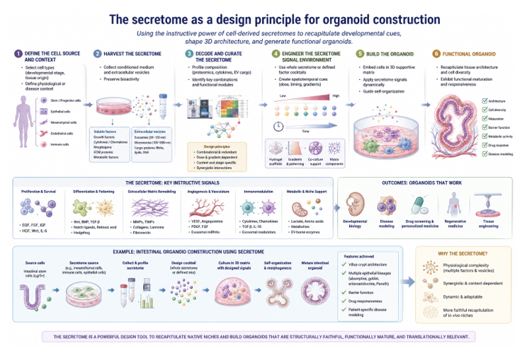
Figure 4: The Secretome as a Design Principle for Organoid Construction.
Cell-derived secretomes—comprising soluble factors and extracellular vesicles—are harvested, molecularly characterized, and rationally curated to define spatiotemporal signaling environments that regulate proliferation, differentiation, patterning, extracellular matrix remodeling, angiogenesis, immune modulation, and metabolic support. By integrating secretome signals with 3D matrices, gradients, co-culture systems, and dynamic delivery, this approach seeks to recapitulate key features of native tissue niches and drive self-organization, maturation, and functional specialization of organoids, with applications in developmental biology, disease modeling, drug screening, regenerative medicine, and tissue engineering.
Applications of technology and biomedicine (Figure 5)
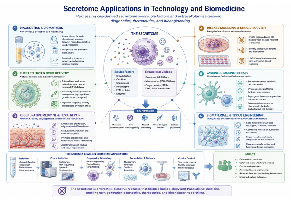
Figure 5: Secretome Applications in Technology and Biomedicine.
Schematic overview of the emerging technological and biomedical applications of the secretome, encompassing soluble factors and extracellular vesicles. Secretome-based approaches are being developed for liquid-biopsy biomarkers and diagnostics, therapeutic delivery, regenerative medicine and tissue repair, disease modeling and drug discovery, vaccines and immunotherapy, and secretome-functionalized biomaterials for tissue engineering. Integrated technologies for secretome isolation, molecular characterization, engineering, formulation, delivery, and quality control provide a translational framework for developing personalized, precise, and scalable biomedical applications.
Cell-free therapy: purified secretome and EVs as an alternative to live-cell transplantation
The recognition that much of the therapeutic activity attributed to transplanted stem and progenitor cells is mediated by their secretome rather than by direct engraftment and differentiation has motivated a shift toward cell-free therapeutic strategies, in which conditioned medium, purified EV fractions, or defined secretome cocktails are administered in place of living cells [95,97]. This approach offers several practical advantages consistent with the organogenic principles discussed in the preceding sections: because the instructive signal, not the cell itself, is the active agent, a cell-free product can in principle be manufactured, dosed, sterilized, and stored with far greater consistency than a population of living, metabolically active cells, while sidestepping risks associated with cell engraftment such as ectopic tissue formation or immune rejection [98]. Early applications have focused on regenerative contexts that mirror the paracrine-dominant mechanisms described earlier — wound healing, ischemic tissue repair, and immunomodulation — rather than contexts that plausibly require direct structural replacement of lost tissue, a distinction that should guide realistic expectations for where cell-free secretome therapy is mechanistically well-suited versus where it is not [99,100].
Secretome-functionalized biomaterials and controlled-release scaffolds
A parallel line of translational work embeds secretome components — either soluble factors or intact EVs — within biomaterial scaffolds engineered for controlled, sustained local release, aiming to recreate the spatially localized, temporally extended paracrine signaling that characterizes endogenous organogenesis rather than the single-bolus exposure typical of direct injection [101,102]. Hydrogel and nanofiber-based delivery systems have been used to tune release kinetics of encapsulated growth factors or EVs, and scaffold stiffness and degradation rate can themselves be engineered to influence the mechanotransductive component of the secretome's action, effectively coupling biochemical and biomechanical instruction within a single implantable material [103-105]. This convergence of tissue engineering and secretome biology reflects a broader design principle emerging from the organogenesis literature: an instructive microenvironment, rather than a cell source alone, may be the more tractable and reproducible unit to engineer.
Secretome profiling as a potency biomarker for cell therapy manufacturing
Perhaps the most immediately consequential application of secretomics is as a quality-control and potency-assessment tool for existing cell therapy manufacturing pipelines. Mesenchymal stromal cell (MSC) therapies have shown highly inconsistent clinical trial outcomes, driven in substantial part by donor-, tissue-source-, and culture-dependent variability in secretome composition, and by the absence of standardized in vitro potency assays that reliably predict in vivo efficacy [106,107]. Secretomic and EV profiling — characterizing the specific protein, cytokine, and vesicular cargo signature released by a given cell batch — has been proposed as a more direct and mechanistically grounded potency metric than the surface marker panels or generic proliferation assays currently used in most manufacturing quality-control workflows, on the reasoning that the secretome, not surface phenotype, is the proximate mediator of therapeutic effect for paracrine-acting cell products [108-110]. Realizing this potential will require establishing which specific secretome components are causally linked to efficacy in a given clinical context — a nontrivial task given the combinatorial complexity — but even partial secretome-based release criteria could meaningfully reduce the batch-to-batch variability currently limiting reproducibility across MSC trial sites.
Engineering the secretome: priming strategies to direct therapeutic composition
Rather than treating the secretome as a fixed cellular output to be characterized post hoc, an emerging body of work aims to actively direct its composition through defined priming conditions applied during cell culture. Hypoxic preconditioning, three-dimensional spheroid or organoid-format culture, and exposure to defined cytokine cocktails have each been reported to shift the secretome profile of stem and progenitor cells toward a more pro-angiogenic, anti-inflammatory, or pro-regenerative composition relative to standard two-dimensional normoxic culture, broadly consistent with the state-dependence of secretome output [111-114]. This priming paradigm reframes the manufacturing problem: instead of accepting a cell population's default secretome and hoping for consistent therapeutic activity, the goal becomes deliberately engineering the culture microenvironment to reproducibly bias secretome output toward a defined, application-specific signature — in effect, applying the organogenic logic, in which local microenvironmental cues instruct paracrine output, as a rational design principle for therapeutic manufacturing rather than treating it solely as a subject of basic developmental inquiry.
Current Challenges and Limitations
Secretome heterogeneity across tissue source, culture condition, and donor age
A recurring theme across both the developmental mechanisms and the translational applications is that secretome composition is highly sensitive to context. Progenitor cells isolated from different tissue sources (e.g., bone marrow, adipose, umbilical cord, or organ-specific niches) display substantially divergent secretome and EV cargo profiles even when nominally defined by the same surface marker panel, and culture conditions — oxygen tension, substrate stiffness, passage number, and confluence — further reshape secretome output in ways that are not fully predictable from cell identity alone [115,116]. Donor age and health status add a further axis of variability: senescent or aged progenitor populations have been reported to secrete a qualitatively altered, often pro-inflammatory profile (sometimes referred to as the senescence-associated secretory phenotype) that differs substantially from the pro-regenerative signature typically desired for therapeutic application [117,118]. This heterogeneity is not merely a technical nuisance; it is a direct extension of the same context-dependence that makes the secretome an effective, flexible developmental signal in vivo, but which becomes a reproducibility liability when a consistent, defined therapeutic product is required.
Establishing causation, not merely correlation, between secretome components and morphogenetic outcomes
Much of the evidence linking specific secretome components to organogenic outcomes, particularly outside of the most extensively studied pathways (Wnt, BMP, Shh, FGF), remains correlative: a factor is observed to be secreted at the right time and place, and its exogenous addition or genetic loss produces a phenotype consistent with an instructive role, but combinatorial redundancy among secreted factors makes it difficult to establish that any single component is necessary, sufficient, or acting independently of the many other molecules co-secreted alongside it [119,120]. This problem is compounded for EV-packaged cargo, where a single vesicle may carry dozens of proteins and RNA species simultaneously, making it substantially more difficult to attribute a downstream effect to any one cargo molecule than for a single soluble factor studied in isolation [121,122]. Resolving this will likely require combinatorial loss-of-function approaches — for example, cargo-specific EV engineering or conditional secretion knockouts — that go beyond the addition/depletion experiments that have generated most of the current evidence base.
Technical limitations in detecting and localizing paracrine signals
Current secretomic methods face a persistent sensitivity trade-off: mass spectrometry-based proteomics and multiplex immunoassays can miss low-abundance but biologically potent signaling molecules, particularly when a bulk tissue or bulk culture supernatant sample dilutes a highly localized paracrine signal below the limit of detection [123,124]. This problem is particularly acute for in vivo studies, where a paracrine signal acting over a short range between two adjacent cell populations must be distinguished from the same molecule's systemic or more distant sources — a distinction that bulk plasma or tissue homogenate sampling cannot resolve, and that even single-cell transcriptomic approaches can only partially address, since transcript abundance for a secreted factor is not a direct measure of local extracellular protein concentration at the site of action [125,126]. Spatial proteomic and in situ EV-detection methods are improving but remain considerably less mature and lower-throughput than their transcriptomic counterparts, leaving a persistent methodological gap between where secretome signaling is presumed to act and where it can currently be directly measured.
The gap between in vitro/organoid fidelity and true in vivo organogenesis
Finally, while organoid systems provide a valuable platform for testing secretome sufficiency in a controlled setting, they remain simplified relative to the in vivo organogenic environment in ways that limit the generalizability of findings derived from them. Organoids typically lack vascularization, the full complement of resident immune cells, and the mechanical and geometric constraints imposed by surrounding tissue in vivo, all of which have been shown to feed back on secretome-mediated signaling in the native developmental context [127-129]. As a result, a secretome combination shown to be sufficient to drive organoid self-organization in vitro should be regarded as a demonstration of sufficiency within that reduced system, not as direct proof that the same combination is necessary or sufficient within the full complexity of in vivo organogenesis — a distinction with direct relevance to how confidently organoid-derived secretome formulations can be expected to translate into in vivo therapeutic contexts (Figure 6).
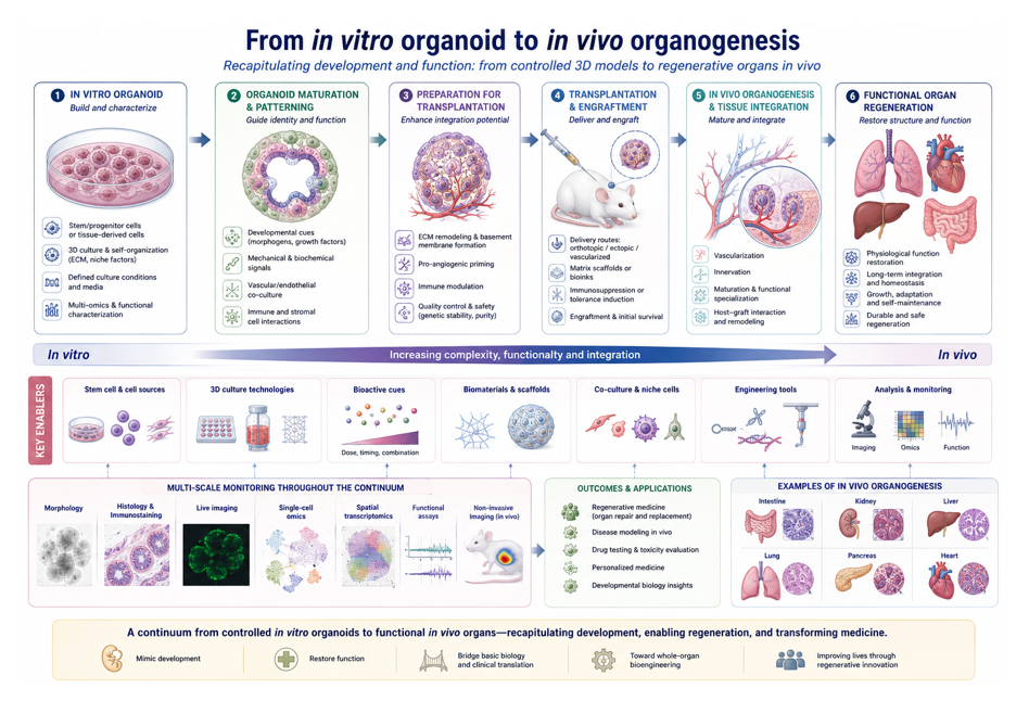
Figure 6: From In Vitro Organoids to In Vivo Organogenesis: Bridging Tissue Engineering and Functional Organ Regeneration.
Stem or tissue-derived cells are assembled into three-dimensional organoids and guided toward maturation and tissue-specific patterning through developmental cues, extracellular matrix, vascular and stromal interactions, and immune modulation; following transplantation, successful engraftment, vascularization, innervation, host–graft integration, and functional maturation enable progressive reconstruction of tissue architecture and physiological function. Multi-scale imaging, single-cell and spatial omics, functional assays, and in vivo monitoring provide quality control and mechanistic insight throughout this continuum, supporting applications in regenerative medicine, disease modeling, drug testing, personalized medicine, and whole-organ bioengineering.
Future Directions (Figure 7)
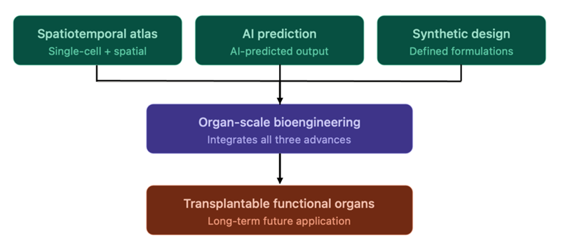
Figure 7: Convergence of future research directions toward organ-scale bioengineering.
Three complementary lines of future work — spatiotemporal secretome atlases integrating single-cell and spatial technologies, computational/AI-based prediction of secretome composition from cell state, and the design of fully synthetic, defined secretome formulations — converge to enable organ-scale bioengineering capability. Realizing this capability is a prerequisite for the long-term goal of transplantable, functional organs, which remains unachieved and depends on first resolving the causal and technical limitations.
Spatiotemporal secretome atlases integrating single-cell and spatial technologies
Addressing the localization problem 3 will likely require systematic integration of single-cell transcriptomic and proteomic data with spatial technologies capable of preserving positional information — spatial transcriptomics, imaging mass cytometry, and in situ sequencing — to construct organ- and stage-specific atlases of secretome ligand-receptor pairs mapped onto their actual anatomical position within a developing organ field [130-132]. Such atlases would allow paracrine circuits to be resolved quantitatively, capturing local ligand concentration, receptor expression gradients, and the effective signaling range of each secretome component. This would provide a substantially more rigorous evidentiary basis than the largely qualitative ligand-receptor pairing evidence that currently dominates the literature.
Computational and machine learning approaches to predict secretome composition from cell state
Given the combinatorial complexity of secretome output, computational approaches that predict secretome composition directly from transcriptomic, epigenomic, or culture-condition input data represent a promising complement to direct secretome measurement, which remains comparatively slow, expensive, and detection-limited [133-135]. Machine learning models trained on paired transcriptome-secretome datasets could in principle allow researchers and manufacturers to predict the therapeutic or morphogenetic secretome profile of a given cell population or priming condition without requiring exhaustive direct secretome characterization for every batch or condition, and could further support in silico screening of candidate priming strategies prior to experimental validation, narrowing the search space for empirical optimization.
Synthetic secretome formulations as a defined alternative to living cell products
The long-term logical extension of cell-free therapeutic strategies is the development of fully synthetic, defined secretome formulations — combinations of recombinant proteins, synthetic or engineered EVs, and small-molecule mimetics assembled to reproduce a validated therapeutic or morphogenetic signaling profile without depending on living, donor-derived cells as the production source at all. By design, such formulations would sidestep the donor-, tissue-, and culture-dependent variability discussed earlier, since their composition would be specified and manufactured directly rather than harvested from a variable biological source.
Realizing this goal, however, depends on first resolving the causation problem: a synthetic secretome can only be rationally designed once the specific components responsible for a given therapeutic or morphogenetic effect have been identified with confidence. This dependency underscores that the mechanistic and translational threads of this review remain fundamentally interdependent.
Conclusion
This review has argued for a specific reframing of stem and progenitor cell biology: that the secretome — the coordinated output of soluble factors, enzymes, and extracellular vesicles a cell releases into its environment — is not a passive byproduct of cellular activity but an active, instructive signal that shapes organ formation. The mechanistic evidence, spanning morphogen gradients, reciprocal epithelial-mesenchymal induction, vesicle-mediated signal delivery, and matrix remodeling, converges on a common principle: organ-level architecture emerges from the spatially and temporally structured exchange of secreted signals among interacting cell populations, rather than from a program encoded entirely within any single progenitor lineage. The organ-specific case studies together with the observation that organoid protocols function largely by empirically reconstituting these same secretome combinations, provide direct, convergent support for this principle across cardiac, renal, neural, and hepatopancreatic development.
This reframing carries practical weight. Once the secretome, rather than the cell itself, is recognized as the proximate mediator of much therapeutic and morphogenetic activity, a coherent translational agenda follows directly: cell-free and biomaterial-based delivery strategies become mechanistically motivated rather than merely convenient alternatives to cell transplantation, and secretome profiling becomes a plausible route to the standardized potency metrics that cell therapy manufacturing has long lacked — a gap this review has connected explicitly to the inconsistent clinical outcomes seen with mesenchymal stromal cell therapies. At the same time, the limitations counsel real caution: secretome heterogeneity, unresolved questions of causation versus correlation, persistent technical detection limits, and the gap between organoid and in vivo fidelity mean that secretome-based strategies are not a shortcut around the rigor that stem cell translation has historically lacked, but rather a more precise target for that rigor to be applied to.
The future directions — spatiotemporal secretome atlases, computational prediction of secretome output from cell state, and ultimately synthetic, defined secretome formulations — each represent incremental steps toward resolving these limitations rather than circumventing them. Taken together, this review suggests that the next phase of progress in regenerative medicine is unlikely to come primarily from identifying new stem cell sources or refining differentiation protocols alone, but from developing the tools to read, predict, and ultimately design the secretome signals that these cells use to build organs in the first place.
Funding Declaration
This research received no external funding.
References
- Mim TJ, Ahmad I, Basher SR, et al. Stem cells in organogenesis and regeneration. Stem Cell Res Ther 17 (2026): 85.
- Bagno LL, Salerno AG, Balkan W, et al. Mechanism of Action of Mesenchymal Stem Cells (MSCs): impact of delivery method. Expert Opin Biol Ther 22 (2022): 449-463.
- Wong RSY, Tan EW, Goh BH. Mesenchymal Stem Cell-Based Therapies: Challenges and Enhancement Strategies. Cell Biochem Biophys 84 (2026): 131-147.
- Bouwmeester T. The Spemann-Mangold organizer: the control of fate specification and morphogenetic rearrangements during gastrulation in Xenopus. Int J Dev Biol 45 (2001): 251-258.
- von Bubnoff A. The Spemann-Mangold organizer discovery and society. Cells Dev 178 (2024): 203906.
- Vizoso FJ, Eiro N, Cid S, et al. Mesenchymal Stem Cell Secretome: Toward Cell-Free Therapeutic Strategies in Regenerative Medicine. Int J Mol Sci 18 (2017): 1852.
- Daneshmandi L, Shah S, Jafari T, et al. Emergence of the Stem Cell Secretome in Regenerative Engineering. Trends Biotechnol 38 (2020): 1373-1384.
- Inomata H. Scaling of pattern formations and morphogen gradients. Develop Growth Differ 59 (2017): 41-51.
- Ségalat L. Loss-of-function genetic diseases and the concept of pharmaceutical targets. Orphanet J Rare Dis 2 (2007): 30.
- Su J, Song Y, Zhu Z, et al. Cell-cell communication: new insights and clinical implications. Signal Transduct Target Ther 9 (2024): 196.
- Ebner-Peking P, Krisch L, Wolf M, et al. Self-assembly of differentiated progenitor cells facilitates spheroid human skin organoid formation and planar skin regeneration. Theranostics 11 (2021): 8430-8447.
- Wang L, Chen A, Zhang D, et al. From Cells to Organoids: Approaches, Regulatory Mechanisms, Applications, and Challenges of Organoids. Cells 14 (2025): 1898.
- Krebs NJ, Neville C, Vacanti JP. Cellular Transplants for Liver Diseases. In: Cellular Transplantation. Academic Press (2007): 215-240.
- Nerger BA, Nelson CM. Engineered extracellular matrices: Emerging strategies for decoupling structural and molecular signals that regulate epithelial branching morphogenesis. Curr Opin Biomed Eng 13 (2020): 103-112.
- Kim HI, Park J, Zhu Y, et al. Recent advances in extracellular vesicles for therapeutic cargo delivery. Exp Mol Med 56 (2024): 836-849.
- Schank MB, Zhao J, Wang L, et al. Extracellular Vesicles: A Comprehensive Review of Their Origins, Functions, and Therapeutic Potential. Biomedicines 14 (2026): 495.
- Dellar ER, Hill C, Melling GE, et al. Unpacking extracellular vesicles: RNA cargo loading and function. J Extracell Biol 1 (2022): e40.
- Kumar MA, Baba SK, Sadida HQ, et al. Extracellular vesicles as tools and targets in therapy for diseases. Signal Transduct Target Ther 9 (2024): 27.
- Yang Q, Shi J, Xu L, et al. Pathways of Protein Secretion in Prokaryotes and Eukaryotes: Molecular Mechanisms, Biological Functions, and Therapeutic Opportunities. MedComm 7 (2026): e70798.
- Schäfer T, Zentgraf H, Zehe C, et al. Unconventional secretion of fibroblast growth factor 2 is mediated by direct translocation across the plasma membrane of mammalian cells. J Biol Chem 279 (2004): 6244-6251.
- Pallotta MT, Nickel W. FGF2 and IL-1β - explorers of unconventional secretory pathways at a glance. J Cell Sci 133 (2020): jcs250449.
- Tricarico C, Clancy J, D'Souza-Schorey C. Biology and biogenesis of shed microvesicles. Small GTPases 8 (2017): 220-232.
- Wang K. Extracellular Vesicle as Biomarker for Liver Diseases. Reference Module in Biomedical Sciences. Elsevier (2019).
- Scott KE, Fraley SI, Rangamani P. A spatial model of YAP/TAZ signaling reveals how stiffness, dimensionality, and shape contribute to emergent outcomes. Proc Natl Acad Sci U S A 118 (2021): e2021571118.
- Cao Y, Eble JM, Moon E, et al. Tumor cells upregulate normoxic HIF-1α in response to doxorubicin. Cancer Res 73 (2013): 6230-6242.
- Palomäki S, Pietilä M, Laitinen S, et al. HIF-1α is upregulated in human mesenchymal stem cells. Stem Cells 31 (2013): 1902-1909.
- Li J, Zhu HJ. Liquid Chromatography-Tandem Mass Spectrometry (LC-MS/MS)-Based Proteomics of Drug-Metabolizing Enzymes and Transporters. Molecules 25 (2020): 2718.
- Ranapangu B, Sandhu M, Paul AT. LC-MS-based proteomics: insights into natural product-directed cellular targeting. Drug Discov Today 30 (2025): 104534.
- Liu J, Allo B, Winnik MA. Development of Multiplexed Bead-Based Immunoassays for Profiling Soluble Cytokines and CD163 Using Mass Cytometry. ACS Meas Sci Au 2 (2022): 629-640.
- Comfort N, Cai K, Bloomquist TR, et al. Nanoparticle Tracking Analysis for the Quantification and Size Determination of Extracellular Vesicles. J Vis Exp (2021): 62447.
- Shen W, Guo K, Adkins GB, et al. A Single Extracellular Vesicle (EV) Flow Cytometry Approach to Reveal EV Heterogeneity. Angew Chem Int Ed Engl 57 (2018): 15675-15680.
- Zhang Y, Meng X, Greening DW, et al. Unveiling Heterogeneity: Innovations and Challenges in Single-Vesicle Analysis for Clinical Translation. J Extracell Vesicles 14 (2025): e70209.
- Rogers KW, Schier AF. Morphogen gradients: from generation to interpretation. Annu Rev Cell Dev Biol 27 (2011): 377-407.
- Mundhe G, Dunsing-Eichenauer V, Philippe JM, et al. Spatial patterning of contractility by a self-organized mechanogen activity gradient underlies Drosophila gastrulation. Nat Commun 17 (2026): 1721.
- Fantetti KN, Fekete DM. Members of the BMP, Shh, and FGF morphogen families promote chicken statoacoustic ganglion neurite outgrowth and neuron survival in vitro. Dev Neurobiol 72 (2012): 1213-1228.
- Lupo G, Novorol C, Smith JR, et al. Multiple roles of Activin/Nodal, bone morphogenetic protein, fibroblast growth factor and Wnt/β-catenin signalling in the anterior neural patterning of adherent human embryonic stem cell cultures. Open Biol 3 (2013): 120167.
- Volckaert T, De Langhe SP. Wnt and FGF mediated epithelial-mesenchymal crosstalk during lung development. Dev Dyn 244 (2015): 342-366.
- Jaeger J, Reinitz J. On the dynamic nature of positional information. Bioessays 28 (2006): 1102-1111.
- Christian JL. Morphogen gradients in development: from form to function. Wiley Interdiscip Rev Dev Biol 1 (2012): 3-15.
- Wedgwood KCA, Ashwin P. Morphogen-directed cell fate boundaries: slow passage through bifurcation and the role of folded saddles. J Theor Biol 549 (2022): 111220.
- Sprinzak D, Lakhanpal A, Lebon L, et al. Cis-interactions between Notch and Delta generate mutually exclusive signalling states. Nature 465 (2010): 86-90.
- Kandachar V, Roegiers F. Endocytosis and control of Notch signaling. Curr Opin Cell Biol 24 (2012): 534-540.
- Iluta S, Nistor M, Buruiana S, et al. Notch and Hedgehog Signaling Unveiled: Crosstalk, Roles, and Breakthroughs in Cancer Stem Cell Research. Life 15 (2025): 228.
- Lang C, Conrad L, Iber D. Organ-Specific Branching Morphogenesis. Front Cell Dev Biol 9 (2021): 671402.
- Yang L, Zhou F, Zheng D, et al. FGF/FGFR signaling: From lung development to respiratory diseases. Cytokine Growth Factor Rev 62 (2021): 94-104.
- Jones MR, Chong L, Bellusci S. Fgf10/Fgfr2b Signaling Orchestrates the Symphony of Molecular, Cellular, and Physical Processes Required for Harmonious Airway Branching Morphogenesis. Front Cell Dev Biol 8 (2021): 620667.
- Karner CM, Das A, Ma Z, et al. Canonical Wnt9b signaling balances progenitor cell expansion and differentiation during kidney development. Development 138 (2011): 1247-1257.
- Rumballe BA, Georgas KM, Combes AN, et al. Nephron formation adopts a novel spatial topology at cessation of nephrogenesis. Dev Biol 360 (2011): 110-122.
- Davis TK, Hoshi M, Jain S. To bud or not to bud: the RET perspective in CAKUT. Pediatr Nephrol 29 (2014): 597-608.
- Mills J, Capece M, Cocucci E, et al. Cancer-Derived Extracellular Vesicle-Associated MicroRNAs in Intercellular Communication: One Cell's Trash Is Another Cell's Treasure. Int J Mol Sci 20 (2019): 6109.
- Menjivar NG, Oropallo J, Gebremedhn S, et al. MicroRNA Nano-Shuttles: Engineering Extracellular Vesicles as a Cutting-Edge Biotechnology Platform for Clinical Use in Therapeutics. Biol Proced Online 26 (2024): 14.
- Takada S, Fujimori S, Shinozuka T, et al. Differences in the secretion and transport of Wnt proteins. J Biochem 161 (2017): 1-7.
- Maggio S, Canonico B, Ceccaroli P, et al. Modulation of the Circulating Extracellular Vesicles in Response to Different Exercise Regimens and Study of Their Inflammatory Effects. Int J Mol Sci 24 (2023): 3039.
- Castaño C, Novials A, Párrizas M. An Overview of Inter-Tissue and Inter-Kingdom Communication Mediated by Extracellular Vesicles in the Regulation of Mammalian Metabolism. Int J Mol Sci 24 (2023): 2071.
- Cruz L, Romero JAA, Iglesia RP, et al. Extracellular Vesicles: Decoding a New Language for Cellular Communication in Early Embryonic Development. Front Cell Dev Biol 6 (2018): 94.
- Edwards DR, Beaudry PP, Laing TD, et al. The roles of tissue inhibitors of metalloproteinases in tissue remodelling and cell growth. Int J Obes Relat Metab Disord 20 (1996): S9-S15.
- Łukaszewicz-Zając M, Mroczko B, Słowik A. Matrix metalloproteinases (MMPs) and their tissue inhibitors (TIMPs) in amyotrophic lateral sclerosis (ALS). J Neural Transm 121 (2014): 1387-1397.
- Kaczorowska A, Miękus N, Stefanowicz J, et al. Selected Matrix Metalloproteinases (MMP-2, MMP-7) and Their Inhibitor (TIMP-2) in Adult and Pediatric Cancer. Diagnostics 10 (2020): 547.
- Vempati P, Popel AS, Mac Gabhann F. Extracellular regulation of VEGF: isoforms, proteolysis, and vascular patterning. Cytokine Growth Factor Rev 25 (2014): 1-19.
- Bonnans C, Chou J, Werb Z. Remodelling the extracellular matrix in development and disease. Nat Rev Mol Cell Biol 15 (2014): 786-801.
- Hrovatin M, Bokalj B, Turk B. Proteolysis at the extracellular matrix interface: Molecular architects and regulators in health and disease. FEBS J 293 (2026): 3758-3787.
- Humphrey JD, Dufresne ER, Schwartz MA. Mechanotransduction and extracellular matrix homeostasis. Nat Rev Mol Cell Biol 15 (2014): 802-812.
- Verbakel J, de Boer J. Engineering stem cell metabolism using biomaterial-induced mechanotransduction. Cell Biomater 1 (2025): 100192.
- Yang F, Bao Z, Li Z, et al. Tailoring mechanical properties of hydrogels for wound repair from perspectives of simulating cellular microenvironment. Cell Rep Phys Sci 7 (2026): 103311.
- Axelrod JD. Delivering the Lateral Inhibition Punchline: It’s All About the Timing. Sci Signal 3 (2010): pe38.
- Iwasaki H, Suda T. Cancer stem cells and their niche. Cancer Sci 100 (2009): 1166-1172.
- Weichselberger V, Moore G, Tlili S, et al. Biophysics of organoids. Dev Cell 61 (2026): 24-41.
- Witman N, Zhou C, Grote Beverborg N, et al. Cardiac progenitors and paracrine mediators in cardiogenesis and heart regeneration. Semin Cell Dev Biol 100 (2020): 29-51.
- Behfar A, Perez-Terzic C, Faustino RS, et al. Cardiopoietic programming of embryonic stem cells for tumor-free heart repair. J Exp Med 204 (2007): 405-420.
- Poelmann RE, Lie-Venema H, Gittenberger-de Groot AC. The role of the epicardium and neural crest as extracardiac contributors to coronary vascular development. Tex Heart Inst J 29 (2002): 255-261.
- Olivey HE, Svensson EC. Epicardial-myocardial signaling directing coronary vasculogenesis. Circ Res 106 (2010): 818-832.
- Protze SI, Lee JH, Keller GM. Human Pluripotent Stem Cell-Derived Cardiovascular Cells: From Developmental Biology to Therapeutic Applications. Cell Stem Cell 25 (2019): 311-327.
- Srinivas S, Wu Z, Chen CM, et al. Dominant effects of RET receptor misexpression and ligand-independent RET signaling on ureteric bud development. Development 126 (1999): 1375-1386.
- Shakya R, Jho E, Kotka P, et al. The role of GDNF in patterning the excretory system. Dev Biol 283 (2005): 70-84.
- Smyth IM. Development of the metanephric kidney. Curr Top Dev Biol 143 (2021): 111-150.
- Cebrian C, Asai N, D'Agati V, et al. The number of fetal nephron progenitor cells limits ureteric branching and adult nephron endowment. Cell Rep 7 (2014): 127-137.
- Davis SN, Grindel SH, Viola JM, et al. Nephron progenitors rhythmically alternate between renewal and differentiation phases that synchronize with kidney branching morphogenesis. bioRxiv (2024): 2023.11.21.568157.
- Lysko DE, Putt M, Golden JA. SDF1 regulates leading process branching and speed of migrating interneurons. J Neurosci 31 (2011): 1739-1745.
- Marín O, Valiente M, Ge X, et al. Guiding neuronal cell migrations. Cold Spring Harb Perspect Biol 2 (2010): a001834.
- Mazur A, Bills EH, DeSchepper KM, et al. Astrocyte-Derived Thrombospondin Induces Cortical Synaptogenesis in a Sex-Specific Manner. eNeuro 8 (2021): ENEURO.0014-21.2021.
- Christopherson KS, Ullian EM, Stokes CC, et al. Thrombospondins are astrocyte-secreted proteins that promote CNS synaptogenesis. Cell 120 (2005): 421-433.
- Rossi JM, Dunn NR, Hogan BL, et al. Distinct mesodermal signals, including BMPs from the septum transversum mesenchyme, are required in combination for hepatogenesis from the endoderm. Genes Dev 15 (2001): 1998-2009.
- Wang J, Rhee S, Palaria A, et al. FGF signaling is required for anterior but not posterior specification of the murine liver bud. Dev Dyn 244 (2015): 431-443.
- Norgaard GA, Jensen JN, Jensen J. FGF10 signaling maintains the pancreatic progenitor cell state revealing a novel role of Notch in organ development. Dev Biol 264 (2003): 323-338.
- van Es JH, Wiebrands K, López-Iglesias C, et al. Enteroendocrine and tuft cells support Lgr5 stem cells on Paneth cell depletion. Proc Natl Acad Sci U S A 116 (2019): 26599-26605.
- Boonekamp KE, Dayton TL, Clevers H. Intestinal organoids as tools for enriching and studying specific and rare cell types: advances and future directions. J Mol Cell Biol 12 (2020): 562-568.
- Luvhengo T, Khan U, Marumo TK. Paneth Cells and Lgr5+ Intestinal Stem Cells in Radiation Enteritis. Appl Sci 13 (2023): 2758.
- Nakamura T, Sato T. Advancing Intestinal Organoid Technology Toward Regenerative Medicine. Cell Mol Gastroenterol Hepatol 5 (2017): 51-60.
- Xu X, Zhang Y, Huang G, et al. Advances and applications of gut organoids: modeling intestinal diseases and therapeutic development. Life Med 4 (2025): lnaf012.
- Brady MV, Vaccarino FM. Role of SHH in Patterning Human Pluripotent Cells towards Ventral Forebrain Fates. Cells 10 (2021): 914.
- Zheng Y, Zhang F, Xu S, et al. Advances in neural organoid systems and their application in neurotoxicity testing of environmental chemicals. Genes Environ 43 (2021): 39.
- Davies JA, Holland I, Gül H. Kidney organoids: steps towards better organization and function. Biochem Soc Trans 52 (2024): 1861-1871.
- Nakanoh H, Tsuji K, Fukushima K, et al. Kidney Organoids: Current Advances and Applications. Life 15 (2025): 1680.
- Behbahan IS, Duan Y, Lam A, et al. New Approaches in the Differentiation of Human Embryonic Stem Cells and Induced Pluripotent Stem Cells toward Hepatocytes. Stem Cell Rev Rep 7 (2011): 748-759.
- Dahl-Jensen S, Grapin-Botton A. The physics of organoids: a biophysical approach to understanding organogenesis. Development 144 (2017): 946-951.
- Zhao Z, Chen X, Dowbaj AM, et al. Organoids. Nat Rev Methods Primers 2 (2022): 94.
- Lin F. Accelerating Organoid Adoption: Set to Transform Cancer Drugs. Genet Eng Biotechnol News 46 (2026): 22-27.
- Lee SJ, Kim DM. Cell-Free Synthesis: Expediting Biomanufacturing of Chemical and Biological Molecules. Molecules 29 (2024): 1878.
- Prado-Yupanqui JW, Ramírez-Orrego L, Cortez D, et al. The Hidden Power of the Secretome: Therapeutic Potential on Wound Healing and Cell-Free Regenerative Medicine-A Systematic Review. Int J Mol Sci 26 (2025): 1926.
- Suhandi C, Wilar G, Elamin KM, et al. The Effect of Stem Cell Secretome on the Improvement of Diabetic Wound Recovery: A Systematic Review and Meta-Analysis of In Vivo Studies. Curr Ther Res Clin Exp 102 (2025): 100778.
- Lumban Gaol LM, Purba A, Diposarosa R, et al. Role of Hypoxic Secretome from Mesenchymal Stem Cells in Enhancing Tissue Repair: Regulatory Effects on HIF-1α, VEGF, and Fibroblast in a Sphincterotomy Rat Model. J Inflamm Res 17 (2024): 7463-7484.
- Rossello-Gelabert M, Escacena N, Igartua M, et al. Translational pathways for MSC secretome therapies: from bioprocessing to delivery. Trends Biotechnol (2026): S0167-7799(25)00501-3.
- Freudenberg U, Liang Y, Kiick KL, et al. Glycosaminoglycan-Based Biohybrid Hydrogels: A Sweet and Smart Choice for Multifunctional Biomaterials. Adv Mater 28 (2016): 8861-8891.
- Cao H, Duan L, Zhang Y, et al. Current hydrogel advances in physicochemical and biological response-driven biomedical application diversity. Signal Transduct Target Ther 6 (2021): 426.
- Farasati Far B, Safaei M, Nahavandi R, et al. Hydrogel Encapsulation Techniques and Its Clinical Applications in Drug Delivery and Regenerative Medicine: A Systematic Review. ACS Omega 9 (2024): 29139-29158.
- Sarsenova M, Kim Y, Raziyeva K, et al. Recent advances to enhance the immunomodulatory potential of mesenchymal stem cells. Front Immunol 13 (2022): 1010399.
- Liu Q, Liu G, Sun D, et al. Pretreated mesenchymal stromal cells and their secretome for kidney disease: mechanisms and applications. Stem Cell Res Ther 17 (2026): 140.
- González-González A, García-Sánchez D, Dotta M, et al. Mesenchymal stem cells secretome: The cornerstone of cell-free regenerative medicine. World J Stem Cells 12 (2020): 1529-1552.
- Da Silva K, Kumar P, Choonara YE. The paradigm of stem cell secretome in tissue repair and regeneration: Present and future perspectives. Wound Repair Regen 33 (2025): e13251.
- Alrushaid N, AlQurashi NA, Alobaidi BS, et al. Cell secretome as a potential anticancer therapeutic agent: composition, mechanisms, preclinical evidence, and translational challenges. Front Oncol 16 (2026): 1729022.
- Ho SS, Hung BP, Heyrani N, et al. Hypoxic Preconditioning of Mesenchymal Stem Cells with Subsequent Spheroid Formation Accelerates Repair of Segmental Bone Defects. Stem Cells 36 (2018): 1393-1403.
- Yang Y, Wu Y, Yang D, et al. Secretive derived from hypoxia preconditioned mesenchymal stem cells promote cartilage regeneration and mitigate joint inflammation via extracellular vesicles. Bioact Mater 27 (2023): 98-112.
- Raik S, Kaur B, Kumar S, et al. Secretome Derived From Mesenchymal Stem Cells Cultured as Monolayer Show Enhanced Bone Regeneration Compared to Secretome From 3D Spheroid – Clues From the Proteome. Adv Healthcare Mater 14 (2025): 2500885.
- Hazeri M, Zaman WSWK, Rezaei MA, et al. Low oxygen preconditioning of umbilical cord MSCs: from biological to mechanistic innovation. J Transl Med 24 (2026): 671.
- Chouaib B, Haack-Sørensen M, Chaubron F, et al. Towards the Standardization of Mesenchymal Stem Cell Secretome-Derived Product Manufacturing for Tissue Regeneration. Int J Mol Sci 24 (2023): 12594.
- Fernández-Pérez AG, Herrera-González A, López-Naranjo EJ, et al. Extracellular Vesicles from Different Mesenchymal Stem Cell Types Exhibit Distinctive Surface Protein Profiling and Molecular Characteristics: A Comparative Analysis. Int J Mol Sci 26 (2025): 3393.
- Farahat M, Brosset S, Chen Y, et al. Human iPSCs-derived mesenchymal stem cells promote skin regeneration and burn wound healing. NPJ Regen Med 10 (2025): 40.
- Choudhery MS, Arif T, Mahmood R. Aging puzzle: A closer look on the complex dilemma of autologous stem cell therapy. World J Stem Cells 18 (2026): 114119.
- Ibrahim R, Mndlovu H, Kumar P, et al. Cell Secretome Strategies for Controlled Drug Delivery and Wound-Healing Applications. Polymers 14 (2022): 2929.
- Da Silva K, Kumar P, Choonara YE. The paradigm of stem cell secretome in tissue repair and regeneration: Present and future perspectives. Wound Repair Regen 33 (2025): e13251.
- Schürz M, Danmayr J, Jaritsch M, et al. EVAnalyzer: High content imaging for rigorous characterisation of single extracellular vesicles using standard laboratory equipment and a new open-source ImageJ/Fiji plugin. J Extracell Vesicles 11 (2022): e12282.
- Chrzanowski W, Wolfram J. Rethinking Extracellular Vesicle Signaling. Adv Mater 38 (2026): e22172.
- Samiminemati A, Aprile D, Siniscalco D, et al. Methods to Investigate the Secretome of Senescent Cells. Methods Protoc 7 (2024): 52.
- Beutgen VM, Shinkevich V, Pörschke J, et al. Secretome Analysis Using Affinity Proteomics and Immunoassays: A Focus on Tumor Biology. Mol Cell Proteomics 23 (2024): 100830.
- Ramelow CC, Dammer EB, Xiao H, et al. Simultaneous profiling of native-state proteomes and transcriptomes of neural cell types using proximity labeling. Nat Commun 17 (2026): 4636.
- Feng R, Spieth L, Liu L, et al. Single-cell spatial transcriptomic profiling defines a pathogenic inflammatory niche in chronic active multiple sclerosis lesions. Immunity 58 (2025): 2989-3005.e10.
- Zhu Z, Cheng Y, Liu X, et al. Advances in the Development and Application of Human Organoids: Techniques, Applications, and Future Perspectives. Cell Transplant 34 (2025): 9636897241303271.
- Fan X, Hou K, Liu G, et al. Strategies to overcome the limitations of current organoid technology - engineered organoids. J Tissue Eng 16 (2025): 20417314251319475.
- Rashidan K, Ayadilord M, Hazrati A, et al. Immune and Immune-Integrated Organoids as NextGeneration Platforms for Disease Modeling. MedComm 6 (2025): e70531.
- Elmentaite R, Kumasaka N, Roberts K, et al. Cells of the human intestinal tract mapped across space and time. Nature 597 (2021): 250-255.
- Yoshida SY, Matsumoto K, Takagi S, et al. Whole-organ and whole-body 3D atlases enable cellome-wide profiling. Cell (2026).
- Li T, Yin J, Hao Y, et al. Single-cell sequencing and organoids: applications in organ development and disease. Mol Biomed 6 (2025): 134.
- Meng F, Uversky VN, Kurgan L. Comprehensive review of methods for prediction of intrinsic disorder and its molecular functions. Cell Mol Life Sci 74 (2017): 3069-3090.
- Poschmann G, Brenig K, Lenz T, et al. Comparative Secretomics Gives Access to High Confident Secretome Data: Evaluation of Different Methods for the Determination of Bona Fide Secreted Proteins. Proteomics 21 (2021): e2000178.
- Macharia TN, Duong TA, Moleleki LN. In silico secretome analyses of the polyphagous root-knot nematode Meloidogyne javanica: a resource for studying M. javanica secreted proteins. BMC Genomics 24 (2023): 296.