Rizwan Ahmad1*, Ashok Kumar Sah2 and Haseeb Ahsan3
1Department of Biochemistry, UCBMSH, Dehradun, India
2Department of Medical lab Technology, Amity University, Haryana, India
3Department of Biochemistry, Faculty of Dentistry, Jamia Millia Islamia, New Delhi, India
*Corresponding Author: Rizwan Ahmad, Department of Biochemistry, UCBMSH, Dehradun, India
Received: 14 October 2016; Accepted: 19 October 2016; Published: 24 October 2016
Advanced glycated end products (AGEs) are molecules formed from the non-enzymatic reaction of reducing sugar with free amino group of proteins, lipids and nucleic acids. The AGEs play crucial role in the pathogenesis of cardiovascular, kidney, Alzheimer’s, neurological, diabetes and joints diseases and aging process. Depletion of cellular antioxidant GSH led to increase binding of glucose derivatives to DNA. DNA can be cross-linked with different substances, some of the nucleotide AGEs are N2-carboxymethyl oxyguanosine, 5-glycolyl deoxycytidine etc. Glycation of DNA evolves from the formation of Amadori products finally culminating into DNA AGEs. Glycation of DNA yields characteristics type of nucleotide adducts which are indicator of many abnormal conditions such as oxidative stress, arthritis etc. The glyoxal and methyl glyoxals are the earlier form of DNA glycation products which are derived from the reducing carbohydrate compounds. The AGEs and their product associated with the mutation, break down of DNA strand, cytotoxicity and various inflammatory conditions. High concentration of accumulated products observed in diseases like diabetes, uremia, atherosclerosis, asthma, arthritis, myocardial infarction, nephropathy, retinopathy, periodontitis, neuropathy and other inflammatory conditions. The decline rate of DNA repair mechanisms and persistence of DNA lesion with increased age of persons enhance the rate of nucleotide glycation.
Glycation; Diabetes; DNA; Nucleotides; AGE; Amadori products
1. Introduction
Glycation is the product of a reducing sugar molecule such as fructose or glucose binding to free amino groups of protein, nucleic acid or lipid molecule without the controlling action of an enzyme sometimes also called non-enzymatic glycosylation [1, 2]. Glycation reaction was first discovered by the French chemist LC Maillard in 1912 [3]. Advanced glycated end products (AGEs) are molecules which are formed from combining with any biomolecules having free amino group with the available reducing sugar molecules without enzymatic. AGEs products yields characteristics type of nucleotide adducts which are indicator of many abnormal conditions such as oxidative stress, arthrits etc [4]. AGEs causes damage to DNA which is associated with important factors for mutagenesis, carcinogenesis and diabetes mellitus [5]. Previously, it was suggested that glycation of DNA resulting in the formation of nucleotide AGEs is associated with an increase in mutation frequency and cytotoxicity [6].
DNA glycation is a toxic compound called as glycotoxin may contribute to the toxicity of several anti-tumor agents and over expression of enzymatic anti-glycation defense with multi drug resistance in major classes of tumors. Improved understanding of glycation may give a direction on decreasing the risk factors and pathophysiological conditions associated with the formation of nucleotide adducts [6]. Heat processed food products are the very important source of exogenous glycation, which literally means 'outside the body', may also be referred to as "dietary" or "pre-formed glycation". Cooked and combined food products like combination of sugars with proteins and fats which enhance the formation of exogenous AGEs are potent carcinogens [2]. Recently it has been showed that the exogenous glycations and AGEs were important contributors to inflammation and diseased states.
Although most of the research work has been done with reference to diabetes, these results are most likely important for many others, as exogenous AGEs are implicated in the initiation of retinal dysfunction, cardiovascular diseases, type 2 diabetes, and many other age related chronic diseases [4]. The food preservatives, flavoring agents, colouring agents and other additives which are present in food and food products are considered as exogenous dietary glycation end product (dAGEs) [7]. It is shown that very high amount of dAGEs is present in donuts, barbecued meats, cake, and dark colored soda pop [8].
1.1 Endogenous glycation
The monosaccharides or simple sugars such as glucose, fructose and galactose are mainly responsible for the glycation in the blood which are digested and absorbed from the small intestine, are endoogenous glycation. The glycation capecity of fructose and galactose is approximately ten times more of glucose, which is main body fuel in humans [4, 9]. Reaction with sugars and free amino group on DNA is the first step in the evolution of these molecules through a complex series of very slow reactions in the body known as Amadori reactions, Schiff base reactions, and Maillard reactions, which may all lead to advanced glycated end products (AGEs). Some AGEs are very useful compounds, but others are very reactive as they are responsible in many age-related chronic diseases such as: cardiovascular diseases (the endothelium, fibrinogen and collagen are damaged), type 2 diabetes mellitus (beta cell damage), Alzheimer's disease (amyloid proteins are side products of the reactions progressing to AGEs), cancer (acrylamide and other side products are released) [9]. These types of diseases are the result of very basic and initial level of glycation which interferes with cellular functioning throughout the body and release of compound which are highly oxidizing such as hydrogen peroxide [10].It effects the nuclic acid by modifying, mutation and breakind the strand of DNA which are the process on glycation [11]. Glycation of DNA gives rise to characteristic nucleotide adduct, some of which have been found to increase in oxidative stress [12-14]. Potent glycating agents in physiological system are a-oxoaldehydeglyoxal, methylglyoxal and 3-deoxyglucosone [15]. The nucleotide most reactive under physiological condition is deoxyguanosine in vivo the major nucleotide AGEs are the imidazopurinone derivatives [16, 17]. Glycation also promotes deamination of cytidine to deoxyuridine [18]. The N(2)-carboxymethyl-2'-deoxyguanosine (CMdG) and N(2)-(1-carboxyethyl)-2'-deoxyguanosine (CEdG) adducts of deoxyguanosine are estimated to be more stable. These adducts are also formed by glycation of DNA with glucose [17, 19] and ascorbic acid [20]; since, glucose and other saccharide derivatives fragment to form glyoxal and methylglyoxal in the early stages of glycation reaction-the Namiki pathway of glycation [21].
The formation of CEdG was associated with depurination of DNA [19]. Glyoxal and methylglyoxal react preferentially with deoxyguanosine, but other nucleotides are formed at high a-oxoaldehyde concentration [18, 22]. At higher concentration of glyoxal and methylglyoxal, interstand cross links formed in duplex DNA for which no structures have been characterized [18, 20]. The physiological significance of the cross link formation is probably very limited (Fig. 1).
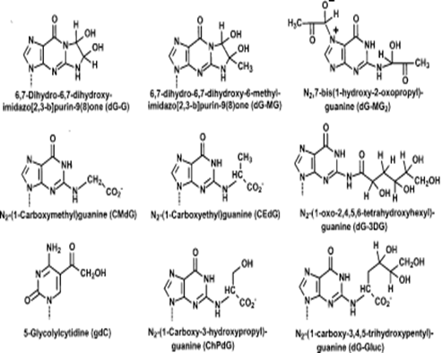
Figure 1: Effect of glycation on nucleotides advanced glycation end products
AGEs are also observed in vitro in similar process while reacting the nucleosides, proteins and lipids with sugar molecules[23-25]. The exocyclic amino group of 2-deoxyguanosine is more prone to glycation reactions leading to the formation of N2-carboxyethyl, N2-carboyxmethyl, N2-(1-carboxy- 3-hydroxypropyl) and N2-(1-carboxy-3,4,5-trihydroxy-pentyl) modifications as well as cyclic dicarbonyl adducts [25-28]. The two glycation product diastereomers of N2-carboxyethyl-2-deoxyguanosine (CEdGA,B) are stable products that are formed from glucose, ascorbic acid, dihydroxyacetone (DHA), or methylglyoxal [27, 29]. First reaction and product of CEdG is formed in vivo was put forward by Schneider et al. [30].
The technique immunoaffinity chromatography coupled with HPLC is very useful for detection of CedG level in human urine[31]. Using the same technique, CEdG level are also detected in laboratory by using different sample such as the genomic DNA of human smooth muscle cells and bovine aorta endothelium cells [15]. Furthermore, AGEs have been observed in many specific cell and tissues of human body such as in the kidney cells from patients with diabetic nephropathy and in the aorta of diabetic and non-diabetic hemodialysis patients. Moreover, CEdG has also been detected in human kidneys and aorta by immunohistochemistry with the monoclonal CEdG antibody.
Thus, CEdG was the only DNA-AGEs which had been detected in vivo so far by the immunoaffinity chromatography coupled with HPLC technique [32].
Glucose 6-phosphate is reducing sugar which directly modifies the DNA molecules and the result can be seen in bacteria and eukaryotic cells [33-35]. Furthermore, CEdG is the major glycation product which affects the DNA under physiological conditions. This chemically defined modification induces damage in DNA such as strand breaks and mutation which are responsible at least in part for the observed reduction of the transformation rate.
The DNA AGEs are potentially genotoxic compounds which could be directly linked to alterations in the DNA structure and functionality. CEdG selectively introduced into the DNA, destabilized the N-glycosidic bond between carboxyethylguanine and the sugar-phosphate DNA backbone, leading to the specific loss of the modified guanine (depurination). Consequently, CEdG-modified is responsible single-strand breaks results DNA mutations [28, 36]. The transformation of bacterial cells by glycated plasmids resulted in an increased mutation frequency caused by insertions, deletions, as well as multiple species [33].
Likewise, when DNA and pre-treated glyoxal or sugars is transected into mammalian cells, single-base substitutions and the transposition of an Alu-containing element were observed [37, 38]. In vivo, the embryonic malformation and teratogenicity is the effect the 3-deoxyglucosone is a glucose degradation product that may be related to DNA AGEs [39], leading to a defect in transcription and the loss of gene function due to mutations [40]. Wani et al [12] have studied the in vitro glycation of DNA. The isolated DNA was incubated with different concentrations of glucose for various time intervals and showed marked changes in the ultraviolet spectroscopic analysis (Table 1). For the measurement of early glycated products in DNA, NBT reduction assay, developed for quantification of fructosamine in serum glycosyl proteins, was performed (Figure 2) [41].
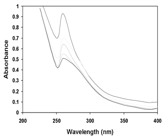
Figure 2: UV absorption of Native DNA (1), DNA incubated with 50 mM (3), 100 mM (5), 150 mM (4) and 200 mM (2) glucose (adapted and modified from Wani et al., 2012).
| Property | Native DNA | Glycated DNA |
|---|---|---|
| Absorbance ratio (A260/280) | 1.7 | 1.32 |
| Melting Temperature (Tm) | 81oC | 85oC |
| % Hypochromicity | - | 51% |
| Amadori products (nmol/mg DNA) |
3 | 8.6 |
Table 1: Characterization of Native and Glycated Human DNA (adapted and modified from Wani et al., 2012)
Aldehydicapurinic/apirimidinidic (AP) sites can be directly induced in DNA by reactive oxygen species [42] or caused by glycosylase enzymes upon removal of oxidized bases [43]. Studies done by other groups have reported that the Amadori product formation in glycated samples is higher than in native DNA samples [41]. Preliminary studies observed that glycation is non-enzymatic product of free sugar and free amnio group on nucleic acids. Furthermore, incubation of DNA with reducing sugars can generate light absorbing molecules that transmit light (chromophores) and fluorescent chemical compounds that re-emit (fluorophores) with structural properties similar to advanced glycated protein products [35, 44]. Ashraf et al. [45] have demonstrated that glucose can cause extensive damage to DNA structure leading to strand breaks and formation of DNA Amadori products and DNA advanced glycation end products (Fig. 3 and 4). The change in absorbance observed is due to changes of molecular structure of glycated DNA which may be due to addition or substitution of molecules of DNA by free radical mediated damage to sugar-phosphate backbone followed by partial unfolding of double helix and hence, exposure of chromophoric bases [46, 47].
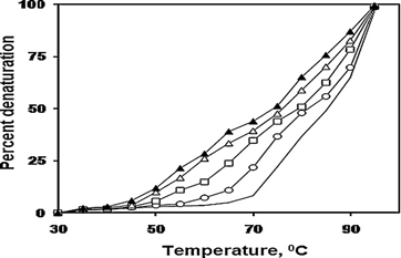
Figure 3: Melting profile of native DNA (-) and one (o), two (▢), three (△) and four weeks old (▴) glycated-DNA (adapted and modified from Ashraf et al., 2012)
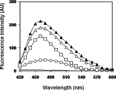
Figure 4: Fluorescence emission spectra of native DNA (-) and one (o), two (▢), three (△) and four weeks old (▴) glycated-DNA (adapted and modified from Ashraf et al, 2012)
strand breaks in DNA [52-53]. Moreover, glycation of DNA evolves from the formation of Amadori products finally culminating into DNA AGEs [45].The formation of hydroxyl radical which is highly reactive compound produced from hydrogen peroxide is known as Fenton reaction. Oxygen radicals are generated in the process of auto oxidation of sugar [54, 55]. The cellular protection against the oxidative damage in DNA is achieved by enzymatic and non-enzymatic antioxidants [56].
The presence of superoxide anion radical was detected by the reaction between sugars and DNA by reduction of NBT (Figure 5) [57]. The superoxide radical formation is suggested to be involved in the Millard reaction of DNA with sugars [58]. Treatment of glycated DNA with scavengers such as glutathione (GSH), catalase and mannitol causes removal of free radicals generated during the glycation reaction (Figure 6). GSH showed scavenging activity in our studies which is in accordance to the previous findings that the depletion of cellular antioxidant GSH led to increased binding of glucose derivatives to DNA [59, 60]. Therefore, the presence of nucleotide AGEs in DNA may be associated with an increased mutation frequency, strand breaks in DNA leading to cytotoxicity [61]. Strains of E. coli that accumulate high levels of glucose- 6-phosphate, which is particularly active in forming AGEs, demonstrate increased levels of transposition, which is the relocation of the transposable genetic element in DNA [62].
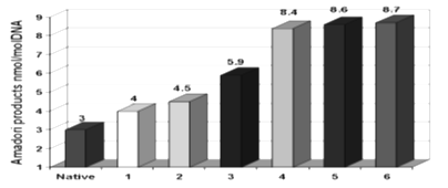
Figure 5: Formation of Amadori products during incubation of glycated DNA with NBT for two days (1), four days (2), one week (3), two weeks (4), three weeks (5) and four weeks (6) (adapted and modified from Wani et al., 2012)
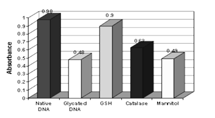
Figure 6: Effect of various free radical scavengers on glycated DNA (adapted and modified Wani et al., 2012) In mammalian cells, it has been found that AGEs formation on DNA is responsible for insertions containing repetitive sequences of the Alu family that have been found to disrupt human genes [63]. The possibility that AGEs may induce genetic rearrangements in vivo has important implications, for example, as a possible cause of congenital malformations in infants of poorly controlled insulin dependent diabetic mothers [64].
1.2 Pathophysiological effects and significance of nucleotide glycation
Nucleotide glycation is more marked in diseases associated with accumulation of glycating agents to a high concentration in diabetes and uremia [21]. It was found that the incubation of endothelial cells under hyperglycemic conditions induced strand breaks in DNA [65]. A similar effect was seen in lymphocytes from diabetic patients with poor glycemic control [66]. The effect may be due to oxidative damage, since increased levels of 8-hydroxydeoxyguanosine (8-OHdG) were found in the lymphocyte DNA of diabetic persons [67]. Increased nucleotide glycation by oxoaldehydes in diabetes has also been implicated in impaired growth of keratinocytes and fibroblasts in skin lesions [68] and perinatal mortality due to teratogenicity [69]. Intake of high amounts of non-fiber carbohydrate compounds that form AGEs which are associated with high risk of colorectal cancer for both men and women [70].
The decline in DNA repair mechanism and persistence of lesions in DNA with increased age [71] may also exacerbate the effect of nucleotide glycation. Many cross-links glycation products are now considered as causative factors and high risk factors in the development of many age-related and diabetic disorders, especially those which are associated with the cardiovascular and renal systems, by causing biochemical and structural alterations on proteins. Accumulation of glycation products in the sub endothelium of plasma proteins such as albumin, low-density lipoprotein (LDL) and immunoglobulin G (IgG) are the major cause of narrowing of arterial lumen [72].It was observed that they get trapped in basement membranes by covalently cross-linking to AGEs on collagen [73].
AGEs are activated by some receptors i.e mononuclear cells (monocytes and macrophages) which were first shown to bear specific receptors for AGEs. These receptors activate macrophages which lead to the production of interlukin -1 insulin like growth factor [74, 75]. These glycated products are llocalized on receptors and suggests that their interaction play significant role in the pathogenesis of diabetic vascular lesions [74].
1.3 Diabetes mellitus
Glycated DNA is also considered to be the pathogenic factor for diabetes mellitus [15]. Diabetes, especially when there is poor control of blood sugar levels, leads to a series of debilitating complications. It affects the eyes, kidneys, heart, and nerves and is a major cause of blindness, renal failure, stroke and heart attack. Diabetes mellitus, a condition characterized mainly by a quantitative deficiency in insulin secretion or a resistance to insulin action, is estimated to affect 5-7% of the population. Microangiopathy, the micro vessel disease in diabetes, includes retinopathy, nephropathy, and neuropathy and in type 1 patient the first signs of these complications may develop even in adolescence (Figure 7). The glycation of hemoglobin is a continuous process occurring throughout the 120 days life span of the red cells [10]. Glycated hemoglobin (HbA1c) concentration proportionally increased in diabetic patients with hyperglycemia and reflects the extent as well as management of diabetic conditions.
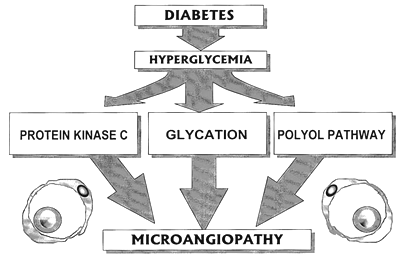
Figure 7: Pathways implicated in hyperglycemia induced diabetic microvascular disease (adapted and modified from Gugliucci, 2000)
Fructosamine (any glycated protein in its primary stage) indicates medium term glycaemia control and AGEs are indicator of long term glycemic control in diabetes mellitus [73]. If AGEs also form on DNA in vivo, deleterious effects on gene expression may occur, and intracellular AGEs formation on cell proteins may thus affect DNA functions [76-81]. The extremely rapid rate of AGEs formation on histone proteins points in this direction. It was shown that AGEs levels was three times greater in histone of rat liver sample taken from one month hyperglycemic condition than those of their age-matched controls [82]. The increased conditions of teratogenesis which is due to glycation play the most important role of pathogenesis of diabetes mellitus [82]. Increasing evidence suggests that glycation and oxidative stress may be linked to the sorbitol pathway, in which glucose is reduced to sorbitol which is then converted to fructose, may also lead to diabetic complications [83-87].
1.4 Atherosclerosis
The AGEs play a major role in the development of atherosclerosis, kidney, vascular and neurological diseases in both diabetes and ageing processes in humans as well as animals [88]. Previous studies have shown that AGEs play a significant role in the formation and progression of atherosclerosis lesions (Figure 8). Increased AGE accumulation in the diabetic vascular tissues has been associated with the changes in endothelial cells macrophages and smooth muscles cell function [63]. AGE-cross link formation results in arterial stiffening with the loss of elasticity of large vessels [89]. The arterial stiffness has recently been shown to be reversed by the administration of another anti-AGE class of compound called AGE-breakers [90]. Anti-glycation agents Carnosine (beta-alanyl L-histidne) a naturally occurring molecule is an aldehyde scavenger and appears to be protective against some crosslinks of carbohydrates with DNA [91]. The cellular protection against glycation of DNA is provided by enzymes that detoxify reactive glycating agents (the glyoxalase system and aldehyde reductase and dehydrogenases) [6, 92, 93].
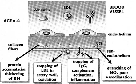
Figure 8: The role of AGE products in microangiopathy and macroangiopathy (adapted and modified from Gugliucci, 2000)
2. Conclusion
The glycation of DNA give rise to characteristic nucleotide adducts, some of which have been found to increase in oxidative stress. Aldehydic apurinic/apirimidinidc (AP) sites can be directly induced in DNA by ROS or caused by glycosylase enzymes upon removal of oxidized bases. The glycation causes damage to DNA which is associated with mutagenesis, carcinogenesis and is also considered to be a pathogenic factor for diabetes mellitus. It has been suggested that glycation of DNA resulting in the form of nucleotide AGEs is associated with increase in mutation frequency and cytotoxicity.DNA glycation is a glytoxin that may contribute to the toxicity of several clinical cytotoxic anti-tumor agents and overexpression of some enzymatic anti-glycation defence which is associated with multi drug resistance in major classes of tumors. Improved understanding of DNA glycation may give direction on decreasing the risk of tumor associated with dietary factors.
Acknowledgement
The authors are thankful to Prof. Rashid Ali for the core concept of this research. Rashid Ali is grateful to his PG students Adil and Shaheena of SBSPGI, Dehradun for the literature review. Rashid Ali is also thankful to the Dean and Vice Dean of Oman Medical College (in academic partnership with WVU, USA) for their help in preparation of this manuscript. Thanks are also due to the management of UCBMSH, Dehradun for the support in the publication of this review.References
- Bailey AJ. Molecular mechanisms of ageing in connective tissues. Mech Ageing Dev 122 (2001): 735-755.
- Brownlee M, Cerami A, Vlassara H. Advanced products of nonenzymatic glycosylation and the pathogenesis of diabetic vascular disease. Diabetes Metab Rev 4 (1988): 437-451.
- Maillard LC. Action of amino acids on sugars. Formation of melanoidins in a methodical way. Compt Rend 154 (1912): 66-68.
- Fu MX, Requena JR, Jenkins AJ, Lyons TJ, Baynes JW, et al. The advanced glycation end product, Nepsilon-(carboxymethyl) lysine, is a product of both lipid peroxidation and glycoxidation reactions. J Biol Chem 271 (1996): 9982-9986.
- Thornalley PJ. Protein and nucleotide damage by glyoxal and methylglyoxal in physiological systems - role in ageing and disease. Clin Lab 45 (1990): 263-273.
- Murata-Kamiya N, Kamiya H, Kaji H, Kasai H. Glyoxal, a major product of DNA oxidation, induces mutations at G:C sites on a shuttle vector plasmid replicated in mammalian cells. Nucleic Acids Res 25 (1997): 1897-902.
- Melpomeni Peppa. Glucose, Advanced Glycation End Products, and Diabetes Complications: What Is New and What Works, Clinical Diabetes 21 (2003): 187.
- Koschinsky T, He CJ, Mitsuhashi T, Bucala R, Liu C, et al. Orally absorbed reactive glycation products (glycotoxins): an environmental risk factor in diabetic nephropathy. Proc Natl Acad Sci 94 (1997): 6474-6479.
- O'Brien J, Morrissey PA. Nutritional and toxicological aspects of the Maillard browning reaction in foods. Crit Rev Food Sci Nutr 28 (1989): 211-248.
- Ulrich P, Cerami A. Protein glycation, diabetes, and aging. Recent Prog Horm Res 56 (2001): 1-21.
- Ames BN. Dietary carcinogens and anticarcinogens. Oxygen radicals and degenerative diseases. Science. 221 (1983): 1256-1264.
- Wani A, Mushtaq S, Ahsan H, Ahmad R. Biochemical studies of in vitro glycation of human DNA. Asian J Biomed Pharma Sci 2 (2012): 23-27.
- Wani A, Mushtaq S, Ahsan H, Ahmad R. Characterization of Human Glycated DNA modified with Peroxynitrite. Helix 1 (2013): 221-225.
- Mistry N, Podmore I, Cooke M, Butler P, Griffiths H, et al. Novel monoclonal antibody recognition of oxidative DNA damage adduct, deoxycytidine-glyoxal. Lab Invest 83 (2003): 241-250.
- Thornalley PJ. The clinical significance of glycation. Clin Lab 45 (1999): 263-273.
- Schneider M, Quistad GB, Casida JE. N2,7-bis(1-hydroxy-2-oxopropyl)-2'-deoxyguanosine: identical noncyclic adducts with 1,3-dichloropropene epoxides and methylglyoxal. Chem Res Toxicol 11 (1988): 1536-1542.
- Papoulis A, al-Abed Y, Bucala R. Identification of N2-(1-carboxyethyl) guanine (CEG) as a guanine advanced glycosylation end product. Biochemistry 34 (1995): 648-655.
- Kasai H, Iwamoto-Tanaka N, Fukada S. DNA modifications by the mutagen glyoxal: adduction to G and C, deamination of C and GC and GA cross-linking. Carcinogenesis 19 (1998): 1459-1465.
- Seidel W, Pischetsrieder M. DNA-glycation leads to depurination by the loss of N2-carboxyethylguanine in vitro. Cell Mol Biol (Noisy-le-grand) 44 (1998): 1165-1170.
- Seidel W, Pischetsrieder M. Immunochemical detection of N2-[1-(1-carboxy) ethyl] guanosine, an advanced glycation end product formed by the reaction of DNA and reducing sugars or L-ascorbic acid in vitro. Biochim Biophys Acta 1425 (1998): 478-484.
- Thornalley PJ, Langborg A, Minhas HS. Formation of glyoxal, methylglyoxal and 3-deoxyglucosone in the glycation of proteins by glucose. Biochem J 344 (1999): 109-116.
- Krymkiewicz N. Reactions of methylglyoxal with nucleic acids. FEBS Lett 29 (1973): 51-54.
- Singh R, Barden A, Mori T, Beilin L. Advanced glycation end-products. Diabetologia 44 (2001): 129-146.
- Lee AT, Cerami A. The formation of reactive intermediates of glucose-6-phosphate and lysine capabale of rapidly reacting with DNA. Mutat Res 179 (1987): 151-158.
- Knerr T, Lerche H, Pischetsrieder M, Severin T. Formation of a novel colored product during the Maillard reaction of D-glucose. J Agric Food Chem 49 (2001): 1966-1970.
- Oschs S, Severin T. Reaction of 2??Deoxyguanosine with Glyceraldehyde. Liebigs Annalen der Chemie 8 (1994): 851-853.
- Larisch B, Pischetsrieder M, Severin T. Formation of guanosine adducts from L-ascorbic acid under oxidative conditions. Bioorg Med Chem Lett 7 (1997): 2681-2686.
- Seidel W, Pischetsrieder M. DNA glycation leads to depurination by the loss of N2-carboxyethylguanine in vitro. Cell Mol Biol (Noisy-le-Grand, France) 44 (1998): 1165-1170.
- Seidel W, Pischetsrieder M. Immunochemical detection of N2-[1-(1-carboxy)ethyl]guanosine, an advanced glycation end product formed by the reaction of DNA and reducing sugars or L-ascorbic acid in vitro. Biochim Biophys Acta 1425 (1998): 478-484.
- Schneider M, Thoss G, Hubner-Parajsz C, Kientsch-Engel R, Stahl P, et al. Determination of glycated nucleobases in human urine by new monoclonal antibody specific for N2-carboxyethyl-2’-deoxyguanosine. Chem Res Toxicol 17 (2004): 1385-1390.
- Schneider M, Georgescu A, Bidmon C, Tutsch M, Fleischmann EH, et al. Detection of DNA bound advanced glycation end products by immunoaffinity chromatography coupled to HPLC-diode array detection. Mol Nutr Food Res 50 (2006): 424-429.
- Li H, Nakamura S, Miyazaki S, Morita T, Suzuki M, et al. N2-carboxyethyl-2’-deoxyguanosine, a DNA glycation marker, in kidneys and aortas of diabetic and uremic patients. Kidney Int 69 (2006): 388-392.
- Bucala R, Model P, Russel M, Cerami A. Modification of DNA by glucose-6-phosphate induces DNA rearrangements in an Escherichia coli plasmid. Proc Natl Acad Sci 82 (1985): 8439-8442.
- Lee AT, Cerami A.Elevated glucose 6-phosphate levels are associated with plasmid mutations in vivo. Proc Natl Acad Sci 84 (1987): 8311-8314.
- Morita J, Kashimura N. The Maillard reaction of DNA with D-fructose 6-phosphate. Agricult Biol Chem 55 (1991): 1359-1366.
- Pischetsrieder M, Seidel W, Munch G, Schinzel R. N(2)-(1-Carboxyethyl)deoxyguanosine, a nonenzymatic glycation adduct of DNA, induces single-strand breaks and increases mutation frequencies. Biochem Biophys Res Commun 264 (1999): 544-549.
- Murata-Kamiya N, Kaji H, Kasai H. Types of mutations induced by glyoxal, a major oxidative DNA-damage product, in Salmonella typhimurium. Mutat Res 377 (1997): 13-16.
- Bucala R, Lee AT, Rourke L, Cerami A. Transposition of an Alu-containing element induced by DNA-advanced glycosylation endproducts. Proc Natl Acad Sci 90 (1993): 2666-2674.
- Eriksson U, Wentzel P, Minhas HS, Thornalley P. Teratogenicity of 3-deoxyglucosone and diabetic embryopathy. Diabetes 47 (1998): 1960-1966.
- Breyer V, Frischmann M, Bidmon C, Schemm A, Schiebel K. Analysis and biological relevance of advanced glycation end-products of DNA in eukaryotic cells. FEBS J 275 (2008): 914-925.
- Johnson RN, Metcalf PA, and Baker JR. Fructosamine: a new approach to the estimation of serum glycosyl protein. An index of diabetic control. Clin Chim Acta 127 (1982): 87-95.
- Breen AP and Murphy JA. Reaction of oxyl radicals with DNA. Free Rad Biol Med 18 (1995): 1033-1077.
- Rosenquist TA, Zharkov DO and Grollman AP. Cloning and characterization of a mammalian 8-oxoguanine DNA glycosylase. Proc Natl Acad Sci 94 (1997): 7429-7434.
- Bucala R, Model P, Cerami A. Modification of DNA by reducing sugars: a possible mechanism for nucleic acid aging and age-related dysfunction in gene expression. Proc Natl Acad Sci 81 (1984): 105-109.
- Ashraf JM, Arif B, Dixit K, Moinuddin, Alam K. Physicochemical analysis of structural changes in DNA modified with glucose. Int J Biol Macromol 51 (2012): 604-611.
- Pethig R, Szent-Gyorgyi A. A. Electronic properties of the casein-methylglyoxal complex. Proc Natl Acad Sci 74 (1977): 226-228.
- Ahmad S, Moinuddin, Dixit K, Shahab U, Alam K, Ali A. Genotoxicity and immunogenicity of DNA-advanced glycation end products formed by methylglyoxal and lysine in presence of Cu2+. Biochem Biophys Res Comm 407 (2011): 568-574.
- Schmitt A, Schmitt J, Munch G, Gasic-Milenkovic J. Characterization of advanced glycation end products for biochemical studies: side chain modifications and fluorescence characteristics. Anal Biochem 338 (2005): 201-215.
- Schmitt A, Gasic-Milenkovic J, Schmitt J. Characterization of advanced glycation end products: mass changes in correlation to side chain modifications. Anal Biochem 334 (2005): 101-106.
- Alam K, Ali A, Ali R. Effect of hydroxyl radical on the antigenicity of native DNA. FEBS Lett 319 (1993): 66-70.
- Habib S, Moinuddin, Ali A, Ali R. Preferential recognition of peroxynitrite modified human DNA by circulating autoantibodies in cancer patients. Cell Immunol 245 (2009): 117-123.
- Mironova R, Niwa T, Handzhiyski Y, Sredovska A, Ivanov I. Evidence for non-enzymatic glycosylation of Escherichia coli chromosomal DNA. Mol Microbiol 55 (2005): 1801-1811.
- Li Y, Cohenford MA, Dutta U, Dain JA. In vitro nonenzymatic glycation of guanosine 5'-triphosphate by dihydroxyacetone phosphate. Anal Bioanal Chem 392 (2008): 1189-1196.
- Kashimura N, Morita J, Komana T. Autoxidation and phagocidal action of some reducing sugar phosphates. Carbohydrate Res 70 (1979): C3-C7.
- Thornally PJ. Monosaccharide autoxidation in health and disease. Environ Health Perspect 64 (1985): 297-309.
- Sies H. Antioxidant in Disease Mechanism and Therapeutic Strategies, 1996. Academic Press, San Diego, CA.
- Nakayama T, Kimura T, Kodama M, Nagata C. Generation of hydrogen peroxide and superoxide anions from active metabolites of naphthalamines and aminoazo dyes: its possible role in carcinogenesis. Carcinogenesis 4(1983): 765-769.
- Morita J and Kashimura N. The Maillard reaction of DNA with D-fructose-6-phosphate. Agric Biol Chem 55 (1991): 1359-1366.
- Shires TK, Tresnak J, Kaminsky M, Herzog SL and Truc-Pharma B. DNA modification in vivo by derivatives of glucose: enhancement by glutathione depletion. FASEB J 4 (1990): 3340-3345.
- Samuni A, Aronovitch J, Godinger D, Chevion M, Czapski G. On the cytotoxicity of vitamin C and metal ions. A site-specific Fenton mechanism. Eur J Biochem 137 (1983: 119-124.
- Lee AT, Cerami A. In vitro and in vivo reactions of nucleic acids with reducing sugars. Mutat Res 238(1990): 185-191.
- Lee AT, Cerami A. Induction of gamma delta transposition in response to elevated glucose-6-phosphate levels. Mutat Res 249 (1991): 125-133.
- Bucala R, Lee AT, Rourke L, Cerami A. Transposition of an Alu-containing element induced by DNA-advanced glycosylation endproducts. Proc Natl Acad Sci 90 (1993): 2666-2670.
- Lee AT, Plump A, DeSimone C, Cerami A, Bucala R. A role for DNA mutations in diabetes-associated teratogenesis in transgenic embryos. Diabetes 44 (1995): 20-24.
- Lorenzi M, Montisano DF, Toledo S, Barrieux A. High glucose induces DNA damage in cultured human endothelial cells. J Clin Invest 77 (1986): 322-325.
- Lorenzi M, Montisano DF, Toledo S, Wong HC. Increased single strand breaks in DNA of lymphocytes from diabetic subjects. J Clin Invest 79 (1987): 653-656.
- Dandona P, Thusu K, Cook S, Snyder B, Makowski J, et al. 1996. Oxidative damage to DNA in diabetes mellitus. Lancet 347 (1996): 444-445.
- Roberts MJ, Wondrak GT, Laurean DC, Jacobson MK, Jacobson EL. DNA damage by carbonyl stress in human skin cells. Mutat Res 522 (2003): 45-56.
- Eriksson UJ, Wentzel P, Minhas HS, Thornalley PJ. Teratogenicity of 3-deoxyglucosone and diabetic embryopathy. Diabetes 47 (1998): 1960-1966.
- Borugian MJ, Sheps SB, Whittemore AS, Wu AH, Potter JD, Gallagher RP. Carbohydrates and colorectal cancer risk among Chinese in North America. Cancer Epidemiol Biomarkers Prev 11 (2002): 187-193.
- Mullaart E, Lohman PH, Berends F, Vijg J. DNA damage metabolism and aging. Mutat Res 237 (1990): 189-210.
- Cai W, He JC, Zhu L, Peppa M, Lu C, Uribarri J, Vlassara H. High levels of dietary advanced glycation end products transform low-density lipoprotein into a potent redox-sensitive mitogen-activated protein kinase stimulant in diabetic patients. Circulation 110 (2004): 285-291.
- He CJ, Koschinsky T, Buenting C, Vlassara H. Presence of diabetic complications in type 1 diabetic patients correlates with low expression of mononuclear cell AGE-receptor-1 and elevated serum AGE. Mol Med 7 (2001): 159-168.
- Vlassara H. The AGE-receptor in the pathogenesis of diabetic complications. Diabetes Metab Res Rev. 17 (2001): 436-443.
- Schmidt AM, Yan SD, Yan SF, Stern DM. The biology of the receptor for advanced glycation end products and its ligands. Biochim Biophys Acta 1498 (2000): 99-111.
- Giardino I, Edelstein D, Brownlee M. Nonenzymatic glycosylation in vitro and in bovine endothelial cells alters basic fibroblast growth factor activity. J Clin Invest 94 (1994): 110-117.
- Giardino I, Edelstein D, Brownlee M. BCL- 2 expression or antioxidants prevent hyperglycemia- induced formation of intracellular advanced glycation endproducts in bovine endothelial cells. J Clin Invest 97 (1996): 1422-1428.
- Giardino I, Edelstein D, Horiuchi S, Araki N, Brownle M. Vitamin E prevents diabetesinduced formation of arterial wall advanced glycation end products. Diabetes 44 (1994): 73A.
- Bucala R, Model P, Cerami A. Modification of DNA by reducing sugars: a possible mechanism for nucleic acid aging and age-related dysfunction in gene expression. Proc Natl Acad Sci 81 (1984): 105-109.
- Bucala R, Model P, Russel M, Cerami A. Modification of DNA by glucose-6-phosphate induces DNA rearrangements in an E. coli plasmid. Proc Natl Acad Sci 82 (1985): 8439-8442.
- Mullokandov EA, Franklin WA, Brownlee M. Damage of DNA by the glycation products of glyceraldehyde-3-phosphate and lysine. Diabetologia 37 (1994): 145-149.
- Gugliucci A, Bendayan M. Histones from diabetic rats contain increased levels of advanced glycation products. Biochem Biophys Res Commun 212 (1995): 56-62.
- Gugliucci A. Glycation as the glucose link to diabetic complications. JAOA 100 (2000): 621-634.
- Kaneto H, Fujii J, T M, Miyazawa N, Islam K, Kawasaki Y, et al. Reducing sugars trigger oxidative modification and apoptosis in pancreatic beta-cells by provoking oxidative stress through the glycation reaction. Biochem J 320 (1996): 855-863.
- Makita Z, Vlassara H, Rayfield E, Cartwright K, Friedman E, Rodby R, et al. Hemoglobin-AGE: a circulating marker for advanced glycosylation. Science 258 (1992).
- McPherson J, Shilton B, Walton D. Role of fructose in glycation and cross-linking of proteins. Biochemistry 27 (1988): 1901-1907.
- Takagi Y, Kashiwagi A, Tanaka Y, Asahina T, Kikkawa R, YS. Significance of fructoseinduced protein oxidation and formation of advanced glycation end product. J Diabetes Complications 9 (1995): 87-91.
- Uribarri J, Peppa M, Cai W, Goldberg T, Lu M, He C, Vlassara H. Restriction of dietary glycotoxins reduces excessive advanced glycation end products in renal failure patients. J Am Soc Nephrol 14 (2003): 728-731.
- Das UN. Metabolic syndrome X: an inflammatory condition? Curr Hypertens Rep 6 (2004): 66-73.
- Halliwell B and Gutteridge JMC. (Eds.) In: Free Radicals in Biology and Medicine 2nd ed., Clarendan Press, Oxford, London 2 (1989): 1-543.
- Suzuki K, Koh YH, Mizuno H, Hamaoka R, Taniguchi N. Overexpression of aldehyde reductase protects PC12 cells from the cytotoxicity of methylglyoxal or 3-deoxyglucosone. J Biochem. 123 (1998): 353-357.
- Feather MS, Flynn TG, Munro KA, Kubiseski TJ, Walton DJ. Catalysis of reduction of carbohydrate 2-oxoaldehydes (osones) by mammalian aldose reductase and aldehyde reductase. Biochim Biophys Acta. 1244 (1995): 10-16.
- Ansari NA, Dash D. Amadori Glycated Proteins: Role in Production of Autoantibodies in Diabetes Mellitus and Effect of Inhibitors on Non-Enzymatic Glycation. Aging Disease 4 (2013): 50-56.