Bizarre Anca Vasculitis in a Young Woman: Aggressive Treatment is Justified
Ermira Aliu1, Erjola Bolleke Likaj2*, Larisa Shehaj3, Alma Idrizi2, Myftar Barbullushi2, Alecya Anyim4, Juna Musa5, Jaclyn Tan-Wohlers6, Grace Lin 6, Erisa Kola7, Elsie Tachie Mensah4, Mohamed Gamal 8, Sedjana Rrustemaj 9 Edlira Lashi 10, Mohamed Subahi 11, Alma Lamo 12 Blerina Kastrati13
1Women’s Center for Radiology, Orlando, USA
2Department of Nephrology, University Hospital Center & Mother Teresa, Tiran, Albania
3Deparment of Nephrology, Salus Hospital, Tiran, Albania
4Department of Internal Medicine, Academic Hospitalist, Mayo Clinic, Rochester, USA
5Department of Endocrinology, Diabetes and nutrition, Mayo Clinic, Rochester, USA
6Department of Laboratory Medicine and Pathology, Mayo Clinic, Rochester, USA
7Department of Pathology, University Hospital Center & Mother Teresa, Tiran, Albania
8Department of Radiology, Mayo Clinic, Rochester, US
9 Post -Baccalaureate Student, Cleveland State University, USA
10 Department of Nursery and Physiotherapy, Faculty of Medical Sciences, Tirana, Albania.
11 Department of Cardiovascular Disease, Mayo Clinic Rochester Minnesota
12 Department of Pharmacy, American Hospital, Tirane, Albania
13 Department of Endocrinology, Hospital and University clinical service of Kosovo, Pristina, Kosovo
*Corresponding Author: Erjola Bolleke Likaj, Department of Nephrology, University Hospital Center & Mother Teresa, Tiran, Albania.
Received: 28 February 2025; Accepted: 07 March 2025; Published: 30 July 2025
Article Information
Citation: Ermira Aliu, Erjola Bolleke Likaj, Larisa Shehaj, Alma Idrizi, Myftar Barbullushi, Alecya Anyim, Juna Musa, Jaclyn Tan-Wohlers, Grace Lynn, Erisa Kola, Elsie Tachie Mensah, Mohamed Gamal, Sedjana Rrustemaj, Edlira Lashi, Mohamed Subahi, Alma Lamo, Blerina Kastrati. Bizarre Anca Vasculitis in a Young Woman: Aggressive Treatment is Justified. Archives of Nephrology and Urology. 8 (2025): 86-88.
DOI: 10.26502/anu.2644-2833100
Share at FacebookAbstract
ANCA vasculitis is a multisystem autoimmune disease characterized by inflammation involving medium and small sized blood vessels. The inflammation is typically caused by self-reacting auto antibodies that bind to neutrophil and make them overly activated. There are 3 subtypes of ANCA mediated vasculitis namely granulomatosis with polyangiitis, microscopic polyangiitis and eosinophilic granulomatosis with polyangiitis. In PR3- ANCA positive vasculitis, the most affected sites are upper respiratory tract, lungs and kidney. A 34-year-old woman presented with cough and dyspnea, and features of AKI among many other symptoms. RT-PCR was negative and pulmonary CT scan showed peripheral ground glass opacities. Kidney Biopsy showed crescentic glomerulonephritis after which COVID-19 infection and SLE were ruled out. PR3-ANCA was positive, and she was diagnosed with granulomatosis with polyangiitis. Her renal and lung function improved eventually after aggressive administration of methylprednisolone followed by cyclophosphamide. Here we present a case of ANCA vasculitis with multisystem involvement, initially COVID-19 confusing the radiological diagnosis.
Keywords
COVID-19; ANCA vasculitis; Blood vessels; RT-PCR; PR3- ANCA; Radiological diagnosis
Article Details
1. Introduction
Antineutrophil cytoplasmic antibody (ANCA) associated vasculitis (AAV) is a group of multi-system autoimmune diseases of characterized by formation of granulomas and inflammation of small arteries, arterioles, venules, and capillaries that can present in any age. These diseases share a common pathology with focal necrotizing lesions such as in the lungs, a capillaritis may cause alveolar hemorrhage; within the glomerulus of the kidney, a crescentic glomerulonephritis may cause acute renal failure; in the dermis, a purpuric rash or vasculitic ulceration may occur which can give rise to a broad array of varied clinical symptoms and signs related to a systemic inflammatory response, end organ microvascular injury, or the mass effect of granulomas. The relative rarity and heterogeneous nature of AAV poses diagnostic challenges and clinical misdiagnosis and mortality rates are high. The major target organs are kidneys affected by AAV and renal involvement may be as high as 80%. To establish the diagnosis, a combination of clinical assessment with serological testing is needed, and a tissue biopsy many times confirms the diagnosis. Without therapy, prognosis is poor but treatments, typically immunosuppressants, have improved survival, albeit with considerable morbidity from glucocorticoids and other immunosuppressive medications. We present here a case of a young woman with several dissociated symptoms that finally resulted in ANCA associated vasculitis and after fortunately treated early the patient aggressively partially recovered; is now on regular follow up.
2. Case Presentation
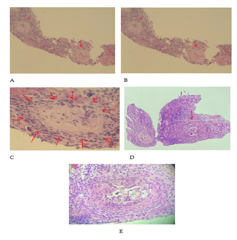
A 34-year-old woman presented to the Emergency Unit October end with a 2-week history of difficulty in breathing, intermittent cough, lower back pain, leg paresthesias, slightly swollen legs and decrease in the urine volume. She also reported history of prolonged sinusitis with epistaxis since February, which was treated with several antibiotics. She has a medical history of severe anemia and arthralgia and was receiving treatment for the same. Physical examination was notable for pallor, bilateral limb edema, and high blood pressure (140/70mmHg) with normal heart rate(72/min) and normal oxygen saturation (98%). Admission lab were significant for low hemoglobin (Hb=5.9 g/dl), raised BUN (161.6 mg/dl), raised creatinine (4.92 mg/dl), raised urea (161.6 mg/dl) and a positive direct Coombs test. Urine analysis showed microscopic hematuria (RBC full for field), positive urinary sediment and 24-hour urine revealed albuminuria (1788 mg/24 hr), thus hinting towards nephritic syndrome and acute renal failure. Peripherally placed ground glass foci of size 1.5-2 cm were identified on the pulmonary CT scan (November 11, 2021) and were initially thought to be due to COVID-19 infection however, the RT/PCR test and serologic tests for IgM and IgG antibodies for SARS-CoV-2 were negative. The management was undertaken by the Nephrology Unit and blood transfusions were immediately initiated. Owing to the multisystemic involvement along with kidney impairment seen at this age, Systemic Lupus Erythematosus was considered as a differential diagnosis. Further lab investigations were unremarkable with a negative extractable nuclear antigen (ENA), double stranded DNA (anti-ds DNA), ANCA myeloperoxidase (2.54 u/ml), HBsAg/anti-HCV, HIV antibodies, anti-TPO, negative C3 and C4, apart from raised rheumatoid factor (35.2 UI/ml) along with 8-fold increase in the PR3-ANCA levels (76.84 U/ml). Abdominal ultrasound showed no abnormalities in the liver, spleen or gallbladder, the kidneys were normal in size (L: 11.8cm, R: 11.4 cm) but showed edema of pyramids in the both kidneys. Her symptoms continued unabated with worsening of her renal function; thus, hemodialysis was initiated, and kidney biopsy was performed at the same time. The patient was initially empirically treated with a 500mg bolus of Methylprednisolone for three consecutive days followed by 0.5mg/Kg. The kidney biopsy showed pronounced inflammatory lymphocytic infiltrate, tubular necrosis and arteriolar hyalinosis. Immunohistochemistry was negative for IgG, IgM, IgG4 and C3d and Crescentic glomerulonephritis in over 50% of the glomerulus. Kidney biopsy along with positive PR3-ANCA confirmed the suspicion for Granulomatosis with polyangiitis. Cyclophosphamide was infused every 2 weeks for 6 cycles along with Mesna for protecting the bladder from hemorrhagic cystitis. The patient also developed two episodes of sanguineous expectoration, probably due to pulmonary hemorrhage and thus was decided to proceed with plasma exchange. Five plasmapheresis sessions were performed every 3 to 5 days and hemodialysis was offered intermittently for coming months. The renal function and anemia improved over several weeks. PR-3 ANCA returned to normal within 5 weeks. There was resolution of microscopic hematuria, and the CT scans showed marked improvement. Hemodialysis was discontinued at creatinine levels of 2 mg/dl and the patient was discharged. She continued to visit the hospital to receive cyclophosphamide. She recovered well and after 6 cycles the medication was switched to Azathioprine (2 mg/kg), and methylprednisolone was being gradually tapered. On this regime, patient developed pancytopenia from herpes infection due to immunosuppression and hence was switched to Mycophenolate Mofetil (2g/day). The patient is completely in remission and is doing well (Figure 1).
Figure 1: ALight microscopy of rapid progressive glomerulonephritis, 7 glomeruli showing global sclerosis with an architectural landmarks and capillaries obliterated. There is marked interstitial inflammation. (Hematoxylin-Eosin staining). Light microscopy showing wide destruction of Bowman’s capsule. Glomerular tuft is collapsed by extracapillary cellular proliferation (crescents). (Hematoxylin-Eosin staining).
3. Discussion
Granulomatous Polyangiitis Vasculitis (GPV) or Wegener Granulomatosis (WG) is an unusual disease mostly affecting middle age groups, involving 3 in 100000 in the United States [1].
The diagnosis of Granulomatous Polyangiitis Vasculitis (GPV) can be challenging due to multisystemic involvement and atypical presentation. Our patient presented with diverse range of vague symptoms such as epistaxis, cough, dyspnea and swelling of legs. This might result in delayed diagnosis, permanent damage to organs which can lead to a fatal outcome. Early evaluation for PR-3ANCA can facilitate in establishment of definitive diagnosis along with timely interventions to halt the progression of disease. However, a positive C-ANCA does not imply diagnosis of GPA. The gold standard is tissue biopsy, although the risk to benefit ratio due to iatrogenic complications and feasibility need to be weighed [2,3]. In our case, multisystemic involvement with anemia and renal symptoms in a young female raised suspicion for SLE, however the negative antibody screen along with positive PR-3 ANCA along with prolonged history of nasal bleed and pulmonary symptoms lead to high index of suspicion for ANCA vasculitis. The suspicion was further confirmed by the kidney biopsy and development of new symptoms like coughing up blood.
The treatment typically includes 2 phases: Induction phase: aims puts the disease into remission and includes treatment with a systemic corticosteroid and immunosuppressant combination. It can last for 3-6 months depending upon the clinical response. The Maintenance Phase aims to consolidate the remission and focuses on minimizing relapse risk. Effective induction therapy with corticosteroid in combination with cyclophosphamide and rituximab have shown to transform 5-year survival rate>80% [4]. Our patient showed remarkable improvement in symptoms and clinical parameters while managed aggressively with the above regimen. The intensity of the approach should be tailor made for each patient as per the type and gravity of the disease as excessive treatment can cause side effects whereas undertreatment may cause early relapse and treatment failure.
Lastly, an open-minded approach with great index of suspicion by the members of multidisciplinary team can aid in early diagnosis and effective treatment.
4. Conclusion
Diagnosing ANCA vasculitis could be challenging because of its rarity and vagueness in its multimodal presentation. Especially amidst of COVID, it poses more confusion because of the alikeness of the sign symptoms between the diseases. Here we present a case of bizarre ANCA vasculitis, which was diagnosed after exclusion and was treated aggressively with steroid and immunotherapy. Our aim with this case report is to bring in attention the ambiguity of ANCA vasculitis and raise concern in its early diagnosis.
References
- Cotch MF, Hoffman GS, Yerg DE, et al. The epidemiology of Wegener's granulomatosis. Estimates of the five-year period prevalence, annual mortality, and geographic disease distribution from population-based data sources. Arthritis & Rheumatism 39 (1996): 87-92.
- Uppal S, Saravanappa N, Davis JP, et al. Pulmonary Wegener's granulomatosis misdiagnosed as malignancy. BMJ 322 (2001): 89.
- van den Heuvel H. Wegener’s granulomatosis – another challenging case. BMJ Case Reports 2011 (2011): bcr0520114284.
- Comarmond C, Cacoub P. Granulomatosis with polyangiitis (Wegener): Clinical aspects and treatment. Autoimmunity Reviews 13 (2014):1121-5.

