Ahmedelmustafa A Musa*, Alaa A Al-Taie
Clinical Imaging Department, Hamad Medical Corporation, Doha, Qatar
*Corresponding Author: Ahmedelmustafa A Musa, Clinical Imaging Department, Hamad Medical Corporation, Doha, Qatar
Received: 30 November 2021; Accepted: 12 January 2022; Published: 25 January 2022
Calcaneal inferior spurs are bony outgrowth that can arise from multiple sites. Compressive and traction forces are theorized to be the etiology behind their genesis. Blunt trauma to the plantar aspect of the foot can lead to calcaneal inferior spur fracture. We present a case of a 38-years-old male presenting inability to bear weight on his left foot with point tenderness after falling from a 3-meters height. Radiographic examination revealed fracture of calcaneal inferior spur, from which the patient was conservatively treated successfully. Fracture inferior calcaneal spur is a rare cause of heel pain. The reviewed literature showed how both conservative and surgical managements are used, the former more frequently.
Calcaneal spur; Heel pain; Spur
The calcaneus is the largest tarsal bone in the foot, where it handles weight-bearing when in an upright position. It contains six surfaces through which articulation with the talus and cuboid bones is provided along with multiple ligamentous and muscles attachments [1]. This sentinel position of the calcaneus makes it vulnerable to different kinds of pathologies: arthritic, neurologic, systemic, and most importantly traumatic [2].
Calcaneal bone spurs, or enthesophytes, are bony outgrowths that arise from the calcaneal tuberosity in relation to the attachment of the Achilles tendon and/or plantar fascia. Although the pathophysiology is poorly understood, studies in the literature postulate a positive relationship to increasing age, athletic activity, obesity, and osteoarthritis as well as possible genetic control [3, 4]. Association of calcaneal spurs to heel pain is a point of debate: Menz et al. [5] observe 4.6 times increased likelihood to find a calcaneal spur when a patient presents with heel pain. On the other hand, Osborne et al. [6] report 46% of the asymptomatic population to have calcaneal spurs.
1. Case report
We report a 38 years old male who presented to our emergency department after falling from 3-meters height and landed on his feet. He also reported hitting his head after landing. He complained of a scalp laceration, a painful left heel, and an inability to bear weight on it. On physical examination, there was point tenderness at the plantar aspect of the calcaneum without superficial changes of inflammation. Range of motion, as well as neurovascular assessment, were preserved. The patient's past medical history was only positive for diabetes mellitus which was controlled with Metformin. The scalp laceration was managed by glue after being cleaned, and a lateral radiograph of the ankle joint was performed. It showed a mildly displaced fracture of an inferior calcaneal spur (Figure 1). This initial radiograph was reported as normal. Hence, the orthopaedic surgeon applied a back slap and discharged the patient on non-steroidal anti-inflammatory drugs and no weight-bearing instructions, with a follow-up appointment at the outpatient clinic in two weeks.
After two weeks, the patient had another lateral radiograph of the ankle that showed similar findings to the previous radiograph (Figure 2). As the patient denied tenderness or significant impact on his activity, the decision to continue conservative management was mutually agreed upon.
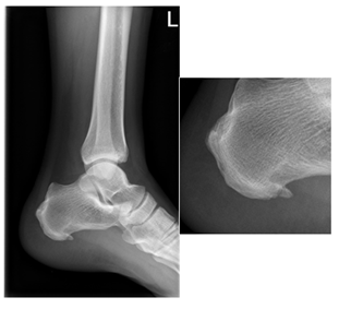
Figure 1: Lateral radiograph of the left ankle. Fracture of inferior/planter calcaneal spur. Zoomed-in image of the fracture.
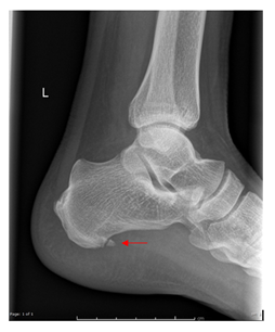
Figure 2: Lateral radiograph of the left ankle. Mildly displaced fracture of inferior calcaneal spur.
2. Discussion
Calcaneal inferior spurs are an entity that is commonly seen in the population. They most commonly arise from the medial aspect of the plantar calcaneal tuberosity. Abreu et al. report that inferior spurs arise from insertions sites of the abductor digiti minimi, flexor digitorum brevis; between these muscles and the plantar fascia; or within the plantar fascia itself (least common) [7]. Although relation to these muscles’ insertions might suggest traction forces to be the culprit, multiple studies have downplayed the rule of traction. Instead, compressive forces admixed with above mentioned positively associated factors are thought to be the main pathophysiology of inferior spurs [8].
Fracture of inferior spurs is a rare cause of heel pain. Few case reports of similar presentation [2, 9-13] are present in the available English language literature. While reviewing these reports, a common theme of blunt trauma applied to the sole (slipping and landing on feet, fall from height, sudden forceful stomping of the foot while attempting to run) can be postulated to be the main factor. As for management, only Subasi et al. [13] have approached their case surgically and excised the fractured spur. The remaining reports, as well as our case, adopted a conservative approach that included: short casts, reduction of physical activity and weight-bearing, employing RICE (rest, ice, compression, and elevation) therapy, and non-steroidal anti-inflammatory drugs. Additionally, others [2, 9] have added local steroid and plasma injections, and extracorporeal shock wave therapy to their management. The importance of such fracture rises from how easily it can be missed as a cause of manageable heel pain such as in our case. Careful assessment of calcaneal spurs in patients presenting with heel pain after blunt trauma to the plantar aspect of the foot will ensure prompt identification, management, and prevention of possible complications (non-union).
References
- Kumar R, Matasar K, Stansberry S, et al. The calcaneus: normal and abnormal. RadioGraphics 11 (1991): 415-440.
- Arican M, Turhan Y, Karaduman ZO. A Rare Cause of Heel Pain: A Calcaneal Spur Fracture. Journal of the American Podiatric Medical Association 109 (2019): 172-173.
- Weiss E. Calcaneal spurs: Examining etiology using prehistoric skeletal remains to understand present day heel pain. The Foot 22 (2012): 125-129.
- Benjamin M, Toumi H, Ralphs JR, et al. Where tendons and ligaments meet bone: attachment sites ('entheses’) in relation to exercise and/or mechanical load. Journal of Anatomy, 208 (2006): 471-490.
- Menz HB, Zammit GV, Landorf KB, et al. Plantar calcaneal spurs in older people: longitudinal traction or vertical compression? Journal of Foot and Ankle Research 1 (2008).
- Osborne H, Breidahl W, Allison G. Critical differences in lateral X-rays with and without a diagnosis of plantar fasciitis. Journal of Science and Medicine in Sport 9 (2006): 231-237.
- Abreu M, Chung C, Mendes L, et al. Plantar calcaneal enthesophytes: new observations regarding sites of origin based on radiographic, MR imaging, anatomic, and paleopathologic analysis. Skeletal Radiology 32 (2003): 13-21.
- Kirkpatrick J, Yassaie O, Mirjalili SA. The plantar calcaneal spur: a review of anatomy, histology, etiology and key associations. Journal of Anatomy 230 (2017): 743-751.
- Nawghare S. Non-union of a Plantar Calcaneal Spur Fracture: A case report. The Foot & Ankle Journal 1 (2008).
- Esmadi M, Ahsan H, Ahmad DS. Fracture of calcaneal Spur. Hongkong J Radiol 15 (2012): 144-6.
- Burks JB, Buk A. Bilateral fractures of the infracalcaneal exostosis. The Journal of Foot [Amp] Ankle Surgery, 42 (2003): 43-44.
- Subasi M, Kapukaya A, Kesemenli C, et al. Nadir görülen kiriklar [Rarely seen fractures]. Ulusal travma dergisi = Turkish journal of trauma & emergency surgery: TJTES, 7 (2001): 282-284.
- Vaish A, Vaishya R. Bilateral broken calcaneal spurs. BMJ Case Reports 13 (2020): 234138.