Michael A Petrou1*, Markella Marcou2, Lawko Brwa Ahmed3
1Scientific Collaborator: Microbiology & Immunology Lecturer, School of Dentistry, European University Cyprus, 6 Diogenous Str., 2404 Egkomi, Nicosia Cyprus
2Medical Microbiologist, Archbishop Makarios III Hospital, 6 Korytsas Street, 2012 Strovolos, Nicosia, Cyprus
3Research Assistant, Scientific Collaborator, European University Cyprus, 6 Diogenous Str., 2404 Egkomi, Nicosia Cyprus
*Corresponding Author: Michael A Petrou PhD. Scientific Collaborator: Microbiology & Immunology Lecturer, School of Dentistry, European University Cyprus, 6 Diogenous Str., 2404 Egkomi, Nicosia Cyprus.
Received: 26 June 2026; Accepted: 29 June 2026; Published: 06 July 2026
Microbial transmission has been studied extensively during and after the SARS-CoV-2 pandemic in the last six years. A new way of transmission of enteric pathogens and enveloped viruses has been suggested, that is infectious aerosols escaping the toilet after flushing, which if true would have serious health consequences. Unlike most enteric pathogens, corona viruses reach the alimentary canal in a similar manner to some parasites in that their life cycle includes the lung from where they are coughed and then swallowed. Many pathogens, bacterial and parasites, can survive the harsh environment of the stomach, however very few enveloped viruses can survive, such as Corona and Hepatitis B viruses, thus the probability of their transmission within the toilet environment, even though unlike bacteria and parasites there is no evidence that they can multiply in the intestines, is justifiable. This study after the addition of bacteria the toilet bowl and stain to the toilet bowl and the cistern used simple routine Microbiological monitoring methods; settle plates and absorbent tissue, to determine whether bacteria or stain containing aerosols detected within the toilet environment escaped from the bowl or the cistern of the toilet after flushing, when the lid was kept open. Many different bacteria and fungi were detected, which were both in numbers and types very similar to those recovered prior to flushing. When bacteria or stain was added to the bowl neither bacteria nor stain was detected, whereas when the stain was added to the cistern aerosols were detected on the white tissue, indicating that they originated from the cistern. In conclusion wash-down low pressure modern gravity-flash toilets do not pose any health threat when the toilet is flushed with the lid open.
SARS-Cov-2; Hepatitis B viruses; Serratia marcescens; Covid 19
SARS-Cov-2 articles; Hepatitis B viruses articles; Serratia marcescens articles; Covid 19 articles.
1. Introduction
It is clear from daily experience that flushing a toilet generates strong turbulence within the bowl and that the speed and flow dynamics of the water in the bowl depend on the type and design of the toilet, with different tank pressure that could be low 10 to 15 psi, pressure assisted 25 to 40 psi or with an electric pump that increases the tank pressure to 60 psi. Anyone who used a squat toilet (also known as squatting, Indian or Turkish toilet) during a sunny day with sunlight entering the room is aware of the presence of rainbows due to water droplets mist ejected in the atmosphere after flushing; the question is: “do the droplets come from the toilet bowl or directly from the cistern?”
The possibility of the toilet being a source of contagion has been studied extensively in the last 70 years [1-4]. The first study to show bioaerosol production during toilet flushing led to improvements in the toilet design not only to prevent escape of microbes but also to reduce the amount of water needed to remove all faecal matter from the bowl during flushing [5]. About 30 years later, using Serratia marcescens and MS2 bacteriophage it was confirmed that high numbers of both the bacteria and the viruses remained in the bowl after flushing, as well as that both the bacteria and the viruses were detected on many surfaces around the domestic toilet [6].
During the Covid 19 pandemic there has been a great lot of information on the way viruses can spread and survive in the environment. Three very valid mechanisms for the transmission of SARS-CoV-2 were proposed that may also apply in the toilet environment [7]. First, inhalation of faecal and/or urinary aerosol from an individual shedding the virus, a mechanism that can also be applied to all enteric pathogens; second, airborne transmission of respiratory aerosols between users face-to-face or during short periods after use, which only applies to respiratory pathogens [8]; and third transmission from fomites via frequent touching that can apply to both enteric and most respiratory pathogens [9, 10].
Though SARS-CoV-2 is an enveloped virus, like Hepatitis B can survive the harsh acidic conditions of the stomach and can cause a variety of gastrointestinal symptoms such as anorexia (39.9%), diarrhoea (2%), vomiting (3.6%), nausea (1%), abdominal pain (2.2%) and gastrointestinal bleeding (4%) [11]. However, neither the viral infective load nor the concentration of the virus in faeces is known, and with diarrhoea that may persist between 4 and 6 days there is a great risk of transmission if aerosols containing the virus escape the toilet bowl during flushing. The possibility of SARS-CoV-2 transmission via floor drains was first investigated in an outbreak in Hong Kong in 2003 [12] and this possibility was also confirmed to exist for SARS-CoV-2 in apartment buildings [13].
The influence of flushing on the spread of SARS-CoV-2 containing aerosol particles was studied using computational fluid dynamics (to explore and visualize the characteristics of fluid flow during toilet flushing) and has demonstrated alarming simulation results [14]. They observed a massive upward transport of particles with 40 – 60% of these particles that reach above the toilet seat and may linger in the air or deposited onto surrounding surfaces. If these computational fluid dynamics are also true for the domestic and public toilet, and the particles escaping after flushing are ejected from the toilet bowl, they may pose serious health risks to anyone using or entering the toilet (breathing or acquiring the microorganism from fomites i.e., by touching) after an infected individual used it.
There are many diarrhoea causing pathogens, bacteria, parasites and viruses that can be spread in the manner described above, such as Enteropathogenic & Enterohaemorrhagic Escherichia coli, Salmonella, Shigella, Vibrio cholerae, Campylobacter, Cryptosporidium but none more than Norovirus with a concentration of »1011 virions/gram of diarrhoeal fluid, and an infective load of only 100 virions, Table 1 [15-17]. The microbial load during diarrhoea as well as in carriers has been studied for many enteric bacterial pathogens and was found to vary considerably among individuals [15, 18]. Microbial load in the infected individual and survival of the microbe in the environment are two of the major factors that can influence spreading and development of disease in the recipient [19].
Table 1: Minimum Infective Doses of some Microorganisms and their concentration in stool
|
Microorganism |
Infective Dose |
Conc. In stool |
|
Enteropathogenic E. coli |
106 |
>108/g |
|
Enterohaemorrhagic E. coli |
10 |
>108/g |
|
Salmonella spp. |
106 |
>109/g |
|
Salmonella spp. In fatty food |
10-103 |
>109/g |
|
Salmonella typhi |
10-102 |
>108/g |
|
Shigella spp. |
10-102 |
>108/g |
|
Vibrio cholerae |
106 |
>108/g |
|
Campylobacter |
10-102 |
106-109/g |
|
Listeria spp. |
> 106 |
>108/g |
|
Listeria spp. in susceptible host |
102 |
>108/g |
|
Cryptosporidium |
10 [1 in HIV patients] |
> 106 oocysts/mL |
|
Norovirus |
10-102 |
>1010 copies/g |
|
Rotavirus |
10-102 |
>1010 particles/mL |
Microorganisms present in faeces have the potential to contaminate surfaces in toilets and bathrooms [6, 9], particularly with the young and very old who are either prone to accidents or exhibit poor hygiene. As mentioned above infectious gastroenteritis causing bacteria and viruses are excreted in very large numbers, 1010 - 1011 /g of faeces during episodes of acute diarrhoea so this study was designed to investigate whether flushing modern wash-down low pressure gravity-flash domestic or of similar design public toilets, will induce turbulent flow that can expel aerosol particles containing microorganisms that have been added into the bowl.
2. Methods
2.1 Toilets
Three different but of similar design wash-down low pressure gravity-flash toilets were used; water pressure 10-15 psi. Two domestic toilets, the first was situated in 5 m2 bathroom (T1) and the second in a 2.3 m2 toilet-room (T2) in the home of one of the authors (MAP). The third in a 2.5 m2 toilet-room which could be considered as public toilet was in Archbishop Makarios III Hospital (T3) for use by staff. The water tanks or cisterns of the three toilets had a reservoir containing 10 Litre of flush water and the toilet bowls contained 1.5 Litre of water. For T1 and T2 the cistern was attached to the bowl whereas for T3 the cistern was mounted approximately 1.5m above and was connected to the toilet with a 5cm diameter plastic pipe. Before each experiment the toilet bowl and all porcelain surfaces were scrubbed with a hypochlorite-containing disinfectant (4.5%) and flushed twice to eliminate traces of the cleaning compound. The same disinfectant was also used to decontaminate the toilets and surfaces after each experiment.
2.2 Toilet Flushing
In most diarrhoea cases the affected person will open the lid, and using the toilet brash will clean everything that was splashed or attached to the sides of the bowl at the same time as flushing the toilet; therefore, the lid was kept open for all experiments.
2.3 Background Bacteria Found in the Toilet Environment
Settle plates were used to determine both the types and amount of background bacteria 2 days before each experiment, by placing Blood, MacConkey and Sabourauds Agar plates around each toilet and its vicinity for 1 min, 10 min, 30 min and 2 hours. The same procedure was repeated during each experiment and the plates were exposed for 2 min (average time an individual remains in the toilet after flushing the toilet), 10 min (assumed maximum time that someone may remain in the toilet) and 30 min (time by which most particles/aerosols will settle onto surfaces). The experiments were also carried out after spraying with disinfectant, with a spray that contained 57.81% ethanol and 0.09% Alkyl-Dimethyl-Benzyl-Ammonium (Dettol©) with the doors closed.
2.4 Plate Distribution in the Toilet Room
Figure 1 shows the distribution of plates on the surfaces around the toilet as well as the plate tree at different heights during each experiment. The plate tree represented people of different heights and was prepared by drilling 1 cm holes into a 180x4 cm pole and inserting 34x1 cm rods leaving 15 cm exposed on each side of the pole. The height increased by 20 cm for each set of plates as shown. Petri dish lids were glued to the edges of rods so that the agar plates could rest securely on them. After the plates were added to the plate tree they were kept closed and the lids were removed seconds before the toilet was flushed. After the 1st timed exposure, lids were added to half of the plates, one on each level and all the plates were closed and removed after completion of the experiment. Except for the first and second set of experiments the entire room, ceiling, surfaces, and the plate tree were sprayed with disinfectant, 30 min prior to each experiment and the surfaces and the plate tree were re-sprayed with the disinfectant and scrapped clean after each use. All experiments were performed with the toilet doors open.
2.5 Bacteria Strains Used for Toilet Seeding
Clinical isolates of Micrococcus spp. (BSL 1) which is non-pathogenic, grows very easily on BA plus its colour make it easy to count the colonies, and a fully sensitive and E. coli (BSL 2) an organism never recovered during the experiments with the Micrococcus spp bacteria were chosen. Both isolates were identified using the Phoenix 100 (Becton Dickinson). Using a 48-hour culture on Blood agar a heavy suspension with a similar concentration to enteric pathogens during diarrhoea, stock solution of >1010 cfu/mL was prepared in sterile saline. The stock solution was checked for viability as well as concentration (semi-quantitatively) by subbing 10 µL onto BA plates; afterwhich was sealed tightly and mixed rigorously before it was gently added to the pre-cleaned toilet bowl in a manner that coated the entire surface of the lower bowl. The viability of the added solution was checked by subbing 10 µL from the toilet bowl onto a BA and MAC plates before the toilet was flushed. Only BA plates, 20 per experiment were used for this isolate and each
experiment was repeated twice for each toilet. The 20 Petri dishes used for each experiment were 10 BA and 10 MCA or 20 MCA plates only (Table 2).
Table 2: The Experiments performed showing the Petri dishes and Microorganism used and the exposure time after flushing
|
Experiment |
Toilet |
Petri Dishes |
Microorganism |
Exposure Time, min |
|
1 |
1 |
BA |
Micrococcus |
2 & 10 |
|
2 |
1 |
BA |
Micrococcus |
2 |
|
3 |
1 |
BA |
Micrococcus |
2 & 10 |
|
4 |
1 |
MAC |
E. coli |
2 & 10 |
|
5 |
2 |
BA&MAC |
E. coli |
10 |
|
6 |
2 |
BA |
Micrococcus |
10 |
|
7 |
3 |
BA&MAC |
E. coli |
10 |
|
8 |
3 |
BA |
Micrococcus |
10 |
BA- Blood Agar, MAC- MacConkey Agar
2.6 Incubation of Plates
All the plates were incubated at 37°C under aerobic conditions and were examined daily for 4 days. Results were recorded after 4 days incubation; afterwhich all the plates were left at Room Temperature and were re-examined after 6 days (total incubation period 10 days).
2.7 Droplet Production after Toilet Flushing
This is a similar experiment to that described previously4 [4] where a dye was added to the bowl or the water tank to determine whether the water droplets ejected and escaped into the air during flushing originated from the bowl or the water tank. Crystal violet dye (Merck) was added either to the bowl (100 mL) or the water tank (300 mL) of the two domestic toilets only. Sheets of white absorbent kitchen paper were placed in the same positions as the Petri dishes (Figure 1) as well as on the top of the bowl and the floor around the toilet as shown in Figure 2. The toilet was flushed, and the room was left undisturbed for 30 min before examining the white paper for visible blue droplets. The experiment was repeated with T1 using cleaning aromatic balls as shown in Figure 2(C). The bowl and the water tank were cleaned with 1.5% Sodium hypochlorite, which removed all traces of the dye between experiments.
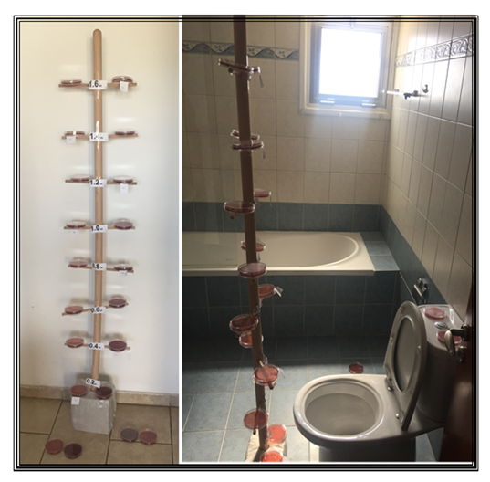
Figure 1: The plate-tree after construction and in position in toilet 1 (T1) after flushing
3. Results
3.1 Background Bacteria Found in the Toilet Environment
Many different types of Bacteria and Fungi were grown and the amount of both increase with an increase in the exposure time. The yellow Micrococcus spp colonies numbers after 10 minutes of exposure were very similar to those in the experiments where Micrococcus spp. was added to the bowl of the toilet, ranging from 1 to 15, average 6 colonies, which increased to an average of 12 and a maximum of 21 colonies at 30 minutes of exposure. Spraying the air and ceiling of the toilet with disinfectant while the door was open and moving around did not alter the numbers of bacteria or fungi recovered when compared to the experiments without spraying. However, spraying the air and surfaces prior to the experiments without disturbance in the toilets, (doors closed) reduced the number of colonies for all bacteria and fungi by >80% when compared to the first two experiments and to the experiments with the doors open and movement; in an additional experiment where the door was opened and closed several times during exposure of the Petri dishes the number of colonies were similar to those where the doors kept open.
3.2 Micrococcus spp Experiments
The organisms recovered and the colony counts are shown in Table 2. Though the highest number of colonies was seen nearest to the floor, recovery of yellow Micrococcus spp was independent of height, with similar numbers to those of the background ranging from 1 to 15 colonies. The numbers recovered after flushing the toilets when compared to the background colonies prior to the experiment proved that there was no statistical difference. It was concluded therefore that this organism was not the ideal indicator, due to the recovery of high numbers of Micrococcus spp. colonies, both before and after seeding the bowl. A single mucoid colony of Enterobacteriaceae was isolated during experiment 3, and was identified as Klebsiella spp. To avoid confusion E. coli, being an organism that was not recovered on any plate during the first three sets of experiments, was selected for further investigations.
3.3 coli Experiments
No E. coli was recovered in any of the three experiments. The numbers of other bacteria and fungi on BA plates were similar to the experiments with Micrococcus spp. Table 2. On MAC plates only moulds and Bacillus spp. were recovered with 75% of plates having no growth.
3.4 Droplet Production
No blue colour was seen on any white tissue surface when the Crystal violet dye was added to the bowl of the toilet before flushing, Figure 2a. Blue stain on the white tissue was seen only on tissues close to the toilet rims and the floor surrounding the toilet as shown on Figure 2b (B1, 2 & 3), when the Crystal violet dye was added to the cistern of the toilet before flushing. The number of droplets increased, particularly on the floor on the side where the cleaning aromatic balls were hanged Figure 2c (C).
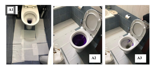
Figure 2: The toilet and white tissue distribution during and after flushing with blue dye.
Figure 2a: Dye added to the toilet bowl (A1, A2 & A3) no drops outside cistern
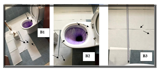
Figure 2b: Dye added to the cistern (B1, B2 & B3) drops 40 cm away from the toilet (B3)
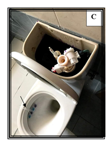
Figure 2c: Dye added to the cistern with cleaning aromatic balls in the toilet (C) drops not shown
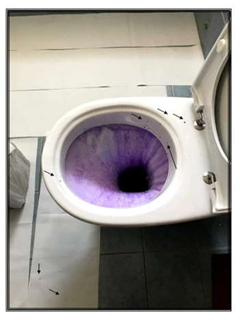
B2 Photo Enlarged
Table 3: The distribution of bacteria and fungi recovered during the eight experiments post flushing after 2- and 10-minutes exposure, N=the total number of plates used in all experiments.
|
Microorganism |
Exposure 2 min |
Exposure 10 min |
||
|
(Plates with Growth N=80) |
Total No of Colonies |
(Plates with Growth N=70) |
Total No of Colonies |
|
|
Micrococcus spp Yellow |
47 |
182 |
46 |
251 |
|
Micrococcus spp Red |
17 |
25 |
12 |
17 |
|
Coag Negative Staphylococcus |
47 |
129 |
52 |
169 |
|
Staphylococcus aureus |
30 |
41 |
36 |
50 |
|
Proteus spp |
1 |
1 |
0 |
0 |
|
Klebsiella spp |
0 |
0 |
1 |
1 |
|
Bacillus spp |
34 |
70 |
37 |
77 |
|
Yellow Aerobic Spore Bearer |
13 |
13 |
10 |
11 |
|
Flat Aerobic Spore Bearer |
21 |
32 |
16 |
33 |
|
Nocardia spp |
7 |
8 |
11 |
13 |
|
Yeast |
3 |
5 |
0 |
0 |
|
Acremonium spp |
1 |
1 |
1 |
1 |
|
Aspergillus flavus |
1 |
1 |
1 |
1 |
|
Mucor spp |
0 |
0 |
1 |
1 |
|
Cladosporium spp |
1 |
2 |
1 |
1 |
|
Penicillium spp |
9 |
30 |
8 |
10 |
|
Chaetomium spp |
2 |
3 |
4 |
5 |
4. Discussion
The greatest risk of environmental contamination with infectious gastroenteritis causing bacteria and viruses is in the toilet/bathroom. The risk increases during acute diarrhoeal illness when faecal material containing billions/trillions of microorganisms is present in the bowl of the toilet both on the porcelain surfaces and the liquid. There is considerable variation both in the design, water pressure and size of modern flush toilets and these in turn will affect the amount of turbulence, splashing, and aerosol production [3, 20]. In a modern wash-down low pressure gravity-flash domestic or public toilet when the flush button is pressed water pours into the toilet bowl where it splits in three; one goes down to the bowl and two travel under the rim one to the right and the other to the left and when they meet on the opposite side create vortices that elevate and fall into the centre of the water contained in the bowl. Depending on the toilet design these vortices may continue upward into the air above the bowl [3, 14]. This elevation may carry small droplets above the toilet rim and seat and if they are dispersed as many studies claimed, they can float in the toilet environment and potentially be inhaled or settle onto surfaces. Our study showed that adding cleaning aromatic balls, or other products hanging under the toilet rim partly blocks the flow of the cistern water thus pushes water upwards that can end up on the toilet rim and floor; this water however contains only the microorganisms present in the cistern and not the bowl.
It has been found that a siphonic toilet produced much lower concentrations of contaminated particles than the older style wash-down pans by 1:4 ratio [2]. It has been reported however that the splashing produced by flushing varied with cistern height and bowl design and was noted that a double trap siphonic toilet, with similar pressures to the single trap, produced more splashes than a wash-down type [3]. This study investigated the infection risk after flushing a toilet contaminated with indicator organisms at concentrations that mimic pathogen shedding during infectious diarrhoea which could be >1010 particles per ml [15]. Using domestic wash-down toilet, a type used widely in Cyprus, the UK and most of the other European Countries, we examined the dynamics of aerosol formation and contamination of environmental surfaces after flushing and found that the only droplets/aerosols escaping after flushing originated from the cistern and not the bowl. Droplets/aerosols from the cistern might be a cause of concern for pathogens like Legionella but seeing the amount escaping and comparing it to that created when a person has a shower it’s insignificant; however, the cistern ought to be cleaned regularly to eliminate biofilms. Couturier and co-workers attributed the Legionella infections of two immunocompromised patients five months apart to toilet flushing; it is evident from our data that the bacteria originated from the cistern where Legionella and other microorganisms found in water form biofilms and the fact that the patients were immunocompromised succumb to infection with a much lower microbial load [21].
Many studies recommend that the toilet lid must be closed before flushing, however, in many countries, including the United States, the toilets in public restrooms are often without lids, particularly the urinals [22] and many suggested that this poses a serious hazard [8]. Most investigators also suggested a better toilet design that would include a lid or better one that closes automatically before flushing. Another factor that maintains the lid open during flushing and contributed to keeping the lid open in our study as mentioned above, is the fact that during diarrhoea faecal material coats most of the internal porcelain of a toilet, thus the person involved will use a brash to clean the area while the water is running in the bowl. Bioaerosol impactor samplers were employed to detect aerosols as well as microorganisms with and without faecal waste in the toilet bowl and the study concluded, like others before them as mentioned above, that microorganisms remaining in the bowl after flushing [23]. Unfortunately, the authors of this study did not provide a list of the microorganisms they suggested escaped the bowl after flushing. The fact that they did not find any difference between flushes with and without waste in the bowl suggests that the microorganisms did not originate from the bowl but from the environment as we found in our study. Their finding of higher concentrations of particles after flushing is not surprising as water flushed at such pressure, just like opening and closing the door, will create turbulence without necessarily any emission of aerosols, a fact that reinforces the suggestions of other studies that all toilets and those available to the public must be adequately ventilated [24]. In recent studies using bio samplers with 100 L/min [25, 26] or 28.3 L/min [27] flow rate has been shown that the microorganisms added to the toilet bowl were detected in the toilet environment, furthermore humidity fluctuations were also noted. The proximity of the air samplers to the toilet might have contributed to their findings; the first Author when looking for the source of Clostridium difficile as well as other monitoring was using the bio-samplers near the entrance thus drawing air out of the room so that the aerosols within the toilet bowl and dust on surfaces were not disturbed.
There are many factors affecting bioaerosol generation during toilet flushing [13, 28]. There are also many interactions between liquid, the air, and the flushing mechanism and above all the structures of toilet28[28]. The description by the Authors [28] of the way the water enters the bowl splashes against the wall as well as their schematic representation in their Figure 2 might represent many types of public and domestic toilets but differs from all three toilets used in this study, the amount of water in the bowl, and in particularly at the point of entry of the water to the bowl. Therefore, the formation of fine droplets or droplet film containing bacteria that can be aerosolized does not apply to the model of toilets used in this study. Any upward airflow vortex produced in the toilets used in this study will be forced down into the bowl thus flushing cannot expel any bioaerosols from the bowl to the air above the toilet. Two studies using commercial toilets with high tank pressures, 60 psi demonstrated both the emission and spread of aerosols [29, 30]. The toilets used in these two studies vary considerably from the domestic toilets used in our study. The high pressure which was four-fold higher than those of simple domestic toilets must be responsible for the emission and dispersal of aerosols. Another study which used similar methods and comparable toilets to our study demonstrated the aerosolization of C. difficile C. difficile and splashing of food colouring [31], the Authors however, pointed out that the single toilet they tested, produced more droplets than the standard wash-down design, furthermore the Authors question the reproducibility of their findings if newer toilet designs were to be tested, in particular those that use less water, as are those used in our study. The first Author when trying to locate the source of C. difficile that colonised all the children in the ward that also housed the Bone Marrow Transplant children in 1981, checked every toilet on the ward (as well all other toilets at Westminster Children’s Hospital) with identical methods used in this study and did not find a single spore to be originating from the toilet bowl. C. difficile was always found during air sampling of the BMT patients’ cubicles and the source was identified as the medal bedpans that contain faecal matter after washing, due to a malfunctioning bedpan washing machine [32]. Households, local authorities, other organisations and in particular personnel looking after public toilets must adhere to regulations and follow simple rules and common sense as clearly outlined [7] to prevent acquisition of potentially fatal infections from domestic and public toilets.
Conclusions and Recommendations
The evidence from many studies indicates that there is a potential risk of acquisition of infectious agents, bacteria, viruses, and protozoa from the toilet and in particular from airborne bioaerosols produced during flushing. Our study has shown that the toilet environment contains many different types of microorganisms mainly skin bacteria, environmental bacteria and fungi as was shown in a review study before [33] however, it was shown that bacteria do not escape from the toilet bowl during flushing. The aerosols detected were from the cistern and not the bowl. Using three different toilets but with similar water flow distribution in the bowl after flushing this study showed that the toilet design is the major factor determining the escape of aerosols from the toilet bowl and whether the lid is closed or open does not necessarily influence microorganisms escaping. Keeping the lid closed has been shown by others to reduce the number of aerosols escaping after flushing and from their distribution of these aerosols in our study we can safely add that the cistern escaping aerosols seen in our study would have been reduced or totally prevented from escaping if the lid had been kept closed [34]. It is evident that many studies ought to be carried out with the many different types of toilets available and in particular squatting toilets, a type of toilet preferred in some countries [34] to establish which types expel bioaerosols during flushing so that the manufacturers can modify them to prevent emission and potential infections. For future improvements the recommendations proposed before are fully supported [15], furthermore disinfection ought to be extended to the cistern.
Limitations
This study used only three different but similar toilets, however as there are many types and shapes of toilets as well as many variables, water pressure, water volume, toilet ventilation etc. our findings might not apply to every type.
Declaration of Interest
There are no relevant declarations of interest for any of the authors. This study was financed in full privately by Dr Markella Marcou and Dr Michael A Petrou. The experiments, result interpretation and writing were evenly shared by all three authors.
Acknowledgements
The authors would like to thank Dr Andreas Neophytou, Scientific Director of Archbishop Makarios III Hospital for his permission to carry out the work at the Hospital. The authors are also grateful to the Microbiology staff of the Hospital, particularly Ms Kalia Meliou, for their assistance in the purchase, storage, and incubation of the plates and control cultures.
References
- Darlow HM, Bale WR. Infective hazards of water-closets. Lancet 1 (1959): 1196-1200.
- Bound WH, Atkinson RI. Bacterial aerosol from water closets. A comparison of two types of pan and two types of cover. Lancet 1966; i:1369-1370.
- Newsom SWB. Microbiology of hospital toilets. Lancet 2 (1972): 700-703.
- Gerba CP, Wallis C, Melnick JL. Microbiological hazards of household toilets: droplet production and the fate of residual organisms. Appl. Microbiol 30 (1975): 229-237.
- Jessen, CU. Luftbårne mikroorganismer. Forekomst og bekæempelse [Airborne Microorganisms: Occurrence and Control].; G.E.C. Gad Forlag, Copenhagen (1955).
- Barker J, Jones MV. The potential spread of infection caused by aerosol contamination of surfaces after flushing a domestic toilet. J Appl Microbiol 99 (2005): 339-347.
- Dancer JS, Li Y, Hart A, Tang JW, Jones DJ. What is the risk of acquiring SARS-CoV-2 from the use of public toilets? Sci Total Environ 792 (2021): 148341.
- Fennelly KP. Particle sizes of infectious aerosols: implications for infection control. Lancet Respir Med 8 (2020): 914-924.
- Abney SE, Higham CA, Amanda M. Wilson AM, et al. Transmission of Viruses from Restroom Use: A Quantitative Microbial Risk Assessment. Food and Environmental Virology 16 (2024): 65-78.
- Boone SA, Betts-Childress ND, Ijaz MK, et al. The impact of an air sanitizer spray on the risk of virus transmission by aerosols generated by toilet flushing. Am J Infect Control 53 (2025): 809-812.
- Tian Y, Rong L, Nian W, et al. Review article: gastrointestinal features in COVID-19 and the possibility of faecal transmission. Aliment Pharmacol Ther 51 (2020): 843-851.
- Gormley M, Aspray TJ, Kelly DA, et al. Pathogen cross-transmission via building sanitary plumbing systems in a full scale pilot test-rig. PLoS One 12 (2017): e0171556.
- Han T, Park H, Jeong Y, et al. COVID-19 Cluster Linked to Aerosol Transmission of SARS-CoV-2 via Floor Drains. J Infect Dis 225 (2022): 1554-1560.
- Li YY, Wang JX, Chen X. Can a toilet promote virus transmission? From a fluid dynamics perspective. Phys Fluids 32 (2020): 065107.
- Abney SE, Bright KR, McKinney J, et al. Toilet hygiene—review and research needs. Journal of Applied Microbiology 131 (2021): 1364-5072.
- Hart CA, Cunliffe NA. Viral gastroenteritis. Curr Opin Infect Dis 12 (1999): 447-457.
- LeBaron CW, Furutan NP, Lew JF, et al. (1990) Viral agents of gastroenteritis public health importance and outbreak management. Morb Mortal Wkly Rep 39 (1990): 1-24.
- Thompson S. The number of bacilli harboured by enteric carriers. J Hyg 52 (1954): 67-70.
- Bhardwaj R, Agrawal A. Likelihood of survival of coronavirus in a respiratory droplet deposited on a solid surface. Phys Fluids 32 (2020):
- Schreck JH, Lashaki MJ, Hashemi J, et al. Aerosol generation in public restrooms. Phys Fluids 33 (2021): 033320.
- Couturier J, Ginevra C, Nesa D, et al. Transmission of Legionnaires’ Disease through Toilet Flushing, Emerging Infectious Diseases 26 (2020): 1526-1528.
- Wang JX, Li YY, Liu XD, et al. Virus transmission from urinals. Phys Fluids 32 (2020): 081703.
- Knowlton SD, Boles CL, Perencevich EN, et al. Bioaerosol concentrations generated from toilet flushing in a hospital-based patient care setting. Antimicrob Resist Infect Control 7 (2018): 16.
- Lee MCJ, Tham KW. Public toilets with insufficient ventilation present high cross infection risk. Sci Rep 11 (2021): e20623.
- Paddy EN, Afolabi OOD, Sohail M. Exploring toilet plume bioaerosol exposure dynamics in public toilets using a Design of Experiments approach. Sci Rep 14 (2024):10665.
- Paddy EN, Sohail M, Afolabi OOD. Evaluating the risk of Clostridioides difficile infection from toilet flushing: a quantitative microbial risk assessment and implications for infection control. Journal of Hospital Infection 159 (2025): 92-99.
- Zhang H, Chen B, Lai ACK. Study on the air gap effect when closing toilet lid on droplet and pathogen escaping from flushing. American Journal of Infection Control 54 (2026): 60-65.
- Lou M, Liu S, Gu C, et al. The bioaerosols emitted from toilet and wastewater treatment plant: a literature review. Environ Sci Pollut Res Int 28 (2021): 2509-2521.
- Crimaldi JP, True AC, Linden KG, et al. Commercial toilets emit energetic and rapidly spreading aerosol plumes. Sci Rep 12 (2022):
- Aithinne KA, Cooper CW, Lynch RA, et al. Toilet plume aerosol generation rate and environmental contamination following bowl water inoculation with clostridium difficile spores. Am. J. Infect. Control 47 (2019): 515-520.
- Best EL, Sandoe JAT, Wilcox MH. Potential for aerosolization of clostridium difficile after flushing toilets: the role of toilet lids introducing environmental contamination risk. J. Hosp. Infect 80 (2012): 1-5.
- Rogers TR, Petrou MA, Lucas C, et al. Spread of Clostridium difficile among patients receiving non-absorbable antibiotics for gut decontamination. Br Med J 283 (1981): 408-409.
- Pan L, Chen SL, Guo YS, et al. Limiting potential COVID-19 contagion in squatting public toilets. Hong Kong Physiotherapy Journal 41 (2021): 119-125.
- Vardoulakis S, Espinoza Oyarce DA, Donner E. Transmission of COVID-19 and other infectious diseases in public washrooms: A systematic review. Science of the Total Environment 803 (2022): 149932.