Blerina Saraçaj¹, Denis Qirinxhi¹, Kledisa Shemsi², Ilir Ahmetgjekaj3*, Elbetel Habtegiorgis⁴, Oltjona Muça⁵, Grace Lin⁵, Edlira Harizi⁶, Evelina Kreko⁷, Eni Muça8, Maria Iskander9, Mohammed Elshahat¹⁰, Fareeha Nasir¹¹, Niki Struga¹², Eliza Rama13, Anila Sani14, Juna Musa5, Erisa Kalemaj 15
¹Department of Radiology, University of Medicine and University Hospital Center "Mother Teresa", Tirana, Albania
²Department of Psychiatry, Ginsberg Hospital, Germany
³Department of Radiology, University Clinical Center of Kosovo, Clinic of Radiology, Pristina, Kosovo
⁴Department of Emergency Medicine, North Memorial Health Hospital, Robinsdale, Minnesota, USA
⁵Department of Laboratory Medicine and Pathology, Mayo Clinic, Rochester, Minnesota, USA
⁶Department of Neurology, Regional Hospital Durrës, Albania
⁷Department of Neonatology and Pediatrics, University Obstetric and Gynecologic Hospital "Koço Glozheni", Tirana, Albania
⁸Department of Biomedical Engineering, Wayne State University, Detroit, Michigan, USA
⁹Boonshoft School of Medicine, Wright State University, Dayton, Ohio, USA
¹⁰Department of Radiology, Mayo Clinic, Rochester, Minnesota, USA
¹¹Primary Care Physician Hemechand Kolli MD Inc.
¹²Medical Student, Our Lady of Good Counsel University, Tirana, Albania
13Student Herlufsholm Skole of Gods Denmark
¹4Department of Neurology, Regional Hospital Hammel Neurocenter, Voldbyvej, Denmark
15Family Doctor, university hospital center “Mother Teresa”, Tirane, Albania
Corresponding Author: Ilir Ahmetgjekaj, Department of Radiology, University Clinical Center of Kosovo, Clinic of Radiology, Pristina, Kosovo.
Received: 29 June 2026; Accepted: 01 July 2026; Published: XX July 2026
Cerebral amyloid angiopathy (CAA) is a small-vessel disease caused by β-amyloid deposition in leptomeningeal and cortical vessels, classically resulting in lobar intracerebral hemorrhage and cerebral microbleeds in older adults. A rare but clinically important subtype, CAA-related inflammation (CAA-RI), arises from an inflammatory response to vascular amyloid and may present with vasogenic edema, subacute hemorrhage, and contrast-enhancing lesions on MRI. Unlike typical CAA, CAA-RI is potentially reversible with immunosuppressive therapy, making early recognition essential. We report a 48-year-old woman with widespread lobar and cortical subcortical microhemorrhages, a subacute cortical hemorrhage, and punctate contrast enhancement on MRI, without marked asymmetric FLAIR hyperintensities or extensive vasogenic edema. Although the patient was younger than the typical age threshold for CAA, the imaging constellation fulfilled criteria for probable CAA and raised strong suspicion for an inflammatory variant. This case highlights the diagnostic value of MRI in identifying CAA-RI, even in atypical presentations, and underscores its role in guiding timely immunosuppressive treatment.
Cerebral amyloid angiopathy (CAA); CAA-related inflammation (CAA-RI); Microhemorrhages; Cerebral microbleeds; MRI; Case report; Boston criteria; Probable CAA
Cerebral amyloid angiopathy (CAA) articles; CAA-related inflammation (CAA-RI) articles; Microhemorrhages articles; Cerebral microbleeds articles; MRI articles; Case report articles; Boston criteria articles; Probable CAA articles.
Introduction
Cerebral amyloid angiopathy (CAA) is a condition in which amyloid proteins build up in the walls of arteries in the brain, causing lobar intracerebral hemorrhage. The protein depositions are less frequent in veins and capillaries [1]. The disease is mostly seen in elderly patients, contributing to cognitive decline. It is radiologically characterized by lobar microhemorrhages as depicted in figure 1, superficial siderosis, and hemorrhages sparing deep structures. A rare inflammatory form, termed cerebral amyloid angiopathy-related inflammation (CAA-RI), occurs when amyloid deposition triggers a perivascular inflammatory response. Radiologically, CAA-RI is characterized by microbleeds together with asymmetric T2/FLAIR hyperintensities consistent with vasogenic edema and contrast-enhancing lesions, and clinically it often presents with subacute encephalopathy, headaches, seizures, or focal deficits. Unlike typical CAA, CAA-RI is a treatable encephalopathy, with many patients improving after corticosteroid or other immunosuppressive therapy, although relapses can occur, highlighting the importance of early recognition [1].
Case Presentation
A 48-year-old woman presented with acute-onset headaches followed over several weeks by progressive cognitive decline and transient focal neurological symptoms, including brief episodes of right-sided weakness and language disturbance. She also reported intermittent confusion and behavioral changes during the preceding weeks. Her past medical history was unremarkable, with no hypertension, diabetes, prior cerebrovascular disease, anticoagulant or antiplatelet use, no history of head trauma, and she did not smoke or use illicit substances. Family history was negative for neurodegenerative or cerebrovascular disorders, and there was no known history of early-onset stroke or dementia in first-degree relatives. On neurological examination, she was alert but exhibited mild disorientation and word-finding difficulty. Cranial nerve examination was normal. Motor strength was preserved in all extremities, though subtle pronator drift was noted on the right side. The sensory and coordination testing were unremarkable.
Magnetic resonance imaging (MRI) demonstrated widespread abnormalities. Susceptibility-weighted imaging (SWI) revealed multiple susceptibility blooming artifacts consistent with microhemorrhages, diffusely distributed in cortical and subcortical white matter of both hemispheres, bilateral thalami, lentiform nuclei (more prominent on the left), brainstem (pons, midbrain), and cerebellum. The lesions ranged in size from 1-10 mm, predominantly chronic in appearance. A 10 mm T1-hyperintense cortical lesion in the temporal lobe was identified, consistent with subacute hemorrhage in the methemoglobin phase. Fluid-attenuated inversion recovery (FLAIR) imaging, depicted in Figure 1, did not show marked asymmetric confluent hyperintensities or extensive vasogenic edema, although mild patchy white-matter signal changes were seen in cortical–subcortical regions corresponding to some of the microhemorrhages [2]. Post-contrast sequences showed punctuation and micronodular enhancement associated with several of the microhemorrhages, without evidence of a rim-enhancing mass, acute ischemia, demyelination, or perilesional edema was detected. The ventricles, cerebrospinal fluid spaces, midline structures, and venous sinuses were preserved. Routine laboratory studies, including complete blood count, metabolic panel, and coagulation profile, were within normal limits. Autoimmune and infectious workups, including Antinuclear antibodies (ANA), Antineutrophil cytoplasmic antibodies (ANCA), HIV, and syphilis serologies, were negative. Cerebrospinal fluid analysis showed mild pleocytosis and elevated protein, with normal glucose and no evidence of infection, supporting an inflammatory process but not specific for any single etiology.
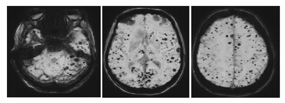
Figure 1: MRI demonstrates numerous lobar, cortical–subcortical, and infratentorial microhemorrhages with a non-hypertensive distribution, consistent with cerebral amyloid angiopathy (CAA).
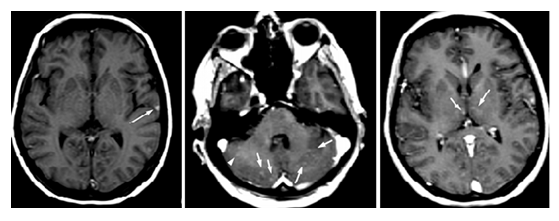
Figure 2: A subacute cortical hemorrhage is evident in a), and the presence of punctate enhancement in several microhemorrhages in b) and d) raises suspicion for an inflammatory variant of CAA, known as CAA-related inflammation (CAA-RI).
Discussion
Cerebral amyloid angiopathy is primarily diagnosed on the basis of neuroimaging, most commonly using the Boston criteria, in which probable CAA represents the highest level of diagnostic confidence achievable without histopathologic confirmation [3]. In this patient, the presence of numerous lobar and cortical–subcortical microhemorrhages with a non-hypertensive distribution strongly supported the diagnosis of probable CAA. Importantly, although classical CAA preferentially spares deep gray nuclei and brainstem structures, atypical distributions and mixed patterns have been described, particularly in younger patients and in inflammatory variants [1]. Patient age posed an important diagnostic challenge. The Boston criteria traditionally apply to patients aged 55 years or older, and our patient, at 48 years of age, falls below this threshold. However, younger age does not exclude CAA, especially in cases of CAA-RI, which has been reported to present at a younger mean age compared with non-inflammatory CAA [1]. Consequently, strict reliance on age criteria alone may delay recognition of this potentially treatable condition. Several imaging features in this case favored an inflammatory subtype. The coexistence of a subacute cortical hemorrhage and punctate contrast enhancement associated with microhemorrhages is atypical for non-inflammatory CAA and raises suspicion for CAA-RI, in which amyloid deposition provokes a perivascular inflammatory response. Clinically, this interpretation was supported by the patient’s subacute headaches, progressive cognitive decline, and transient focal neurological deficits, symptoms that are commonly reported in CAA-RI but are less characteristic of typical CAA [1-7]. Classically, CAA-RI is associated with asymmetric T2/FLAIR hyperintensities reflecting vasogenic edema; however, such findings were not prominent in this patient. This observation highlights the spectrum of radiologic presentations in CAA-RI and emphasizes that the absence of extensive edema does not exclude the diagnosis, particularly in early or atypical cases. Recognition of this variability is essential to avoid misdiagnosis as neoplastic, demyelinating, or infectious processes. Cerebrospinal fluid analysis plays an important supportive role in the diagnostic workup [1-6]. While CSF findings in CAA-RI are nonspecific, mild pleocytosis and elevated protein levels, as observed in this case, are consistent with an inflammatory process and help exclude alternative infectious etiologies. Ultimately, the diagnosis of CAA-RI rests on the integration of clinical presentation, imaging findings, laboratory data, and therapeutic response. The distinction between typical CAA and CAA-RI has significant therapeutic implications. Unlike non-inflammatory CAA, which is largely managed with supportive care and risk-factor modification, CAA-RI is potentially reversible. Numerous reports describe clinical and radiologic improvement following corticosteroid or other immunosuppressive therapy, although relapses can occur and require close follow-up [1-7]. In our patient, clinical improvement and reduction of contrast enhancement on follow-up MRI after corticosteroid treatment further supported the diagnosis of CAA-RI and mirrored outcomes described in prior case series. In summary, this case underscores that CAA-RI should be considered even in patients younger than the conventional age cutoff and in the absence of classic extensive vasogenic edema.
Conclusion
This case highlights the classical radiologic features of cerebral amyloid angiopathy (CAA) along with additional clinical and MRI findings suggestive of CAA-related inflammation. It underscores the importance for radiologists and clinicians to recognize this variant, as CAA-RI, unlike typical CAA, is a treatable condition in which timely immunosuppressive therapy can lead to favorable clinical and radiologic outcomes and may prevent long-term cognitive and functional decline.
Learning Points
- CAA typically presents with lobar microhemorrhages, sparing deep structures affected by hypertensive angiopathy.
- The presence of contrast enhancement and subacute hemorrhage should raise suspicion for CAA-related inflammation (CAA-RI).
- Recognition of CAA-RI is essential, as it can mimic neoplastic, infectious, or demyelinating diseases but is potentially reversible with immunosuppressive therapy.
Patient Consent
Written informed consent was obtained from the patient for publication of this case report and accompanying images.
References
- Chung KK, Anderson NE, Hutchinson D, et al. Cerebral amyloid angiopathy-related inflammation: three case reports and a review. J Neurol Neurosurg Psychiatry 82 (2011): 20-26.
- Kinnecom C, Lev MH, Wendell L, et al. Course of cerebral amyloid angiopathy-related inflammation. Neurology 68 (2007): 1411-1416.
- Greenberg SM, Charidimou A. Diagnosis of cerebral amyloid angiopathy: Evolution of the Boston criteria. Stroke 49 (2018): 491-497.
- Scolding NJ, Joseph F, Kirby PA, et al. Aβ-related angiitis: Primary angiitis of the central nervous system associated with cerebral amyloid angiopathy. Brain 128(2005): 500-515.
- Eng JA, Frosch MP, Choi K, et al. Clinical manifestations of cerebral amyloid angiopathy-related inflammation. Ann Neurol 55 (2004): 250-256.
- Linn J, Halpin A, Demaerel P, et al. Prevalence of superficial siderosis in patients with cerebral amyloid angiopathy. Neurology 74 (2010): 1346-1350.
- Piazza F, Greenberg SM, Savoiardo M, et al. Anti-amyloid beta autoantibodies in cerebral amyloid angiopathy-related inflammation: Implications for amyloid-modifying therapies. Ann Neurol 73 (2013): 449-458.