Ross A. Hauser1, Morgan Griffiths2, Ashley Watterson3, Danielle Matias1, Benjamin R. Rawlings1*
1Caring Medical Florida, 9738 Commerce Center Court, Fort Myers, FL. 33908 USA.
2Independent Researcher, 9738 Commerce Center Court, Fort Myers, FL. 33908 USA.
3Division of Behavioral and Organizational Sciences, Claremont Graduate University, 150 East 10th Street, Claremont, CA. 91711 USA.
*Corresponding Author: Benjamin R. Rawlings, Caring Medical Florida, 9738 Commerce Center Court, Fort Myers, FL. 33908 USA.
Received: 11 May 2026; Accepted: 18 May 2026; Published: 12 June 2026
DOI: 10.26502/fjor.2644-002400116
SharePurpose: Evaluate objective neurovascular and cervical spine test findings and their potential associations in a cohort of patients presenting to an outpatient neck center with complaints of blurry vision in the absence of known preexisting cervical or ocular pathology.
Methods: Single center, retrospective observational study assessing objective test findings of 145 consecutive patients reporting chronic blurry vision without known preexisting etiology at an outpatient neck center from January 1—June 30, 2022. A retrospective chart review was conducted to collect demographic, clinical, and diagnostic data, including digital motion x-ray (videofluoroscopy) and upright cone beam computed tomography of the cervical spine, ultrasound of the carotid sheath and the eye, tonometry, and pupillometry.
Results: Nearly all patients demonstrated concurrent forward head posture, reduced depth of curve, ligamentous cervical instability at C1-C2, and decreased cross-sectional areas of the internal jugular veins (IJVs) and vagus nerves. A paired t-test and a Wilcoxon signed-rank test both confirmed a statistically significant difference between IJV C1 supine and IJV C4-C5 supine, (p <0.001). Pearson correlation analysis identified a significant positive relationship between pupil diameter and IOP (r = 0.27, p<0.001).
Discussion: Preliminary findings indicate a high prevalence of ligamentous cervical instability, internal jugular vein compression, and reduced vagus nerve cross-sectional area in a cohort of patients with chronic blurry vision, supporting potential neurovascular and autonomic contributions to blurry vision in this cohort. Further investigation is needed to clarify their role in the pathophysiology of blurry vision.
Blurry vision; Cervical spine; Internal jugular vein; Vagus nerve; autonomic nervous system; Cerebrospinal fluid; Ligamentous cervical instability; Optic nerve sheath diameter
Blurry vision articles; Cervical spine articles; Internal jugular vein articles; Vagus nerve articles; autonomic nervous system articles; Cerebrospinal fluid articles; Ligamentous cervical instability articles; Optic nerve sheath diameter articles
1. Introduction
Blurry vision is a decrease in visual sharpness or clarity that may appear in one or both eyes, and which may arise from many different mechanisms. While blurry vision is a common visual symptom associated with conditions with known ocular pathology such as cataracts, macular degeneration, glaucoma, refractive errors, and ocular hypertension, its etiology is less clear with other associated conditions, including functional visual disorder, migraine-associated visual symptoms, anxiety-related visual disturbance, mild traumatic brain injury, dysautonomia, intracranial hypertension, and computer vision syndrome, as well as musculoskeletal conditions such as neck pain, cervical spondylosis (degeneration), headaches, and text neck [1-8]. While various associations have been noted in cases of blurry vision of unknown etiology, including amount of screen time and poor posture, a mechanistic explanation for its occurrence remains elusive [9].
Chronic blurry vision is a common symptom in such conditions as whiplash-associated disorder, post-concussion, text neck, and computer vision syndrome, occurring 40-90% of the time in these populations [10-13]. Some recognized contributors to chronic blurry vision include digital eye strain, convergence insufficiency, accommodative dysfunction, and ocular muscular fatigue, however no unifying theory has adequately explained the high occurrence, leading us to speculate that some cases of unresolved blurry vision may have overlapping mechanisms related to the neck [14]. Furthermore, there is a notable absence of advanced diagnostic frameworks for cases of blurry vision that fall outside established diagnostic criteria, contributing to persistent diagnostic uncertainty and unresolved clinical presentations.
It is known that patients with chronic neck pain have a higher prevalence of troublesome visual symptoms [14]. The condition has even been named “cervicogenic visual disturbance” [15]. Various mechanisms have been put forth to explain this association, including sympathetic hyperfunction, retinal cell dysfunction—a type of neuroinflammation along the brain-ocular pathways—and altered sensorimotor control between the cervical afferents and efferents within the central nervous system [16,17]. We previously reported a multi-symptom cohort including 1 of 6 eye symptoms which outlined the potential mechanistic link between cervical spine pathology and eye symptoms [18]. The present study focuses specifically on blurry vision, which allows for a more targeted assessment of how objective test findings relate just to patients with blurry vision, and may guide evaluation and treatment strategies when this symptom is a primary or unresponsive complaint.
The significant cervical structural pathologies found in this cohort included ligamentous cervical instability—especially at the atlantoaxial (C1-C2) facet joint—increased C6-atlas interval (C6AI, measurement of forward head), and decreased depth of cervical curve. The head is naturally positioned on the most mobile segments of the spine (C1-C2) to allow for optimal function and range of motion, but we are not meant to be looking down or leaning forward for a majority of the day. When looking down, the neck muscles relax, and the vertebrae spread apart while the posterior cervical ligaments stretch [19]. A flexed neck looking down at a cell phone (40-45°) endures triple the force from the weight of the head, averaging 10 kg greater force than in an upright position, and is a known risk factor for neck pain and symptoms, which often include visual changes [20-22]. Extended time in this facedown/forward head posture can lead to injury of the posterior ligament complex, contributing to ligamentous cervical instability, a degenerative condition of the ligamentous integrity of the cervical spine which can progress to a breakdown of the normal cervical lordotic curve (cervical dysstructure), with net effects of a forward-shifted atlas in the sagittal plane, causing stretch and compression of the carotid sheath, including the internal jugular veins and vagus nerves [23,24]. See Figure 1.
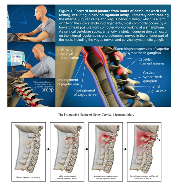
Figure 1: Forward head posture from hours of computer work and texting, resulting in cervical ligament laxity, ultimately compressing the internal jugular veins and vagus nerve. “Creep,” which is a term signifying the slow stretching of ligaments, most commonly occurs by a forward head posture from computer work or looking at a smartphone. As cervical vertebrae sublux anteriorly, a stretch compression can occur on the internal jugular veins and autonomic nerves in the anterior part of the neck, including the vagus nerves and cervical sympathetic ganglion [18].
This retrospective study was conducted to explore the hypothesis that a high percentage of patients who reported to an outpatient neck center with chronic blurry vision may have underlying objective structural neck issues, including documented ligamentous cervical instability and structural forward head posture, along with associated objective test findings, including abnormal ultrasound measurements of the cross-sectional areas of the internal jugular veins and vagus nerves, and abnormal ophthalmological findings, including elevated optic nerve sheath diameter and excessive pupillary dilation. The objective of the study was to identify potential reproducible cervical spine and neurovascular patterns in patients with otherwise unexplained blurry vision. Findings may suggest a potential new avenue for diagnostic evaluation in patients with chronic unexplained blurry vision and may warrant consideration of cervical structural treatment as an option for their care. See Figure 2.
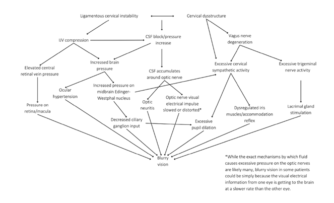
Figure 2: Ligamentous Cervical Instability Etiologies for Blurry Vision.
2. Methods
2.1 Study Design
This retrospective observational study was approved by the WCG Institutional Review Board (Study #1364545, Protocol #20235176). Data were retrospectively obtained from clinical chart reviews at a single-center outpatient neck clinic from January 1—June 30, 2022. The study cohort consisted of patients presenting to an outpatient neck clinic for evaluation of a potential cervical spine contribution to persistent and/or unexplained symptoms. All patients reported subjective blurry vision persisting for more than one year. The cohort represents a treatment-refractory population; Although many patients reported prior conventional medical assessment and management, prior treatment histories were not systematically documented. Blurry vision and the absence of confirmatory findings on ophthalmologic examination were self-reported, though ophthalmologic results were not available for independent review in this study.
All patients underwent the standard clinical diagnostics, including dynamic cervical structural analysis and neck vitals analysis, performed by certified radiologic technologists or medical sonographers for clinical, not research purposes. Assessors were not blinded to clinical status, as imaging was performed following routine clinical care.
The study population included patients aged 20-50 years reporting blurry vision with onset at least one year prior and without a previously identified cause during initial intake. Patients were excluded if they had a previously known diagnosis that could explain their symptoms (such as previously confirmed ophthalmic conditions like refractive errors or visual field defects, or ocular disease issues, including those that necessitated current usage of eye medication or surgery) or a known preceding traumatic event (motor vehicle accident, sporting incident, injury, or surgery). See Supplementary Figure 1.
2.2 Dynamic Cervical Structural Testing
To evaluate cervical spine structure, patients received 2 scans of the cervical spine, performed by a radiology technologist: an upright digital motion x-ray (DMX, videofluoroscopy) and an upright cervical cone beam CT (CBCT) scan. DMX provides a high degree of accuracy for identifying vertebral instability, as it allows for real-time motion imaging in the upright position, which can aid in the diagnosis of pathology that static imaging in the supine position could miss [25]. CBCT is a valid tool for measuring in vivo cervical spine kinematics, including the upper cervical spine, and for assessing bony structures with submillimeter accuracy [26]. Scanning in the upright position allows for better visualization of how gravity affects the cervical spine and can reveal abnormalities that may be missed during a lying-down position [27]. To evaluate for ligamentous cervical instability, measurements were obtained in the lower cervical spine in flexion and extension, and in the upper cervical spine (C1-C2) in lateral flexion with open mouth views (to evaluate for facet joint instability). The open mouth lateral bending position used to evaluate LCI C1-C2 demonstrates good-to-excellent inter-examiner reliability, with intraclass correlation coefficients ranging from 0.56-0.97 [28]. To evaluate structural forward head and neck posture, C6AI was measured as the horizontal distance between the posterior border of the C6 vertebral body and a line drawn perpendicular from the anterior arch of the atlas in the sagittal view. C6AI objectively identifies forward head and neck posture by measuring the atlas’s position relative to the lower cervical spine in the sagittal plane [29].
Depth of curve (DOC) was measured as the distance from the posteroinferior aspect of the C4 vertebra to a line drawn from the posteroinferior aspect of the C6 vertebral body to the peak of the dens of C2. DOC is used to objectively identify the state of cervical lordosis [18]. See Figure 3.
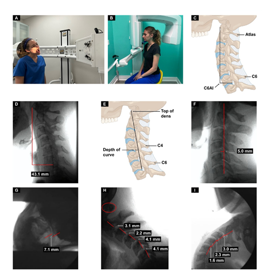
Figure 3: Upright digital motion (fluoroscopic) x-ray (DMX) and cone beam CT (CBCT) scan with structural measurements. A. DMX positioning for open mouth lateral flexion. B. CBCT setup. C. Forward head (C6AI*) illustration. D. C6AI measurement. E. Depth of curve** illustration. F. Depth of curve using DMX. G. C1-C2 instability. H. Flexion, lower cervical instability. I. Extension, lower cervical instability [18].
*C6AI = horizontal distance in the sagittal plane of the posterior inferior C6 vertebra to anterior atlas (optimal is <10 mm).
**Depth of curve = horizontal distance in the sagittal plane from posterior inferior C4 vertebra to line drawn from posterior inferior C6 vertebra to top of dens (optimal is 7-17 mm).
2.3 Neck Vitals Analysis
A neck vitals analysis was performed on each patient by a medical ultrasonographer, which included cervical and ocular ultrasounds and measurements. See Figure 4.
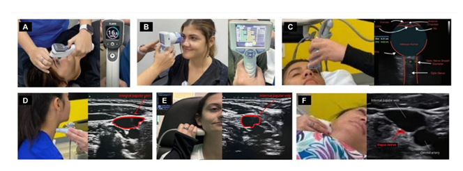
Figure 4: Neck vitals analysis. A. Tonometry. B. Pupillary light reflex. C. Optic nerve sheath diameter. D. Internal jugular vein (IJV) cross-sectional area (CSA) at C4-C5. E. IJV CSA at C1. F. Vagus nerve CSA [18].
Cross-sectional areas of the vagus nerves (mid-neck, approximately C4) and internal jugular veins (supine position at C1 and at C4-C5) were measured using Canon Aplio a550 ultrasound with a 7-MHz linear probe. Ultrasound evaluation of the vagus nerves’ cross-sectional areas at the level of the thyroid gland (mid-cervical region, C5) has good inter-rater agreement with intraclass correlation coefficients from 0.72-0.97 for inter-rater reliability [30-32]. Ultrasound measurement of the internal jugular veins’ cross-sectional areas demonstrates good-to-high inter-rater reliability, with intraclass correlation coefficients over 0.82-0.93 [33-35]. Pupil diameter and light reflex percentages were measured using a NeurOptics NPi®-200 pupillometer. The NeurOptics NPi®-200 pupillometer demonstrates excellent inter-rater reliability for pupillary measurements, with studies reporting intraclass correlation coefficient values greater than 0.90 for the Neurological Pupil Index, maximum diameter, and minimum diameter [36]. Intraocular pressure (IOP) was measured using an iCare ic200 tonometer. The iCare ic200 provides highly reliable and repeatable IOP measurements across operators, with multiple studies demonstrating excellent intra-operator intraclass correlation coefficient values ≥0.99 and inter-operator intraclass correlation coefficient values ≥0.99 [37]. Optic nerve sheath diameter (ONSD) was measured using Canon ultrasound on the ocular setting. Ultrasonic measurement of optic nerve sheath diameter demonstrates good-to-excellent inter-rater reliability, with an intraclass correlation coefficient of 0.89 [38].
2.4 Statistical Analysis
Data were analyzed using RStudio version 2024.04.2+764. Continuous variables were summarized using means and standard deviations, and categorical variables (e.g., number of symptoms) were summarized using counts and valid percentages. Statistical significance was defined as p <0.05 for all tests.
The analytic sample consisted of 145 patients. No univariate outliers were removed. All recorded values were reviewed and determined to be clinically valid and accurately documented. Retaining the full dataset preserved the natural clinical variability present in this patient population.
Missing data were handled using a pairwise deletion approach, meaning each statistical test included all available cases for the variables involved. This method preserved the maximum number of observations per analysis, and the corresponding sample size for each test is reported accordingly.
Assumptions for parametric testing were evaluated before analysis. Skewness and kurtosis values for continuous variables fell within acceptable ranges (±3 and ±10, respectively). Normality of difference scores for paired comparisons was assessed using Shapiro–Wilk tests and visual inspection of histograms and Q–Q plots. These comparisons indicated some deviations from normality. To ensure robustness of the findings, non-parametric Wilcoxon signed-rank tests were conducted alongside paired t-tests. Linearity of relationships for correlation analyses was evaluated using scatter plots. Paired t-tests were used to compare IJV CSA across positions (C1 supine, C4–C5 supine, and C1 supine on Denneroll®). Pearson correlation analyses were used to examine linear relationships between pupil diameter and IOP. A Bonferroni correction was applied to adjust for multiple comparisons. No covariates were included in the analysis, and subgroup or interaction effects were not examined, given the exploratory nature of the study. All analyses were conducted using raw, complete-case data without sampling weights or design-based corrections.
3. Results
3.1 Demographics and Objective Test Results
A total of 145 patients aged 20-50 years reported blurry vision with no known cause at an outpatient neck center between January 1, 2022 and June 30, 2022 to be included in this retrospective study. The average age was 37.8 +/- 8.5 years, with 72 (49.7%) female and 73 (50.3%) male.
The mean and SD of the dynamic cervical structural tests, as well as the total (bilateral sum) IJV and vagus nerve CSAs, pupillary diameters, IOPs, ONSDs, and light reflex percentages of the 145 patients with blurry vision are summarized in Table 1. This approach of bilateral sum reporting reflects the typical clinical presentation in our patient population, with cervical-related findings in which unilateral measurements tend to be highly symmetrical; data are therefore reported this way to streamline results.
|
Testing Parameters |
Mean |
SD |
|
C6AI |
41.67 mm |
14.12 |
|
Depth of curve |
2.68 mm |
3.86 |
|
Flexion instability total* |
4.36 mm |
3.18 |
|
Extension instability total* |
4.27 mm |
3.35 |
|
C1-C2 facet joint instability** |
7.19 mm |
2.98 |
|
IJV C4-C5 supine** |
131.79 mm2 |
83.66 |
|
IJV CSA C1 supine** |
68.94 mm2 |
37.95 |
|
IJV CSA C1 supine with Denneroll® (n = 131)** |
111.20 mm2 |
48.30 |
|
Vagus nerve CSA** |
2.69 mm2 |
0.82 |
|
Pupil diameter** |
10.34 mm |
1.55 |
|
Intraocular pressure** |
35.71 mmHg |
8.33 |
|
ONSD** |
15.35 mm |
1.71 |
|
Percent change (light reflex)** |
74.70% |
10.41 |
|
*Sum totals C2-C6 |
||
Table 1: Neck Vitals Analysis Summary (n = 145).
Percentages of significant abnormal cervical structural and objective eye testing findings are described in Table 2. All 145 (100%) patients had C6AI greater than 10 mm (normal <10 mm) in the sagittal plane, the average being 41.7 mm (1.7 inches). A total of 128 (89%) patients were found to have decreased DOC (<7 mm), with the average cervical lordosis depth being decreased at 2.68 mm. Ideal cervical lordosis depth, evaluated by the Borden method, is between 7-17 mm [39-40]. Many patients, 127 (88%), had documented ligamentous upper cervical (C1-C2) facet joint instability (>4 mm total), with the average bilateral total being 7.19 mm +/- 2.98 mm. Decreased IJV CSA (total IJV CSA supine <180 mm2) at the level of C1 (atlas) was documented in 98.6%. Elevated ONSD was documented in 98.6% (total bilateral >12.2 mm). Decreased vagus nerve CSA (vagus nerve CSA total <4.2 mm) was documented in 138 (95%) patients [41]. In addition, 138 (95%) exhibited higher-than-expected pupillary light reflex (percent change in pupil diameter >60% total), and 134 (92%) had above-average pupil diameters at rest (total >8 mm) [42-44].
|
Testing Parameters |
% of Patients with Abnormal Findings (Number of Patients) |
|
|
C6AI |
100% (145) |
<10 mm |
|
Depth of curve (n = 144) |
89% (128) |
7-17 mm |
|
C1-C2 facet joint instability* |
88% (127) |
<4 mm |
|
Flexion/extension instability C2-C6** |
88% (127) |
<4 mm |
|
Vagus nerve CSA* |
95% (138) |
>4.2 mm |
|
IJV CSA C1 supine* |
99% (143) |
>180 mm |
|
IJV C4/C5 supine* |
76% (110) |
>180 mm |
|
IJV CSA C1 supine with Denneroll® (n = 131)* |
90% (118) |
>180 mm |
|
Pupil diameter* |
92% (134) |
<8 mm |
|
ONSD* |
98% (142) |
<12.2 mm |
|
Percent change (light reflex)* |
95% (138) |
30-60% |
|
Intraocular pressure* |
19% (28) |
<42 mmHg |
|
*Bilateral totals. Data are collected unilaterally but reported here as bilateral totals (except for C6-atlas interval) due to clinical bilateral symmetry, and to streamline results. **Total flexion and extension instability at each level from C2 through C6 (as C6-C7 was not consistently seen on DMX nor cone beam CT scan). Normal is <4 mm [46]. |
||
Table 2: Summary of cervical structural and objective testing parameters that were abnormal in a cohort of 145 patients with blurry vision.
3.2 Comparisons and Linear Relationships
A paired t-test revealed a statistically significant difference between IJV C1 supine and IJV C4–C5 supine (t[144] = –11.75, p <0.001, d = 0.98), with a mean difference of –62.85 units (95% CI [–73.42, –52.28]). A Wilcoxon signed-rank test confirmed this finding (V = 592.5, p <0.001), indicating that IJV C1 supine values were significantly lower than IJV C4–C5 supine.
A paired t-test also indicated a statistically significant difference between IJV C1 supine and IJV C1 measured with the Denneroll® device (t[130] = –10.81, p <0.001, d = 0.95), with a mean difference of –38.07 units (95% CI [–45.04, –31.10]). This finding was similarly confirmed using a Wilcoxon signed-rank test, indicating that IJV C1 supine values were significantly lower than those obtained with the Denneroll®. (See Figures 5, 6).
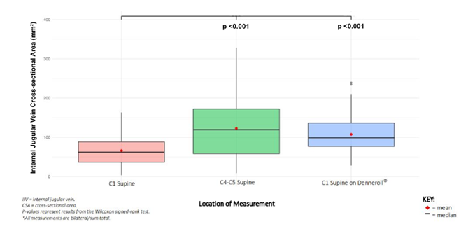
Figure 5: Comparison of C1 supine vs. C4-C5 and C1 supine, lying on Denneroll®. The figure represents a Wilcoxon signed-rank test comparing IJV CSA supine at C1 with IJV CSA at C4-C5 and IJV CSA at C1 with Denneroll®. There was a statistically significant difference at p <0.001 between IJV CSA at C1 while lying supine with and without a Denneroll®, and between IJV CSA at C1 and C4-C5 while lying supine.
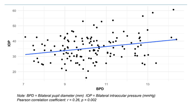
Figure 6: Scatter plot of pupil diameter and intraocular pressure.
4. Discussion
In this retrospective observational study, we analyzed the cervical structural findings and objective test results of 145 patients reporting blurry vision without a previously identified cause during their initial intake at an outpatient neck center. To our knowledge, this is the first study to analyze objective cervical structural and ocular pathologies together in relatively young patients (average age 37.8) with no obvious cause for their blurry vision. All patients demonstrated abnormal cervical structural findings, primarily bilateral ligamentous C1-C2 facet joint instability (LCI C1-C2), reduced cervical lordosis (decreased depth of curve), and a forward-shifted atlas (C1) in relation to the lower cervical spine in the sagittal plane (increased C6AI). These findings demonstrate frequent co-occurrence of cervical structural and neurovascular observations in this cohort.
Blurry vision is a possible symptom of almost all ocular diseases, including glaucoma, macular degeneration, dry eye, and retinal detachments, but in this young (aged 20-50) patient population, age-related factors are unlikely and patients with previously known diseases or other identified ocular conditions were excluded. Given these criteria, we hypothesize that this young patient population group has likely accumulated many years of screen time, which has been propounded to influence cervical biomechanics [47-50]. Increased time spent using electronic devices is being associated with visual symptoms and neck pain [51]. We further propose that prolonged and repetitive screen use contributes to cumulative mechanical loading of the cervical spine, resulting in structural and functional alterations that may influence visual symptoms. Common maladies such as computer vision and text neck syndrome share many eye, visual, and musculoskeletal symptoms with LCI. See Figure 7.
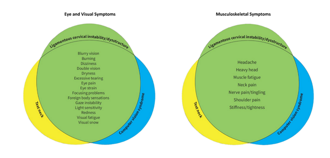
Figure 7: Venn diagrams demonstrating the commonality of eye, visual, and musculoskeletal symptoms in ligamentous cervical instability, text neck, and computer vision syndrome [61].
Blurry vision is becoming a more prevalent complaint in clinical practice, as 90% of people who spend at least 3 hours a day using a computer have computer vision syndrome, with 30-60% experiencing blurry vision as their primary complaint [52-54]. A significant association is found between hours of use (screen time) in not only vision-related problems, but also neck disability [55,56]. It is clear that digital devices are contributors to some vision-related problems, including text neck syndrome, which is associated with not only visual changes, but also headaches and neck and shoulder pain, although the mechanisms leading to these symptoms are yet to be defined [57]. While there are many hypotheses as to what may be causing the blurry vision and other visual symptoms due to ever-prevalent electronic device use, such as blue light, glare, quality of the screen, and ergonomics, a clear consensus has not yet been established [58-60].
The overarching hypothesis detailing how eye symptoms related to electronic device use and poor posture may share underlying ligamentous cervical instability etiology is further explained in our prior work, Cervical Oculopathy: The Cervical Spine Etiology of Visual Symptoms and Eye Diseases—A Hypothesis Exploring Mechanisms Linking the Neck and the Eye [61].
4.1 Ligamentous Cervical Instability
Clinical ligamentous cervical instability is an inability of the cervical ligaments to maintain individual, adjacent, or global vertebral alignment, especially at the facet joints, when subjected to increased forces by various postures, positions, and/or motions that alter bony, soft tissue, and/or neurovascular alignment and function such that symptoms result [62,63]. LCI was assessed by DMX (videofluoroscopy), which allows for a detailed and uninterrupted examination of the cervical spine (C0-C7) during various movements and in multiple dimensions, including frontal and sagittal, as well as rotational, planes [65]. LCI is characterized by more than 2 mm of vertebral translation in one direction, examined in various positions, and is defined by the degree of overhang by an adjacent vertebra, but some consider as little as 1 mm to be significant [46,64].
The DMX studies show the functional integrity of the ligaments in the cervical spine with a high degree of accuracy in the upright position, specifically the anterior and posterior longitudinal, supraspinous, interspinous, transverse, alar, facet capsular, and ligamentum flavum ligaments [65]. The capsular ligaments through the cervical spine provide structural support for the 4 major movements of the head and neck: flexion, extension, axial rotation, and lateral flexion [66]. We documented weakened capsular ligaments in the upper cervical spine by the overhang of the inferior articular pillar of the atlas on the superior articular pillar of C2, and in the lower cervical spine the motion is seen in flexion and extension.
The most mobile joint in the cervical spine is the C1-C2 joint, which sits between 2 more stable areas (C0-C1 and C2-C7) and is responsible for a large amount of rotation and flexibility due to the peglike dens structure of C2 around which the arch of C1 rotates. The 2 lateral C1-C2 facet joints are to be differentiated from the atlantodens (median atlantoaxial) joint. Ligamentous atlantoaxial instability primarily involves the transvers ligament and is typically diagnosed using non-motion MRI or CT imaging, with extreme cases warranting surgical intervention [67,68]. See Figure 8. Upright and motion scanning allow for structural deviations to be seen during movements and amongst different positions, which can document vertebral translations that could be putting strain on vital structures in the area surrounding the excess motion, such as the IJV and vagus nerve, especially in the area of C1 [69,70].
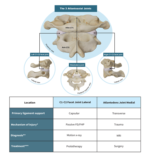
Figure 8: Ligamentous Atlantoaxial (C1-C2) Facet vs. Medical Atlantodens Joint Instability.
Facedown/forward head posture (FD/FHP).
*While both types of instability can occur passively from a facedown/forward head lifestyle in patients who have connective tissue disorders such as Ehlers-Danlos syndrome or genetic disorders, the force to produce transverse vs. capsular ligament injury is greater, and thus trauma is typically the inciting event in atlantodens instabilities.
**In adults, a digital motion x-ray demonstrating C1 facet joint overhang on the C2 facet joint with an open mouth while the patient does lateral flexion is utilized to document ligamentous C1-C2 facet joint instability, whereas medial atlantodens instability is assessed by comparing the dens location to various other bony structures at the craniocervical junction in the neutral, flexion, or extension position with x-ray, but more commonly with CT scan or MRI.
***Treatment regimens depend on many different factors, including the types and severity of symptoms, but while the posterior capsular ligaments are accessible by injection, the transverse ligament at its bony attachments is not. Prolotherapy can be utilized for ligamentous C1-C2 facet joint instability, while cases of ligamentous atlantodens instability from severe transverse ligament injury or disruption necessitate surgical stabilization.
In this study, 137 (94%) reported neck pain. Neck pain and muscular tension are often discussed regarding forward head posture, but what may be overlooked is ligament pathology. While the etiology of chronic neck pain can be multifactorial, it is plausible that neck pain reported in 137/145 (94%) of this patient population relates to chronic neck tension induced by the ligamento-muscular reflex from the upper and lower cervical instability seen, presumably similar to muscle strain felt from the resultant forward head posture and loss of lordosis [71,72]. The ligamento-muscular reflex is a protective mechanism to instill stability in a joint that is unstable to protect not only the joint but adjacent neurovascular structures, in this case the carotid sheath [73,74]. See Figure 9. Cervical structural changes, LCI, increased C6AI, and decreased DOC may lead to unfavorable outcomes for the neck (causing pain) and for the visual system by causing stretch and compression of vital structures such as the IJV and vagus nerves, potentially resulting in blurry vision and other visual disturbances by a variety of potential mechanisms [61].
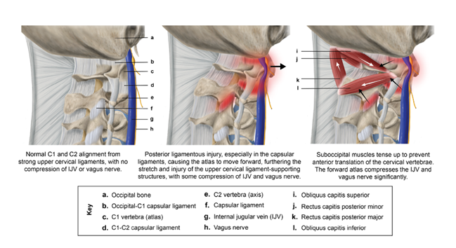
Figure 9: Ligamento-Muscular Reflex in Upper Cervical Region [18].
Static cervical structure was evaluated by upright CBCT to assess loss of lordotic cervical curvature. We measured the DOC and C6AI (sagittal x-ray used if C6 vertebra was not seen with CBCT). Forward head posture is the most common cervical postural abnormality in the sagittal plane, but it typically refers to forward placement of the head in relation to the shoulder, when a clinical shift to prioritizing objective cervical radiographic measurements should be considered 75,76]. Forward head posture causes known pathologic changes in both the upper and lower cervical spine, including an increase in upper cervical angle (increased extension) and flexion in the lower cervical spine [77,78]. To better clarify what exactly is going on in the entire cervical region in the sagittal plane, we measured C6AI, which compares the anatomy in the upper cervical (specifically, the atlas) to the lower cervical spine (C6) [79]. C6AI is the amount of forward displacement of the atlas (C1) compared to the lower cervical vertebra (C6) in the sagittal plane, a measurement that quantifies structural forward head posture that can be easily replicated to measure changes or improvements [80-82]. In addition to leading to a forward-shifted atlas which may sometimes go unrecognized, forward head posture makes one prone to ligamentous cervical instability [83].
4.2 IJV Compression and Blurry Vision
In 87% of patients, IJV CSA C1 supine was significantly lower than IJV CSA C4-C5 supine (mean difference -62.85 mm), determined by a paired t-test and a Wilcoxon signed-rank test. The average total IJV C1 supine was 68.94 mm2 compared to 131.79 mm2 at C4-C5 (ideal is >180 mm2). IJV measurements are typically taken only mid-neck, at the C4-C5 level, leaving room for error when assessing pathological IJV compression [84]. IJV CSA is easily measured under B-mode ultrasound. See Figure 10. In the supine position, normal unilateral IJV CSA is >90-100 mm, collapsing to about 25 mm in the upright position due to negative pressure in the thoracic cavity with respiration, to maintain intracranial pressure in the upright position [85-87]. This study documents that measuring IJV at the level of the atlas (C1) in the supine position is a more sensitive test when evaluating for IJV compression with ultrasound, compared to the mid-cervical region (C4-C5), which is where IJV CSA is typically measured [88]. Although the prevalence of upper cervical IJV compression in asymptomatic individuals remains unclear, one study reported an incidental finding in 26% of 164 patients without multiple sclerosis [89]. It is also important to note that most studies report CSA of the IJV exceeding 100 mm², with some values reaching as high as 165 mm². In contrast, our patient cohort demonstrates markedly smaller CSA measurements, even at the mid-cervical level.
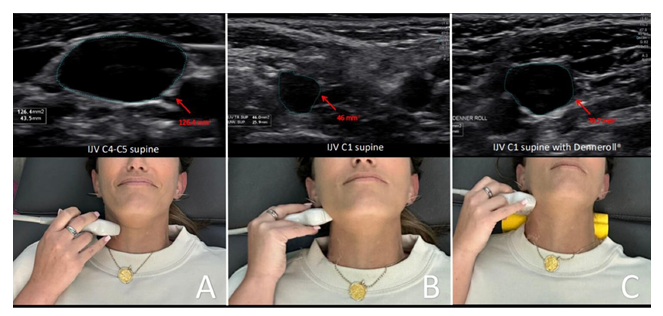
Figure 10: Internal jugular vein (IJV) cross-sectional area (CSA) measurement using ultrasound while supine. A. Supine measurement at the C4-C5 level. B. Supine measurement at the atlas (C1) level. C. Supine measurement at the C1 level while lying on a Denneroll®. When there is a breakdown of the cervical curve (dysstructure) and/or ligamentous cervical instability, the atlas is displaced anteriorly, thus causing the IJV CSA to be decreased when measuring it at C1 and open when the cervical curve is improved with the Denneroll® orthotic device [18].
It must be remembered that at any given time, most of the blood volume in the brain is in the venous system, trying to get out [91]. While 32 quarts of fluids flow through the brain each day, 70-80% of them are contained in the brain’s venous system [ibid]. In 94% of healthy people, the IJV is the main venous system through which blood exits the brain, while only 6% of people drain less than one-third of the brain’s fluids through the IJV [92].
Consequences of IJV compression include elevated CSF pressure, as well as increased cerebral venous sinus and episcleral venous pressures, potentially causing increased IOP and ICP [93-96]. Outflow from the orbits of the eyes travels through the episcleral veins into the superior ophthalmic veins, which lead to the cavernous sinus and the petrosal sinus and exit the head through the IJVs [97-98]. We hypothesize that compression at the IJVs can potentially cause upstream effects of built-up pressure, possibly all the way back up to the episcleral veins, hindering the outflow from the eye, which would cause an increase in intraocular pressure. According to the modified Goldman equation, episcleral venous pressure accounts for about 47-60% of IOP in humans [96].
In this study, IJV CSA C1 while using a cervical orthotic, the Denneroll®, was significantly higher than IJV CSA supine (mean 42.3 mm, t[=130]). This observation supports our theory that correcting the cervical curve and restoring cervical structural integrity would lead to increased IJV CSA at C1 supine, relieving compression, possibly improving cerebral drainage, and could hypothetically lead to improved vision in some cases. Similarly to the documented IJV compression, all but one patient had elevated ONSD. Blurry vision is a main clinical sign of increased intracranial pressure, often identified by ONSD measurements [99].
4.3 Optic Nerve Sheath Diameter and Blurry Vision
The most frequent objective ocular pathology found was the elevated bilateral total ONSD, averaging 15.35 mm. Only one patient in this cohort did not have findings of elevated ONSD. Ultrasound ONSD measurement, utilized in this study, is a valid indicator of increased intracranial pressure, and easily accessible and replicable in an in-office setting for detecting and monitoring intracranial hypertension [100-102]. While some studies utilize unilateral measurements as low as 5.0-5.2 mm, we chose 6.1 mm (total bilateral 12.2 mm) as our cut-off for establishing elevated ONSD, which has a high sensitivity and specificity for intracranial hypertension (>20 mmHg) [103-110]. The ONSD should be measured by identifying the hypoechoic linear structure with a hyperechoic border (nerve sheath) that emerges from the posterior part of the globe and should measure 3 mm behind the posterior rim of the globe, including the outer rim. Generally, in healthy adults, ONSD should measure 4.9-5.3 mm [111]. See Figure 11.
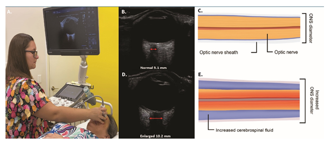
Figure 11: Ocular ultrasound demonstrating a normal and an enlarged optic nerve sheath diameter (ONSD). A. Technique. B. Normal ultrasound image. C. Graphic pictorial of normal ONSD. D. High ONSD image. E. Illustration of extra cerebrospinal fluid around the optic nerve. A high ONSD is a noninvasive test for intracranial hypertension (brain pressure >20 mmHg) [61].
Blurry vision and other visual and eye symptoms are common with intracranial hypertension [112-113]. Cerebral and cervical venous outflow disorders are a known cause of increased brain pressure [114-115]. Blurry vision as a result of idiopathic intracranial hypertension is frequently discussed in literature as resulting from CSF accumulating around the optic nerve, but still as idiopathic—meaning without a known cause—for the increased intracranial pressure [116]. Nonetheless, blurry vision with intracranial hypertension is known to be associated with elevated CSF pressure, papilledema, increased ONSD measurements, and even visual loss, all of which have been demonstrated to improve immediately following reduction of intracranial pressure, and there is general agreement that abnormal cerebrospinal fluid flow dynamics and increased venous sinus pressure are likely involved [117-118]. Despite CSF removal being a common procedure to reduce intracranial pressure and thereby reducing ONSD, it has not proven to be a long-term solution nor thought to be a particularly useful therapeutic method [119].
Perhaps some cases of intracranial hypertension associated with blurry vision are not actually idiopathic, but rather from cervical structural issues impeding venous fluid outflow from the brain, such as with IJV compression [120]. Additionally, we find it important to consider that blurry vision, along with headaches and neck pain, is frequently reported in patients with restricted cervical movement and is also associated with extensive computer or cell phone use, with which the correlation remains unclear, insinuating there could be a shared etiology amongst many overlapping diagnoses [121-122].
Abnormalities in the optic nerve are a common pathology found in almost all eye conditions, including glaucoma, macular degeneration, hypertension, and eye symptoms such as blurry vision, eye pain, visual field defects, and visual distortions [123-125]. None of the patients in our study were found to have papilledema on ultrasound examination, despite the high prevalence of elevated ONSD [126]. This discrepancy likely reflects our unique patient population, which was comprised primarily of young people (aged 20-50) with normal or below average body weight, therefore making it likely that lifestyle factors other than age or body weight play a causative role. We hypothesize that our patients may experience intermittent or dynamic CSF flow alterations that affect ONSD without having progressed to visible papilledema (but which may progress in the future).
CSF surrounds the optic nerve through the subarachnoid space, all the way up to the back of the globe, playing a significant role in determining the retrolaminar pressure, as evidenced by Morgan, et al, and cerebral drainage is proven to be hindered by IJV compression [127-128]. As CSF accumulates around the optic nerve, the increase in pressure could cause impaired electric impulse conduction or mechanical stress, which could impair visual signaling, potentially causing transient blurry vision before presenting as papilledema, and/or before degenerative changes that could happen to the optic nerve over time [129-130]. See Figure 12.
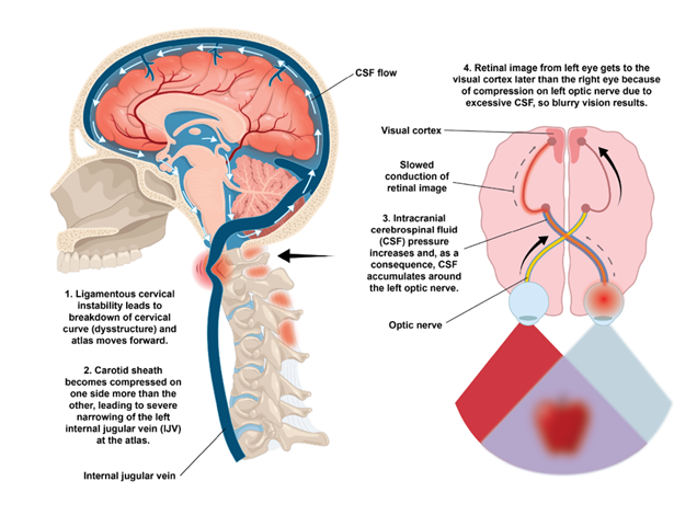
Figure 12: Ligamentous Cervical Instability Potential Etiology of Blurry Vision [61].
4.4. Intraocular Pressure
Ocular hypertension, defined as increased IOP >21 mmHg unilaterally with normal optic nerve findings, was identified in only 28 (19%) patients [131]. A Pearson correlation coefficient analysis demonstrated a strong positive relationship between pupil diameter and intraocular pressure (r = 0.27, p< 0.001), though ocular hypertension was not a prevalent finding, therefore glaucomatous changes were not suspected nor present in this cohort. In this patient population, we suspect increased CSF due to venous outflow obstruction at the IJVs and possible intracranial hypertension to be more significant contributing factors.
The optic nerve is exposed to IOP from within the eye, and to ICP via CSF within the subarachnoid space. A change in these 2 pressurized zones is known to cause damage to retinal ganglion cell axons, causing diseases such as glaucoma [132].
If both CSF pressure and IOP become elevated, the eye could become subject to compressive pressures from the inside and outside, creating a “double eye squish,” posing a particular threat to the retina, which is also subject to both forces individually. The result of pressure on the retina could be detrimental, leading to not only blurry vision but also double vision and focusing issues, and could hypothetically progress to glaucoma, as well as macular and retinal degeneration. See Figure 13. We hypothesize that many patients may experience fluctuating intracranial pressure, causing early manifestations, and that we could be capturing early or intermittent changes resulting in blurry vision before degenerative signs are evident in this particular patient population. If future studies prove this proposal to be true, clinical treatment can aim to correct the cause of abnormal pressure dynamics at the earliest possible stage to prevent progressive symptoms or degenerative changes.
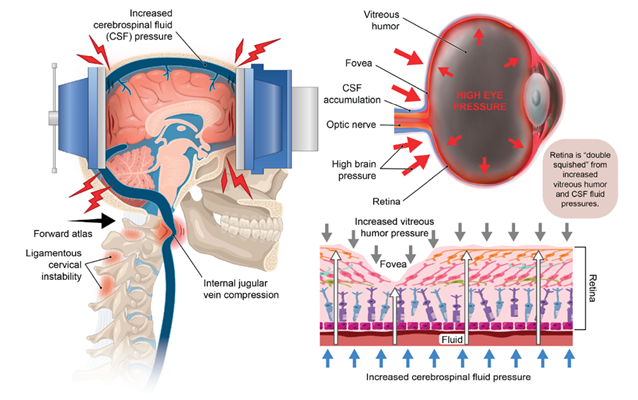
Figure 13: The Structural Pressures on the Retina. When brain pressure increases from narrowing of the internal jugular vein from ligamentous cervical instability, the posterior globe of the eye gets compressed, cerebrospinal fluid accumulates around the optic nerve, and there is a decrease in fluid flow out of the eye, the net effect being an elevation in eye pressure (ocular hypertension). The reina is then "double squished" between the high pressure on the outside of the eye and the elevated pressure within it, potentially leading to such symptoms and diseases as blurry vision, double vision, focusing issues, and glaucoma, as well as macular and retinal degeneration [61].
4.5 Vagus Nerve
As the vagus nerve travels alongside the internal jugular vein within the carotid sheath, it was not surprising to also find an extremely high rate of abnormally small vagus nerve CSA (95%) in this patient population. The vagus nerves can be easily seen with cervical ultrasound. See Figure 14.
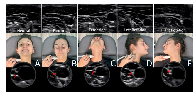
Figure 14: Ultrasound of vagus nerve in mid-cervical region with various neck positions. A. Neck in neutral position. B. Neck flexed. C. Neck extended. D. Neck rotated left. E. Neck rotated right. As can be seen, the vagus nerve within the carotid sheath undergoes various structural tensions, depending on neck positions [61].
While structural neck postures and disorders can impair vagus nerve function, dysfunctional neck issues (including instability) could also potentially negatively affect the sympathetic ganglia and fibers in the cervical spine, especially the superior cervical sympathetic ganglia (SCSGs), another potential etiology for autonomic dysfunction and chronic symptoms [133-136]. The anterior locations traversing the neck make both the nodose ganglion of the vagus nerve and the SCSG easily compressed or aggravated by LCI and cervical dysstructure, such as the forward shift of the upper cervical spine seen with increased C6AI, all profoundly documented in our patient population. See Figure 15. Vagus nerve dysfunction brought on by cervical pathology is explained in detail in a previous publication, Cervicovagopathy: ligamentous cervical instability and dysstructure as a potential etiology for vagus nerve dysfunction in the cause of human symptoms and disease [137].
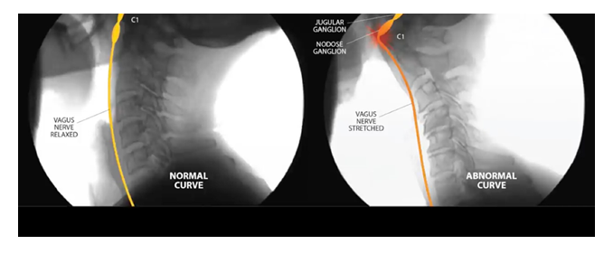
Figure 15: Comparison of normal lateral c-spine x-rays to cervical dysstructure. In the normal, healthy lordotic nerve, the vagus nerve does not encounter any resistance. In an abnormal curve caused by instability due to ligament incompetence, the vagus nerve is under stretch and compression, causing injury.
The autonomic nervous system (ANS) is involved with many physiological functions of the eye, including regulation of intraocular pressure, pupil dimensions and function, lens accommodation, and circulation within the eyes. Pupillary metrics, including size and dynamic responses to light as seen on a pupillometer, are recognized as reliable indicators of ANS dysfunction, not just for the eye, but systemically for the body [138-139]. The chronically dilated pupils seen with nervous system dysfunction can cause blurry vision by hindering the ability to focus (decreased depth of field) and by allowing more light to enter the eye and hit the retina, either directly or peripherally, which can cause ocular aberrations [140].
Since approximately 75-80% of the nerve fibers in the parasympathetic nervous system originate from the vagus nerve, injury to the vagus nerve may cause low vagal tone, an increasingly recognized cause of dysautonomia, specifically parasympathetic nervous system dysfunction, which would illicit a sympathetic state [141-143]. Parasympathetic nervous system dysfunction may be a contributing factor in suspected ocular dysfunction in this patient population, as 92% had total pupil diameters on the higher end or greater than normal (>8 mm), averaging 10.29 mm bilateral total, and 95% had abnormal pupillary light constriction (>60% bilateral total), averaging 75% total [42-44]. Autonomic nervous system dysfunction is increasingly recognized as causing or occurring in ocular diseases and is most often related to parasympathetic nervous system dysfunction [144-145].
The pupillary light reflex is under the control of the ANS and is defined as the constriction and subsequent dilation of the pupil in response to light. The pupillary light reflex can be affected by a wide range of disorders and by parasympathetic or sympathetic stimulation or inhibition [44,146]. The pupillary light response is commonly used in clinical settings to assess dysautonomia, which is characterized by dysregulation of the ANS, an imbalance between the sympathetic and parasympathetic nervous systems [147-149].
The suspected ocular autonomic dysfunction seen in our patient population could be explained by cervical structural effects of the forward-shifted atlas on the vagus nerve, as the 2 vagus nerve ganglia—the nodose ganglion and the jugular ganglion—sit just anterior to the atlas (C1), as well as on the superior cervical sympathetic ganglion, which is located just anterior to the axis (C2). Cervical sympathetic ganglion stimulation has been shown to increase IOP, demonstrating involvement in regulating ocular functions [150]. The vagus nerve is the main inhibitory mechanism to sympathetic stimulation, as it has direct communication with the SCSG [151]. See Figure 16. Both the vagus nerve and the SCSG are not only interconnected with each other, but also with many other structures in the upper cervical region, including the trigeminal, facial, glossopharyngeal, accessory, and hypoglossal nerves (cranial nerves V, VII, IX, XI, and XII, respectively), along with the connections to the cervical sympathetic trunks and C1-C3 spinal nerve roots [152-155].
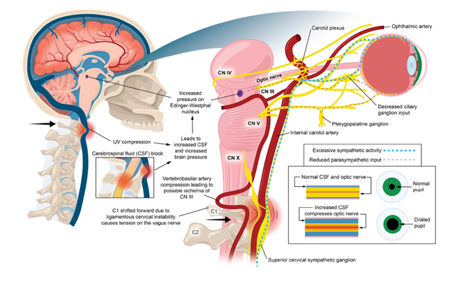
Figure 16: Ocular Dysautonomia with Pupillary Dilation. Ligamentous cervical instability leads to internal jugular vein (IJV) compression and ocular dysautonomia with resultant pupillary dilation [18].
Though not tested in our patient population, accommodation dysfunction is also a possible etiology of blurry vision (typically diagnosed during a comprehensive eye exam), potentially affected by automimic dysfunction produced by LCI, as pupillary diameter and ocular accommodation are each controlled by eye muscles in the iris and ciliary body, receiving innervation by the ciliary (parasympathetic) and postganglionic fibers in the upper cervical (sympathetic) ganglion [156]. Vagus nerve dysfunction could be an underlying contributor to abnormal pupillary dynamics brought on even intermittently by an altered sympathetic/parasympathetic state due to ligamentous cervical instability or cervical structural changes, causing stretch and compression of the cervical nerves.
5. Limitations and Future Directions
The findings from this study suggest a possible association between cervical structural findings and unexplained blurry vision, with proposed neurovascular and autonomic mechanisms remaining theoretical. The presence of blurry vision was based solely on patient self-report without objective ophthalmologic confirmation. This study was performed in a neck center cohort of patients with blurry vision, and the findings should therefore be considered representative of this population, rather than the general population. The absence of a control group, particularly individuals with normal neck anatomy or neck symptoms without blurry vision, further limits interpretation. Accordingly, the observed cervical and neurovascular findings reflect characteristics of a symptomatic neck center population in the context of investigating possible neck-related contributors to blurry vision.
Additional limitations include the retrospective design, lack of standardized comprehensive ophthalmologic and retinal testing, and absence of longitudinal follow-up to assess symptom progression or response to intervention. Relevant variables such as screen exposure, posture, and other environmental or behavioral factors were not formally quantified. Prior ophthalmologic evaluations were self-reported by patients and were not independently verified.
All methods used in this study are reproducible and may be applied in future research to further investigate associations between cervical structural findings and unexplained blurry vision. Hypothetically, if some cases of blurry vision are related to cervical structural changes, then the blurry vision might improve or resolve with relieving the neurovascular pathways at play by following a cervical structural program that restores cervical lordosis and improves stability aimed at alleviating traction, tension, or compression of vital nerves and blood vessels (in this case, the vagus nerve and IJV). A dynamic structural medicine protocol could include recommendations such as postural ergonomic changes (computer setup and prism glasses to view cell phones), physical therapy, therapeutic exercises, gentle chiropractic or osteopathic adjustments, and regenerative therapies to assist in cervical curve stabilization by strengthening the ligaments with Prolotherapy [46,49,157]. Prolotherapy to the neck specifically targets the posterior ligament complex to induce healing, tightening, and strengthening of the ligaments [158-159]. At present, any therapeutic implications remain hypothetical and cannot be inferred from this retrospective dataset.
The study identifies associations between ligamentous cervical instability, objective findings, and ocular symptoms, but does not establish causality. All mechanistic interpretations remain speculative and are intended to guide future investigation, rather than establish causality. These relationships may be influenced by confounding factors, including systemic conditions affecting both the cervical spine and the eyes. Future studies should employ prospective, controlled designs with appropriate comparison groups, including patients with similar cervical symptoms without visual complaints. Formal ophthalmologic assessment, cervical imaging, and autonomic testing will be necessary to further evaluate the proposed associations. Interdisciplinary collaboration will be essential to determine whether these findings represent a distinct clinical phenotype or a nonspecific association within a neck-symptomatic population. As this analysis is based on initial presentations, further longitudinal and controlled studies, particularly those assessing structural cervical changes alongside symptomatic and diagnostic improvements, are needed to clarify the relationships.
6. Conclusions
Frequent co-occurrences of cervical structural and neurovascular findings were observed in this cohort of patients with chronic blurry vision evaluated at an outpatient neck center. These findings suggest a potential role for cervical factors in a subset of patients with unexplained blurry vision and may help inform future diagnostic approaches, while supporting interdisciplinary investigation across musculoskeletal and cervical spine disorders, ophthalmology, neurology, and autonomic domains. Further studies are needed to clarify potential associations between unexplained blurry vision and cervical spine dysfunction.
7. Abbreviations
The following abbreviations are used in this manuscript:
ANS Autonomic nervous system
C6AI C6-atlas interval
CBCT Cone beam computed tomography
CSA Cross-sectional area
CSF Cerebrospinal fluid
CT Computed tomography
DOC Depth of curve
IJV Internal jugular vein
IOP Intraocular pressure
LCI Ligamentous cervical instability
MRI Magnetic resonance imaging
ONSD Optic nerve sheath diameter
SCSG Superior cervical sympathetic ganglion
8. Author Contributions
Conceptualization: RH; Data curation: AW; Formal analysis: AW; Funding acquisition: None; Investigation: RH, DM; Methodology: RH; Project administration: RH, MG, BR; Resources: RH, DM; Software: None; Supervision: RH, BR; Validation: RH; Visualization: RH, MG, BR; Writing – original draft: RH, MG; Writing – review & editing: RH, MG, BR.
Acknowledgements:
The authors would like to thank Kerstyn Gay for contributing graphic designs to this article.
Conflicting Interest:
The authors declare no conflicts of interest.
Funding statement:
This research received no external funding.
Institutional Review Board Statement:
The study was conducted in accordance with the Declaration of Helsinki and approved by the Institutional Review Board (or Ethics Committee) of WCG (study code 1364545, protocol code 20235176 on November 13, 2023).
Informed Consent Statement:
Written informed consent was obtained from all subjects involved in the study.
Data availability statement:
Data supporting reported results can be provided upon request.
References
- Ophthalmology Pavel IA, Bogdanici CM, Donica VC, et al. Computer Vision Syndrome: An Ophthalmic Pathology of the Modern Era. Medicina (Kaunas) 59 (2023): 412.
- Uddin O, Light J, Henderson A. Headaches and Blurry Vision. J Neuroophthalmol 44 (2024): e526.
- Hu CX, Zangalli C, Hsieh M, et al. What do patients with glaucoma see? Visual symptoms reported by patients with glaucoma. Am J Med Sci 348 (2014): 403-409.
- Sun Y, Muheremu A, Tian W. Atypical symptoms in patients with cervical spondylosis: Comparison of the treatment effect of different surgical approaches. Medicine (Baltimore) 97 (2018): e10731.
- Leung KKY, Chu EC, Chin WL, et al. Cervicogenic visual dysfunction: an understanding of its pathomechanism. Med Pharm Rep 96 (2023): 16-19.
- Raviskanthan S, Wendt S, Ugoh PM, et al. Functional vision disorders in adults: a paradigm and nomenclature shift for ophthalmology. Surv Ophthalmol 67 (2022): 8-18.
- van Dongen RM, Haan J. Symptoms related to the visual system in migraine. F1000Res 8 (2019): F1000 Faculty Rev-1219.
- Ortiz-Peregrina S, Ortiz C, Casares-López M, et al. The Relationship between Anxiety, Visual Function, and Symptomatology in University Students. J Clin Med 12 (2023): 6595.
- Ellis Sandoval N, Peña Martinez MI, Fernandez Cea AB, et al. Effects on Prolonged Screen Time on Postural Health and Visual Health in Children and Adolescents: A Scoping Review. Orthop Res Rev 17 (2025): 553-562.
- Burke JP, Orton HP, West J, et al. Whiplash and its effect on the visual system. Graefes Arch Clin Exp Ophthalmol 230 (1992): 335-339.
- Bell CA, Grossman SN, Balcer LJ, et al. Vision as a piece of the head trauma puzzle. Eye (Lond) 37 (2023): 2385-2390.
- Barzegari S, ArabKermani Z, Mahmoudvand Z, et al. Prevalence and contributing factors of computer vision syndrome among university students in Iran: a cross-sectional study. BMC Ophthalmol 25 (2025): 501.
- Almutairi H, Alhammad L, Aldossari B, et al. Prevalence and Interrelationships of Screen Time, Visual Disorders, and Neck Pain Among University Students: A Cross-Sectional Study at Majmaah University. Healthcare (Basel) 12 (2024): 2067.
- Treleaven J, Takasaki H. Characteristics of visual disturbances reported by subjections with neck pain. Manual Therapy 19 (2014): 203-207.
- Leung KKY, Chu EC, Chin WL, et al. Cervicogenic visual dysfunction: an understanding of its pathomechanism. Med Pharm Rep 96 (2023): 16-19.
- Treleaven J. Dizziness, unsteadiness, visual disturbances, and sensorimotor control in traumatic neck pain. J Orthop Sports Phys Ther 47 (2017): 492-502.
- Chen Q, Zheng W, Chen X, et al. Whether Visual-related Structural and Functional Changes Occur in Brain of Patients with Acute Incomplete Cervical Cord Injury: A Multimodal Based MRI Study. Neuroscience 393 (2018): 284-294.
- Hauser RA, Griffiths M, Watterson A, et al. Objective test findings in patients with chronic eye symptoms seeking care at an outpatient neck center: ligamentous cervical instability etiology? Front Neurol 16 (2025): 1576315.
- Lu G, Li J, Mao H, et al. Correlation analysis between neck muscles and lifestyle habits in patients with cervical instability. BMC Musculoskelet Disord 26 (2025): 484.
- Anderst W. In vivo cervical spine kinematics arthrokinematics and disc loading in asymptomatic control subjects and anterior fusion patients. University of Pittsburgh (2014).
- Bonney RA, Corlett EN. Head posture and loading of the cervical spine. Appl Ergon 33 (2002): 415-417.
- Lee S, Kang H, Shin G. Head flexion angle while using a smartphone. Ergonomics 58 (2015): 220-226.
- Hauser RA, Matias D, Rawlings B. The ligamentous cervical instability etiology of human disease from the forward head-facedown lifestyle: emphasis on obstruction of fluid flow into and out of the brain. Front Neurol 15 (2024): 1430390.
- Guan X, Fan G, Wu X, et al. Photographic measurement of head and cervical posture when viewing mobile phone: a pilot study. Eur Spine J 24 (2015): 2892-2898.
- Freeman MD, Katz EA, Rosa SL, et al. Diagnostic Accuracy of Videofluoroscopy for Symptomatic Cervical Spine Injury Following Whiplash Trauma. Int J Environ Res Public Health 17 (2020): 1693.
- Wan Z, Wang W, Li C, et al. Validation and application of a novel in vivo cervical spine kinematics analysis technique. Sci Rep 11 (2021): 24266.
- Gupta V, Khandelwal N, Mathuria SN, et al. Dynamic magnetic resonance imaging evaluation of craniovertebral junction abnormalities. J Comput Assist Tomogr 31 (2007): 354-359.
- Hariharan KV, Terhorst L, Maxwell MD, et al. Inter-examiner reliability of radiographic measurements from Open-mouth lateral bending cervical radiographs. Chiropr Man Therap 28 (2020): 32.
- Hauser RA, Griffiths M, Watterson A, et al. Characterizing Initial Cervical Spine and Neurovascular Findings in 84 Consecutive Patients with Hypermobile Ehlers-Danlos Syndrome: A Retrospective Study. J Clin Med 15 (2026): 2212.
- Dong S, Shen B, Jiang X, et al. Comparison of vagus nerve cross-sectional area between brain-first and body-first Parkinson’s disease. npj Parkinsons Dis 10 (2024): 231.
- Hegde RR, Dham JK, Okodaso H. Ultrasonographic Reference Values for Vagus Nerve Cross-Sectional Area and Associated Variables in Healthy Individuals. J Diag Med Sonogr 42 (2025): 3.
- Belau E, Weise JP. Reference values for the cross-sectional area of the vagus nerve in healthy subjects – a high-resolution ultrasound study. Clin Neurophysiol 128 (2017): 339.
- Parenti N, Scalese M, Palazzi C, et al. Role of Internal Jugular Vein Ultrasound Measurements in the Assessment of Central Venous Pressure in Spontaneously Breathing Patients: A Systematic Review. J Acute Med 9 (2019): 39-48.
- Donahue SP, Wood JP, Patel BM, et al. Correlation of sonographic measurements of the internal jugular vein with central venous pressure. Am J Emerg Med 27 (2009): 851-855.
- Chayapinun V, Koratala A, Assavapokee T. Seeing beneath the surface: Harnessing point-of-care ultrasound for internal jugular vein evaluation. World J Cardiol 16 (2024): 73-79.
- Rattan Y, Girgla KK, Mahajan G, et al. Repeatability and reproducibility of dynamic pupillary parameters using an automated quantitative pupillometer. Biomedicine 43 (2023): 1826-1831.
- Li N, Chen Y, Qiu K, et al. Agreement and repeatability of intraocular pressure measurements between SK-5500B and Icare IC200 tonometers in sitting and supine positions: a comparative study. Front Physiol 16 (2026): 1746123.
- Kilic TY, Eyler Y, Güllüpınar B, et al. Inter-Rater Reliability of Ultrasonographic Measurements of Optic Nerve Sheath Diameter Performed by Emergency Nurses. J Emerg Nurs 51 (2025): 480-486.
- Borden AGB, Rechtman AM, Gershon-Cohen J. The normal cervical lordosis. Radiology 74 (1960): 806-809.
- Hou SB, Sun XZ, Liu FY, et al. Relationship of Change in Cervical Curvature after Laminectomy with Lateral Mass Screw Fixation to Spinal Cord Shift and Clinical Efficacy. J Neurol Surg A Cent Eur Neurosurg 83 (2022): 129-134.
- Abdelnaby R, Elsayed M, Mohamed KA, et al. Sonographic Reference Values of Vagus Nerve: A Systematic Review and Meta-analysis. J Clin Neurophysiol 39 (2022): 59-71.
- Kiel M, Grabitz SD, Hopf S, et al. Distribution of Pupil Size and Associated Factors: Results from the Population-Based Gutenberg Health Study. J Ophthalmol 2022 (2022): 9520512.
- Couret D, Boumaza D, Grisotto C, et al. Reliability of standard pupillometry practice in neurocritical care: an observational, double-blinded study. Crit Care 20 (2016): 99.
- Hall CA, Chilcott RP. Eyeing up the Future of the Pupillary Light Reflex in Neurodiagnostics. Diagnostics (Basel) 8 (2018): 19.
- Hauser RA, Griffiths M, Watterson A, et al. Characterizing Initial Cervical Spine and Neurovascular Findings in 84 Consecutive Patients with Hypermobile Ehlers-Danlos Syndrome: A Retrospective Study. J Clin Med 15 (2026): 2212.
- Alvarez AP, Anderson A, Farhan SD, et al. The Utility of Flexion-Extension Radiographs in Degenerative Cervical Spondylolisthesis. Clin Spine Surg 35 (2022): 319-322.
- Betsch M, Kalbhen K, Michalik R, et al. The influence of smartphone use on spinal posture - A laboratory study. Gait Posture 85 (2021): 298-303.
- Anderson M, Jiang J. Teens, social media & technology 2018. Pew Research Center (2018).
- Screen time statistics: 2024 data and trends. DemandSage (2024).
- Almutairi H, Alhammad L, Aldossari B, et al. Prevalence and Interrelationships of Screen Time, Visual Disorders, and Neck Pain Among University Students: A Cross-Sectional Study at Majmaah University. Healthcare (Basel) 12 (2024): 2067.
- Tsang SMH, Cheing GLY, Lam AKC, et al. Excessive use of electronic devices among children and adolescents is associated with musculoskeletal symptoms, visual symptoms, psychosocial health, and quality of life: a cross-sectional study. Front Public Health 11 (2023): 1178769.
- Erdinest N, Berkow D. Computer Vision Syndrome. Harefuah 160 (2021): 386-392.
- Adane F, Alamneh YM, Desta M. Computer vision syndrome and predictors among computer users in Ethiopia: a systematic review and meta-analysis. Trop Med Health 50 (2022): 26.
- Bogdănici CM, Săndulache DE, Nechita CA. Eyesight quality and Computer Vision Syndrome. Rom J Ophthalmol 61 (2017): 112-116.
- Teo C, Giffard P, Johnston V, et al. Computer vision symptoms in people with and without neck pain. Appl Ergon 80 (2019): 50-56.
- Sen A, Richardson S. A study of computer-related upper limb discomfort and computer vision syndrome. J Hum Ergol (Tokyo) 36 (2007): 45-50.
- Tsantili AR, Chrysikos D, Troupis T. Text Neck Syndrome: Disentangling a New Epidemic. Acta Med Acad 51 (2022): 123-127.
- Singh S, Keller PR, Busija L, et al. Blue-light filtering spectacle lenses for visual performance, sleep, and macular health in adults. Cochrane Database Syst Rev 8 (2023): CD013244.
- Abudawood GA, Ashi HM, Almarzouki NK. Computer Vision Syndrome among Undergraduate Medical Students in King Abdulaziz University, Jeddah, Saudi Arabia. J Ophthalmol 2020 (2020): 2789376.
- Blehm C, Vishnu S, Khattak A, et al. Computer vision syndrome: a review. Surv Ophthalmol 50 (2005): 253-262.
- Hauser RA, Griffiths M, Matias D, et al. Cervical Oculopathy: The Cervical Spine Etiology of Visual Symptoms and Eye Diseases-A Hypothesis Exploring Mechanisms Linking the Neck and the Eye. Diagnostics (Basel) 15 (2025): 2650.
- Vij N, Tolson H, Kiernan H, et al. Pathoanatomy, biomechanics, and treatment of upper cervical ligamentous instability: A literature review. Orthop Rev (Pavia) 14 (2022): 37099.
- Olson KA, Joder D. Diagnosis and treatment of cervical spine clinical instability. J Orthop Sports Phys Ther 31 (2001): 194-206.
- Daffner RH. Imaging of Vertebral Trauma. 3rd ed. Cambridge University Press, Cambridge, UK (2011): 163.
- Freeman MD, Katz EA, Rosa SL, et al. Diagnostic Accuracy of Videofluoroscopy for Symptomatic Cervical Spine Injury Following Whiplash Trauma. Int J Environ Res Public Health 17 (2020): 1693.
- Bogduk N. Functional anatomy of the spine. Handb Clin Neurol 136 (2016): 675-688.
- Radcliff K, Kepler C, Reitman C, et al. CT and MRI-based diagnosis of craniocervical dislocations: the role of the occipitoatlantal ligament. Clin Orthop Relat Res 470 (2012): 1602-1613.
- Dickman CA, Mamourian A, Sonntag VKH, et al. Magnetic resonance imaging of the transverse atlantal ligament for the evaluation of atlantoaxial instability. J Neurosurg 75 (1991): 221-227.
- Liao S, Jung MK, Hörnig L, et al. Injuries of the upper cervical spine-how can instability be identified? Int Orthop 44 (2020): 1239-1253.
- Freeman MD, Rosa S, Harshfield D, et al. A case-control study of cerebellar tonsillar ectopia (Chiari) and head/neck trauma (whiplash). Brain Inj 24 (2010): 988-994.
- Azar NR, Kallakuri S, Chen C, et al. Strain and load thresholds for cervical muscle recruitment in response to quasi-static tensile stretch of the caprine C5-C6 facet joint capsule. J Electromyogr Kinesiol 19 (2009): e387-e394.
- Patwardhan AG, Khayatzadeh S, Havey RM, et al. Cervical sagittal balance: a biomechanical perspective can help clinical practice. Eur Spine J 27 (2018): 25-38.
- Steilen D, Hauser R, Woldin B, et al. Chronic neck pain: making the connection between capsular ligament laxity and cervical instability. Open Orthop J 8 (2014): 326-345.
- Solomonow M. Sensory-motor control of ligaments and associated neuromuscular disorders. J Electromyogr Kinesiol 16 (2006): 549-567.
- Mahmoud NF, Hassan KA, Abdelmajeed SF, et al. The Relationship Between Forward Head Posture and Neck Pain: a Systematic Review and Meta-Analysis. Curr Rev Musculoskelet Med 12 (2019): 562-577.
- Oakley PA, Moustafa IM, Haas JW, et al. Two Methods of Forward Head Posture Assessment: Radiography vs. Posture and Their Clinical Comparison. J Clin Med 13 (2024): 2149.
- Poursadegh M, Azghani MR, Chakeri Z, et al. Postures of the head, upper, and lower neck in forward head posture: Static and quasi-static analyses. Middle East J Rehabil Health Stud 10 (2024): e136377.
- Shaghayegh Fard B, Ahmadi A, Maroufi N, et al. Evaluation of forward head posture in sitting and standing positions. Eur Spine J 25 (2016): 3577-3582.
- Deniz Y, Pehlivan E, Cicek E. Biomechanical variances in the development of forward head posture. Phys Ther Korea 31 (2024): 104-113.
- Hauser RA. Hauser’s Laws on the Ligamentous Structural Causes of Chronic Disabling Symptoms of Human Diseases. On J Neur & Br Disord 7 (2024): 642-689.
- Teo AQA, Thomas AC, Hey HWD. Sagittal alignment of the cervical spine: do we know enough for successful surgery? J Spine Surg 6 (2020): 124-135.
- Patel PD, Arutyunyan G, Plusch K, et al. A review of cervical spine alignment in the normal and degenerative spine. J Spine Surg 6 (2020): 106-123.
- Chu ECP, Lo FS, Bhaumik A. Plausible impact of forward head posture on upper cervical spine stability. J Family Med Prim Care 9 (2020): 2517-2520.
- Jeon JC, Choi WI, Lee JH, et al. Anatomical Morphology Analysis of Internal Jugular Veins and Factors Affecting Internal Jugular Vein Size. Medicina (Kaunas) 56 (2020): 135.
- Gisolf J, van Lieshout JJ, van Heusden K, et al. Human cerebral venous outflow pathway depends on posture and central venous pressure. J Physiol 560 (2004): 317-327.
- Tartière D, Seguin P, Juhel C, et al. Estimation of the diameter and cross-sectional area of the internal jugular veins in adult patients. Crit Care 13 (2009): R197.
- Yoon HK, Lee HK, Jeon YT, et al. Clinical significance of the cross-sectional area of the internal jugular vein. J Cardiothorac Vasc Anesth 27 (2013): 685-689.
- Buch K, Groller R, Nadgir RN, et al. Variability in the Cross-Sectional Area and Narrowing of the Internal Jugular Vein in Patients Without Multiple Sclerosis. AJR Am J Roentgenol 206 (2016): 1082-1086.
- Diehn FE, Schwartz KM, Hunt CH, et al. Prevalence of incidental narrowing of the superior segment of the internal jugular vein in patients without multiple sclerosis. Clin Neuroradiol 24 (2014): 121-127.
- Yoon HK, Lee HK, Jeon YT, et al. Clinical significance of the cross-sectional area of the internal jugular vein. J Cardiothorac Vasc Anesth 27 (2013): 685-689.
- Chen S, Chen Y, Xu L, et al. Venous system in acute brain injury: Mechanisms of pathophysiological change and function. Exp Neurol 272 (2015): 4-10.
- Doepp F, Schreiber SJ, von Münster T, et al. How does the blood leave the brain? A systematic ultrasound analysis of cerebral venous drainage patterns. Neuroradiology 46 (2004): 565-570.
- Manfré L, Lagalla R, Mangiameli A, et al. Idiopathic intracranial hypertension: orbital MRI. Neuroradiology 37 (1995): 459-461.
- Simon MJ, Iliff JJ. Regulation of cerebrospinal fluid (CSF) flow in neurodegenerative, neurovascular and neuroinflammatory disease. Biochim Biophys Acta 1862 (2016): 442-451.
- Ghate D, Kedar S, Havens S, et al. The Effects of Acute Intracranial Pressure Changes on the Episcleral Venous Pressure, Retinal Vein Diameter and Intraocular Pressure in a Pig Model. Curr Eye Res 46 (2021): 524-531.
- Lee SS, Robinson MR, Weinreb RN. Episcleral Venous Pressure and the Ocular Hypotensive Effects of Topical and Intracameral Prostaglandin Analogs. J Glaucoma 28 (2019): 846-857.
- Ngnitewe Massa R, Minutello K, Mesfin FB. Neuroanatomy, Cavernous Sinus. StatPearls Publishing (2024).
- Kiel JW. The Ocular Circulation. Morgan & Claypool Life Sciences, San Rafael, CA (2010).
- Malky IE, Aita WE, Elkordy A, et al. Optic nerve sonographic parameters in idiopathic intracranial hypertension, case-control study. Sci Rep 15 (2025): 1788.
- Wang LJ, Chen LM, Chen Y, et al. Ultrasonography Assessments of Optic Nerve Sheath Diameter as a Noninvasive and Dynamic Method of Detecting Changes in Intracranial Pressure. JAMA Ophthalmol 136 (2018): 250-256.
- Hylkema C. Optic Nerve Sheath Diameter Ultrasound and the Diagnosis of Increased Intracranial Pressure. Crit Care Nurs Clin North Am 28 (2016): 95-99.
- Mathieu E, Gupta N, Ahari A, et al. Evidence for Cerebrospinal Fluid Entry Into the Optic Nerve via a Glymphatic Pathway. Invest Ophthalmol Vis Sci 58 (2017): 4784-4791.
- Raffiz M, Abdullah JM. Optic nerve sheath diameter measurement: a means of detecting raised ICP in adult traumatic and non-traumatic neurosurgical patients. Am J Emerg Med 35 (2017): 150-153.
- Maissan IM, Dirven PJ, Haitsma IK, et al. Ultrasonographic measured optic nerve sheath diameter as an accurate and quick monitor for changes in intracranial pressure. J Neurosurg 123 (2015): 743-747.
- Malky IE, Aita WE, Elkordy A, et al. Optic nerve sonographic parameters in idiopathic intracranial hypertension, case-control study. Sci Rep 15 (2025): 1788.
- Kishk NA, Ebraheim AM, Ashour AS, et al. Optic nerve sonographic examination to predict raised intracranial pressure in idiopathic intracranial hypertension: The cut-off points. Neuroradiol J 31 (2018): 490-495.
- Hassen GW, Al-Juboori M, Koppel B, et al. Real time optic nerve sheath diameter measurement during lumbar puncture. Am J Emerg Med 36 (2018): 736.e1-736.e3.
- Li Z, Zhang XX, Yang HQ, et al. Correlation between ultrasonographic optic nerve sheath diameter and intracranial pressure. Zhonghua Yan Ke Za Zhi 54 (2018): 683-687.
- Robba C, Santori G, Czosnyka M, et al. Optic nerve sheath diameter measured sonographically as non-invasive estimator of intracranial pressure: a systematic review and meta-analysis. Intensive Care Med 44 (2018): 1284-1294.
- Yu ZY, Xing YQ, Li C, et al. Ultrasonic optic disc height combined with the optic nerve sheath diameter as a promising non-invasive marker of elevated intracranial pressure. Front Physiol 14 (2023): 957758.
- Ertl M, Knüppel C, Veitweber M, et al. Normal Age- and Sex-Related Values of the Optic Nerve Sheath Diameter and Its Dependency on Position and Positive End-Expiratory Pressure. Ultrasound Med Biol 46 (2020): 3279-3285.
- Li M, Sun Y, Chan CC, et al. Internal jugular vein stenosis associated with elongated styloid process: five case reports and literature review. BMC Neurol 19 (2019): 112.
- Toshniwal SS, Kinkar J, Chadha Y, et al. Navigating the Enigma: A Comprehensive Review of Idiopathic Intracranial Hypertension. Cureus 16 (2024): e56256.
- Fargen KM, Midtlien JP, Margraf CR, et al. Idiopathic intracranial hypertension pathogenesis: The jugular hypothesis. Interv Neuroradiol (2024): 15910199241270660.
- Tuță S. Cerebral Venous Outflow Implications in Idiopathic Intracranial Hypertension-From Physiopathology to Treatment. Life (Basel) 12 (2022): 854.
- Berberat J, Pircher A, Gruber P, et al. Case Report: Cerebrospinal Fluid Dynamics in the Optic Nerve Subarachnoid Space and the Brain Applying Diffusion Weighted MRI in Patients With Idiopathic Intracranial Hypertension-A Pilot Study. Front Neurol 13 (2022): 862808.
- Sarrami AH, Bass DI, Rutman AM, et al. Idiopathic intracranial hypertension imaging approaches and the implications in patient management. Br J Radiol 95 (2022): 20220136.
- Bozdoğan Z, Şenel E, Özmuk Ö, et al. Comparison of Optic Nerve Sheath Diameters Measured by Optic Ultrasonography Before and After Lumbar Puncture in Idiopathic Intracranial Hypertension Patients. Noro Psikiyatr Ars 60 (2023): 117-123.
- Zaic S, Krajnc N, Macher S, et al. Therapeutic effect of a single lumbar puncture in idiopathic intracranial hypertension. J Headache Pain 25 (2024): 145.
- Dinkin M, Oliveira C. Men Are from Mars, Idiopathic Intracranial Hypertension Is from Venous: The Role of Venous Sinus Stenosis and Stenting in Idiopathic Intracranial Hypertension. Semin Neurol 39 (2019): 692-703.
- Singh S, McGuinness MB, Anderson AJ, et al. Interventions for the Management of Computer Vision Syndrome: A Systematic Review and Meta-analysis. Ophthalmology 129 (2022): 1192-1215.
- Zamysłowska-Szmytke E, Adamczewski T, Ziąber J, et al. Cervico-ocular reflex upregulation in dizzy patients with asymmetric neck pathology. Int J Occup Med Environ Health 32 (2019): 723-733.
- D'Angelo A, Vitiello L, Lixi F, et al. Optic Nerve Neuroprotection in Glaucoma: A Narrative Review. J Clin Med 13 (2024): 2214.
- Wilhelm H, Schabet M. The Diagnosis and Treatment of Optic Neuritis. Dtsch Arztebl Int 112 (2015): 616-625.
- Bennett JL. Optic Neuritis. Continuum (Minneap Minn) 25 (2019): 1236-1264.
- Sinnott JR, Mohebbi MR, Koboldt T. Papilledema: Point-of-Care Ultrasound Diagnosis in the Emergency Department. Clin Pract Cases Emerg Med 2 (2018): 125-127.
- Morgan WH, Yu DY, Cooper RL, et al. The influence of cerebrospinal fluid pressure on the lamina cribrosa tissue pressure gradient. Invest Ophthalmol Vis Sci 36 (1995): 1163-1172.
- Mohammed NY, Di Domenico G, Gambaccini M. Cerebral venous drainage through internal jugular vein. Veins Lymphatics 8 (2019).
- Margolin E. The swollen optic nerve: an approach to diagnosis and management. Pract Neurol 19 (2019): 302-309.
- Xue X, Zhou C, Gao Y, et al. Optic nerve sheath fenestration for visual impairment in cerebral venous diseases. Front Neurol 14 (2023): 1065315.
- Jonas JB, Wang N, Wang YX, et al. Ocular hypertension: general characteristics and estimated cerebrospinal fluid pressure. The Beijing Eye Study 2011. PLoS One 9 (2014): e100533.
- Siaudvytyte L, Januleviciene I, Daveckaite A, et al. Literature review and meta-analysis of translaminar pressure difference in open-angle glaucoma. Eye (Lond) 29 (2015): 1242-1250.
- Tominaga Y, Maak TG, Ivancic PC, et al. Head-turned rear impact causing dynamic cervical intervertebral foramen narrowing: implications for ganglion and nerve root injury. J Neurosurg Spine 4 (2006): 380-387.
- Chen HS, van Roon L, Ge Y, et al. The relevance of the superior cervical ganglion for cardiac autonomic innervation in health and disease: a systematic review. Clin Auton Res 34 (2024): 45-77.
- Dieguez HH, Romeo HE, González Fleitas MF, et al. Superior cervical gangliectomy induces non-exudative age-related macular degeneration in mice. Dis Model Mech 11 (2018): dmm031641.
- Watanuki A. The effect of the sympathetic nervous system on cervical spondylosis. Nihon Seikeigeka Gakkai Z 55 (1981): 371-385.
- Hauser RA, Matias D, Rawlings BR. Cervicovagopathy: ligamentous cervical instability and dysstructure as a potential etiology for vagus nerve dysfunction in the cause of human symptoms and diseases. Front Neurol 16 (2025): 1572863.
- Park HL, Jung SH, Park SH, et al. Detecting autonomic dysfunction in patients with glaucoma using dynamic pupillometry. Medicine (Baltimore) 98 (2019): e14658.
- Jain D, Arbogast K, McDonald C. Objective eye tracking metrics of vision and autonomic dysfunction distinguish adolescents with acute concussion and those with persistent post-concussion symptoms from uninjured controls. Neurology 98 (2022).
- Manion GN, Stokkermans TJ. The Effect of Pupil Size on Visual Resolution. StatPearls Publishing (2025).
- Huckemann S, Mueller K, Averdunk P, et al. Vagal cross-sectional area correlates with parasympathetic dysfunction in Parkinson's disease. Brain Commun 5 (2023): fcad006.
- Goldberger JJ, Arora R, Buckley U, et al. Autonomic Nervous System Dysfunction: JACC Focus Seminar. J Am Coll Cardiol 73 (2019): 1189-1206.
- Zanin A, Amah G, Chakroun S, et al. Parasympathetic autonomic dysfunction is more often evidenced than sympathetic autonomic dysfunction in fluctuating and polymorphic symptoms of "long-COVID" patients. Sci Rep 13 (2023): 8251.
- Murakami A. Autonomic nervous system dysfunction in ocular diseases. Juntendon Med J 62 (2016): 377-380.
- Kaido M, Arita R, Mitsukura Y, et al. Variability of autonomic nerve activity in dry eye with decreased tear stability. PLoS One 17 (2022): e0276945.
- Bittner DM, Wieseler I, Wilhelm H, et al. Repetitive pupil light reflex: potential marker in Alzheimer's disease? J Alzheimers Dis 42 (2014): 1469-1477.
- Hamrakova A, Ondrejka I, Sekaninova N, et al. Central autonomic regulation assessed by pupillary light reflex is impaired in children with attention deficit hyperactivity disorder. Physiol Res 69 (2020): S513-S521.
- Karahan M, Demirtaş AA, Hazar L, et al. Autonomic dysfunction detection by an automatic pupillometer as a non-invasive test in patients recovered from COVID-19. Graefes Arch Clin Exp Ophthalmol 259 (2021): 2821-2826.
- Tsitsi P, Nilsson M, Waldthaler J, et al. Pupil light reflex dynamics in Parkinson's disease. Front Integr Neurosci 17 (2023): 1249554.
- Gallar J, Liu JH. Stimulation of the cervical sympathetic nerves increases intraocular pressure. Invest Ophthalmol Vis Sci 34 (1993): 596-605.
- Mitsuoka K, Kikutani T, Sato I. Morphological relationship between the superior cervical ganglion and cervical nerves in Japanese cadaver donors. Brain Behav 7 (2017): e00619.
- Chawla JC, Falconer MA. Glossopharyngeal and vagal neuralgia. Br Med J 3 (1967): 529-531.
- Cardinali DP, Vacas MI, Gejman PV. The sympathetic superior cervical ganglia as peripheral neuroendocrine centers. J Neural Transm 52 (1981): 1-21.
- Mul Fedele ML, Galiana MD, Golombek DA, et al. Alterations in Metabolism and Diurnal Rhythms following Bilateral Surgical Removal of the Superior Cervical Ganglia in Rats. Front Endocrinol (Lausanne) 8 (2018): 370.
- Anterieu P, Vassal F, Sindou M. Vagoglossopharyngeal neuraligia revealed through predominant digestive vagal manifestations. Case report and literature review. Neurochirurgie 62 (2016): 174-177.
- Khalid K, Padda J, Pokhriyal S, et al. Pseudomyopia and Its Association With Anxiety. Cureus 13 (2021): e17411.
- Moustafa IM, Diab AA, Hegazy F, et al. Demonstration of central conduction time and neuroplastic changes after cervical lordosis rehabilitation in asymptomatic subjects: a randomized, placebo-controlled trial. Sci Rep 11 (2021): 15379.
- Hackett GS, Hendersen DG. Joint stabilization; an experimental, histologic study with comments on the clinical application in ligament proliferation. Am J Surg 89 (1955): 968-973.
- Hauser RA, Matias D, Woznica D, Rawlings B, Woldin BA. Lumbar instability as an etiology of low back pain and its treatment by prolotherapy: A review. J Back Musculoskelet Rehabil. 2022;35(4):701-712.
Supplementary Figure:
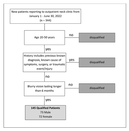
Supplementary Figure: Flow diagram of participant inclusion and exclusion.