Sara Aversa1,2,3*, Bargiacchi Lavinia2, Linda Ricci3, Veronica Sorrentino1,2, Romano Antonio4, Vincenzo Petrozza2,5*, Cristiana Bellan3
1Department of Anatomical, Histological, Forensic and Orthopaedic Sciences, Section of Human Anatomy, Sapienza University of Rome, Rome, Italy.
2Pathology Unit, I.C.O.T. Hospital, Latina, Italy.
3Department of Medical Biotechnologies, University of Siena, Siena, Pathology Unit, Siena, Italy
4U.O.C Otolaryngology, University Hospital "Le Scotte" Siena, Italy
5Department of Medico-Surgical Sciences and Biotechnologies, Sapienza University, Rome, Italy .
*Corresponding Author(s): Sara Aversa, Department of Anatomical, Histological, Forensic and Orthopaedic Sciences, Section of Human Anatomy, Sapienza University of Rome, Rome, Italy (1), Pathology Unit, I.C.O.T. Hospital, Latina, Italy (2)
Vincenzo Petrozza, Department of Medico-Surgical Sciences and Biotechnologies, Sapienza University, Rome, Italy (5). Pathology Unit, I.C.O.T. Hospital, Latina, Italy (2)
Received: 09 January 2021; Accepted: 22 January 2021; Published: 22 February 2021
Nasal polyp is a non neoplastic lesion of the respiratory mucosa. In few cases, it can be possible to detech the presence of cholesterolgranuloma. We present the case of a 55 year-old patient affected by a nasal lesion, localized in sphenoethmoidal recess, with particular microscopic feature and a review of the literature.
Nasal polyp; Lesion; Cholesterolgranuloma
1. Introduction
Nasal polyp is a benign lesion of the respiratory mucosa.
This condition is present in 40% of the population and it has a chronic course in about 4% of cases [1]. They are more common in adults in particular localized in ethmoid sinuses [2]. It has been observed that the pediatric population frequently presents this pathology with a preference for the antrocoanal localization [3]. Cholesterolgranuloma in the paranasal sinuses is rare. Here we report the case of a patient with a sphenoethmoidal localization of the lesion with a particular microscopic feature. A review of the literature follows.
2. Material and Methods
A 55-year-old patient presented to clinicians observation complaining difficulty breathing and a lost sense of smell. Remote pathological anamnesis detected an allergy to grasses. The patient first underwent nasal endoscopy, which did not detect any alteration. Subsequently CT scan of the facial mass was carried out reveling an isolated neoformation of the right nasal fossa of the sphenoethmoid recess. It is decided to proceed with the surgical removal of the lesion. Surgical Sample consisted of a fragment polyploid in shape with a maximum diameter of 2,3 cm. The neoformation showed a smooth fleshy, gray-pink surface. At microscopic examination the lesion was composed by myxoid stroma with increased vasculature (Figure 1) covered by respiratory epithelium admixed with glandular structures. Into edematous lamina propria, weak lymphocytic infiltration, constituting of small lymphocytes, could be observed. At the apex of the lesion a gigantocellular granulomatous inflammatory reaction was appreciated, surrounding optically empty spaces reachable by cholesterol needles (Figure 2). Final diagnosis was: Paranasal sinus cholesterol granuloma.
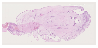
Figure 1: An entire section of the nasal polyp that allow to show its histologic architecture. Hematoxilin eosin 1,25 X.
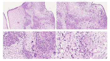
Figure 2: (A) Hematoxilin eosin 5 X; (B) Hematoxilin eosin 10 X; (C) Hematoxilin eosin 20 X; (D) Hematoxilin eosin 40 X.
3. Discussion and Conclusion
Nasal polyps are soft, painless, noncancerous growths on the lining of nasal passages or sinuses [4]. They are associated with a persistent local inflammation condition due to asthma, recurring infection, allergies, drug sensitivity or certain immune disorders [5]. Groups of nasal polyps or lager ones cause a swelling of the nasal mucosa and a reduction of the respiratory space and lead to breathing problems such as progressive nasal obstruction, frontal headache, alteration of smell. Small nasal polyps may not cause symptoms [6]. They appear as traslucent mass that histologically showes edematous lamina propria with variable inflammatory infiltrate including eosinophils [7]. Different subtypes of this lesion has been describe: angiectatic (angiomatous), cystic, edematous, fibrous, glandular. Our case is caractherized by the presence of cholesterol needles at the top of the lesion. Macrophage cells rupture or necrosis cause cholesterol release, which crystallizes in the form of empty needles shapes. There is granulation tissue with foreign bodytype giant cells that surround the needles created by the cholesterol crystals. Repeated bleeding that occurs in a mucosa degenerated by chronic inflammation could explain the onset of such lesions. Correlation between the neoformation size and dimension of paranasal sinus concerned can be an important parameter to consider. An exophitic lesion localized in a “small chamber” lined with a non-functioning mucosa is not optimally vascularized. This could allow a local microtrauma which favor the precipitation of cholesterol with the formation of crystals [8]. The site of the reaction (the “peak” of neoformation) favors both the hypothesis: the most peripheral portion of a lesion is the site that most easily undergoes degeneration. Cholesterol granulomas have been found mainly in the neoformations of the frontal and maxillary sinuses [9]. Few cases of nasal lesions with granulomatous reaction have been described in ethmoidal and antrocoanal sinus [2, 3]. At the current state there is no difference in diagnostic tests and therapy reguarding cholesterol granulomatous polyps [10, 11]. A histologic evaluation of the lesion is always advisable [12]. An accurate analysis can allow to highlight aspects that should be taken into consideration and may promote better overview of underlying disease.
References
- Newton JR, Ah-See KW. A review of nasal polyposis. Ther Clin Risk Manag 4 (2008): 507-512.
- Hellquist H, Lundgren J, Olofsson J. Cholesterol granuloma of the maxillary and frontal sinuses. ORL J Otorhinolaryngol Relat Spec 46 (1984): 153-158.
- Alzahrani M, Morinière S, Duprez R, et al. Cholesterol granuloma of the maxillary sinus. Rev Laryngol Otol Rhinol (Bord) 131 (2010): 309-311.
- Val-Bernal JF, Martino M, Castaneda-Curto N, et al. Cholesterol granuloma in an antrochoanal polyp. A rare lesion in children. Rev Esp Patol 51 (2018): 262-266.
- Zhu LX, Kong WJ, Gong SS, et al. Twenty four cases of cholesterol granuloma of the paranasal sinuses. Zhonghua Er Bi Yan Hou Tou Jing Wai Ke Za Zhi 40 (2005): 517-520.
- Schaberg MR, Shah GB, Evans JJ, et al. Concomitant transsphenoidal approach to the anterior skull base and endoscopic sinus surgery in patients with chronic rhinosinusitis. J Neurol Surg B Skull Base 74 (013): 241-246.
- Sivasubramaniam R, Harvey RJ. How to Assess, Control, and Manage Uncontrolled CRS/Nasal Polyp Patients. Curr Allergy Asthma Rep 17 (2017): 58.
- Yeh DH, Wong J, Hoffbauer S, et al. The utility of routine polyp histopathology after endoscopic sinus surgery. Int Forum Allergy Rhinol 4 (2014): 926-930.
- DeConde AS, Mace JC, Levy JM, et al. Prevalence of polyp recurrence after endoscopic sinus surgery for chronic rhinosinusitis with nasal polyposis. Laryngoscope 127 (2017): 550-555.
- Stevens WW, Schleimer RP, Kern RC. Chronic Rhinosinusitis with Nasal Polyps. J Allergy Clin Immunol Pract 4 (2016): 565-572.
- Schleimer RP. Immunopathogenesis of Chronic Rhinosinusitis and Nasal Polyposis. Annu Rev Pathol 12 (2017): 331-357.
- Shvili I, Hadar T, Shvero J, et al. Cholesterol granulomas in antrochoanal polyps: a clinicopathologic study. Eur Arch Otorhinolaryngol 262 (2005): 821-825.
- Leon ME, Chavez C, Fyfe B, et al. Cholesterol granuloma of the maxillary sinus. Arch Pathol Lab Med 126 (2002): 217-219.