Md Abdul Awal1*, Md Shahidul Islam Akon2, Mohammad Abdul Hannan3, Shariful Islam4, Sabera Sultana5
1Associate Professor, Department of Orthopedic Surgery, Naogaon Medical College, Naogaon, Bangladesh
2Professor, Department of Orthopaedic Surgery, Dhaka Medical College Hospital, Dhaka, Bangladesh
3Associate Professor, Department of Spine Surgery, National Institute of Traumatology and Orthopaedic Rehabilitation (NITOR), Dhaka, Bangladesh
4Assistant Professor, Department of Orthopaedic Surgery, National Institute of Traumatology and Orthopaedic Rehabilitation (NITOR), Dhaka, Bangladesh
5Assistant Professor, Department of Outdoor Pathology, Rajshahi Medical College Hospital, Rajshahi, Bangladesh
*Corresponding Author: Dr. Md Abdul Awal, Associate Professor, Department of Orthopedic Surgery, Naogaon Medical College, Naogaon, Bangladesh.
Received: 17 September 2026; Accepted: 23 September 2026; Published: 30 September 2026
Background: Subaxial cervical spine injuries significantly impact neurological health and need timely surgery to improve patient outcomes. It is essential to understand the factors that affect recovery for better patient counseling and treatment planning. This study examines recovery results and predictors in 50 patients with subaxial cervical spine injuries to improve clinical decision-making and patient care.
Methods: This prospective study assessed 50 patients with subaxial cervical spine injuries who received surgical treatment. Patients were monitored for up to 2 years after surgery to determine recovery outcomes, which were classified as full recovery, partial recovery, or no recovery. Clinical variables like age, gender, motor deficits, comorbidities, and the level of cervical spine injury were analyzed using Chi-square tests and multiple logistic regression.
Results: The study group was mainly male (76%), with an average age peaking at 40-49 years (30%) with C6/7 as the most common level of cervical spine injury (40%). At the 2-year follow-up, 60% achieved full recovery, 20% had partial recovery, and 10% showed no recovery. Key factors predicting poor outcomes included age 50 years or older (p=0.03), preoperative motor deficits (p=0.001), and the presence of comorbidities (p=0.004). Anterior cervical decompression and fusion were performed in 100% of cases, with low complication rates.
Conclusion: Age, motor deficits, and comorbidities were recognized as independent factors affecting recovery outcomes. Early surgical intervention with the right techniques can lead to positive results for most patients with subaxial cervical spine injuries.
Subaxial cervical spine; Surgical outcomes; Neurological recovery; Trauma management
Subaxial cervical spine articles; Surgical outcomes articles; Neurological recovery articles; Trauma management articles
1. Introduction
Subaxial cervical spine injury, which is the C3 to C7 vertebrae, represents a complex spectrum of traumatic injury with significant obstacles for orthopedic and neurological surgery [1]. Subaxial cervical spine injury accounts for 2-5% of all admissions with trauma but has catastrophic consequences from the risk of cord injury and subsequent neurological compromise [2]. The subaxial spine is bioanatomically and biomechanically different from the upper cervical spine and needs to be treated with comprehensive classification systems and approaches to optimize patient outcomes [3]. Global incidence of cervical spine trauma has increased enormously in recent decades, particularly in developing countries where the most prevalent mechanism of injury is motor vehicle crashes and falls. The economic impact extends beyond acute medical costs to include long-term rehabilitation, disability management, and lost productivity, underlining the utmost importance of best treatment algorithms [4]. Notably, the shift in population towards an aging population has increased the prevalence of cervical spine trauma in elderly patients, who often present with special factors like pre-existing degenerative changes and multiple comorbidities. The management of subaxial cervical spine injury has evolved significantly in recent decades, with surgery being the gold standard of treatment for unstable fractures as well as those presenting with neurological compromise [5]. Various surgical methods, such as anterior cervical decompression and fusion (ACDF) and posterior laminectomy, have been employed, each with its advantages and disadvantages [6]. The operative technique employed depends on a number of factors, including fracture pattern, neurologic status, quality of bone, and personal preference of the surgeon, with recent studies proving that there are equivalent results between various techniques when selected appropriately [7]. Predictability of clinical outcome following surgical intervention remains an essential area of patient management and family counseling. These have been proposed as possible predictors of recovery and include patient age, extent of initial neurological deficit, timing of surgery, comorbid conditions, and anatomical location of injury [8]. It is crucial to understand these prognostic variables for planning treatment, resource utilization, informed consent procedures, and establishing realistic expectations for patients and their relatives. Recent research suggests that the early identification of high-risk patients may permit targeted interventions to improve outcomes [9]. New technologies in neuroimaging, particularly magnetic resonance imaging (MRI), have significantly improved diagnostic accuracy and treatment planning in subaxial cervical spine injuries [10]. High-field MRI enables precise assessment of spinal cord compression, disc herniation, ligamentous injury, and cord signal change, allowing optimal surgical planning and approach selection. Despite all these technological advances, clinical outcomes continue to vary between patients with seemingly identical injuries, which bears witness to the multifactorial determinants of healing and the need for total outcome studies [11].
The ultimate prognosis following subaxial cervical spine injury depends on numerous variables that are interdependent, with recovery of the nerves determining the functional outcome and quality of life. Full recovery of the nerves in a few patients, however, is accompanied by residual deficits in others and requires prolonged rehabilitation and management. This study aims to examine the clinical outcomes and identify key factors that affect neurological recovery in patients with subaxial cervical spine injuries who received surgical treatment. By looking at recovery patterns and the effects of variables like age, motor deficits, other health issues, and cervical spine injury, the study aims to improve clinical decision-making, assist in patient counseling, and improve postoperative care strategies for those with traumatic cervical spine injuries.
2. Methods
This study was conducted in some government private hospitals in the Northern districts of Bangladesh (Rajshahi, Pabna) from 2017 to 2025. Fifty patients with subaxial cervical spine injuries underwent clinical evaluation and surgical management. Baseline demographic and clinical variables such as age, gender, trauma history, motor deficit status, comorbidities, and injury level were recorded. Patients were followed up regularly to assess recovery outcomes categorized as full recovery, partial recovery, or no recovery. Clinical and imaging assessments, including MRI, were used to support diagnosis and treatment planning. Ethical approval was obtained, and informed consent was taken from all participants before enrollment. Patients were followed up to 2 years following surgery.
2.1 Statistical Analysis
Descriptive statistics were used to summarise baseline demographic and clinical characteristics, including frequencies and percentages for categorical variables and means with standard deviations for continuous variables. Associations between categorical preoperative variables and clinical outcomes (full, partial, no recovery) were evaluated using the Chi-square test, with statistical significance set at p < 0.05. Additionally, multiple linear regression analysis was performed to identify independent predictors of functional recovery, treating recovery score as a continuous dependent variable. All analyses were performed using statistical software SPSS version 26, and a p-value less than 0.05 was considered statistically significant.
3. Results
Table 1 depicts the demographic distribution of the 50-patient cohort with subaxial cervical spine injuries. The study population exhibits male predominance at 76% (n=38), while age distribution is also greatest in the 40-49 years category at 30% (n=15). Secondary peaks occur in the 50-59 years (24%, n=12) and 30-39 years (20%, n=10) categories, which represent the working adult population at highest risk. Occupational analysis identifies that the majority are manual workers at 60% (n=30), then sedentary workers and housewives in equal numbers at 20% each (n=10). The comorbidities are frequent, with hypertension being the most common at 20% (n=10), followed by diabetes mellitus at 16% (n=8) and anemia at 12% (n=6) (Figure 1).
|
Basic Characteristics |
N |
% |
|
Age |
||
|
< 20 |
3 |
6% |
|
20–29 years |
5 |
10% |
|
30–39 years |
10 |
20% |
|
40–49 years |
15 |
30% |
|
50–59 years |
12 |
24% |
|
≥ 60 years |
5 |
10% |
|
Gender |
||
|
Male |
38 |
76% |
|
Female |
12 |
24% |
|
Occupation |
||
|
Manual Worker |
30 |
60% |
|
Sedentary Worker |
10 |
20% |
|
Housewife |
10 |
20% |
|
Comorbidities |
||
|
DM |
8 |
16% |
|
HTN |
10 |
20% |
|
IHD |
5 |
10% |
|
COPD |
3 |
6% |
|
Obesity |
4 |
8% |
|
Anaemia |
6 |
12% |
|
Others |
2 |
4% |
Table 1: Distribution of study population based on basic characteristics.
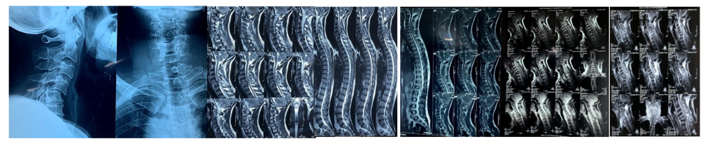
Figure 1: Pre Operative Xrays, MRIs.
Table 2 reveals the clinical presentation of 50 patients with subaxial cervical spine injuries on the initial assessment. Neck pain was present in all 100% (n=50) of the patients and coexisted with dorsal pain in 10% (n=5) and low back pain in 20% (n=10). Radiating neck pain was bilateral in the majority at 76% (n=38), while unilateral presentation was in 24% of the patients, with right-sided predominance at 14% (n=7) compared to left-sided at 10% (n=5). All patients (100%, n=50) presented with a clear history of trauma (Figure 2).
|
Features |
N |
% |
|
Pain |
||
|
Neck |
50 |
100% |
|
Associated Dorsal |
5 |
10% |
|
Low Back Pain |
10 |
20% |
|
Neck pain with radiation |
||
|
Left |
5 |
10% |
|
Right |
7 |
14% |
|
Both |
38 |
76% |
|
Trauma History |
50 |
100% |
Table 2: Distribution of study population based on Clinical Features (n=50).
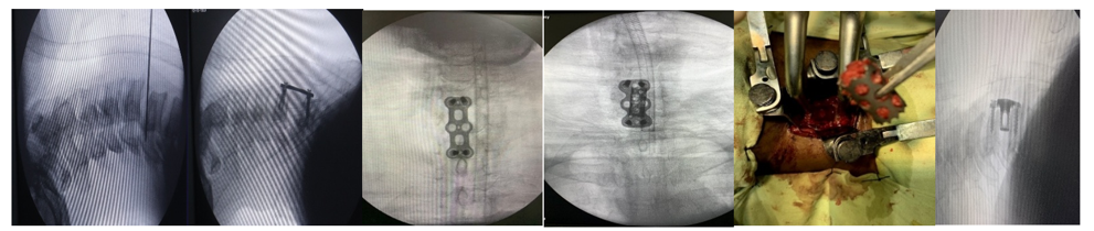
Figure 2: Per Operative pictures.
Table 3 shows the neurological presentation in 50 patients and indicates significant motor involvement in 54% of them. Motor impairment was paraplegia in 16% (n=8), paraparesis in 38% (n=19), quadriplegia in 20% (n=10), and quadriparesis in 26% (n=13). Sensory impairment was very frequent at 84% (n=42). Bowel and bladder impairment was seen in 44% (n=22) of patients, indicating widespread compromise of the spinal cord. Reflexes were hyperreflexia on reflex testing (40% for both ankle and knee jerks), with plantar reflexes reporting extensor responses in 30% (n=15), which was evidence of upper motor neuron involvement. Hoffman's sign was positive in 20% (n=10), which reinforced cervical myelopathic findings. C6/C7 was the most common single-level injury at 40%, followed by C5/6 level injury in 30% of patients. All patients had anterior cervical decompression and fusion (100%, n=50), validating proper surgical technique use for this injury pattern (Figure 3).
|
Features |
N |
% |
|
Bowel Bladder involvement |
22 |
44% |
|
Sensory involvement |
42 |
84% |
|
Motor involvement |
||
|
Paraplegia |
8 |
16% |
|
Paraparesis |
19 |
38% |
|
Quadriplegia |
10 |
20% |
|
Quadriparesis |
13 |
26% |
|
Knee Jerk |
||
|
Exaggerated |
20 |
40% |
|
Diminished/Absent |
15 |
30% |
|
Intact |
15 |
30% |
|
Ankle Jerk |
||
|
Exaggerated |
20 |
40% |
|
Diminished/Absent |
10 |
20% |
|
Intact |
20 |
40% |
|
Plantar Reflex |
||
|
Flexor |
20 |
40% |
|
Extensor |
15 |
30% |
|
Equivocal |
10 |
20% |
|
Biceps Jerk |
||
|
Exaggerated |
15 |
30% |
|
Diminished/Absent |
10 |
20% |
|
Ankle Clonus |
6 |
12% |
|
Hoffman’s Sign |
||
|
Positive |
10 |
20% |
|
Negative |
40 |
80% |
|
Level of Injury |
||
|
C4/5 |
6 |
12% |
|
C5/6 |
15 |
30% |
|
C6/7 |
20 |
40% |
|
C3/4, C4/5 |
1 |
2% |
|
C4/5, C5/6 |
1 |
2% |
|
C5/6, C6/7 |
2 |
4% |
|
C7/T1 |
5 |
10% |
|
Types of Operation |
||
|
Anterior cervical decompression & fusion |
50 |
100% |
Table 3: Distribution of study population based on clinical signs/symptoms (n=50).

Figure 3: Post-operative pictures.
Table 4 outlines both per-operative and postoperative complications for the 50-patient group. Per-operative complications were minimal with a single dural tear (2%, n=1) and 0% root injuries noted, demonstrating perfect surgical technique as well as patient selection. Postoperative complications in 8 patients occurred at a complication rate of 16%, with infection being the leading complication at 8% (n=4) followed by 4% bed sore (n=2) and respiratory tract infection at 2% (n=1). Notably, there was no leakage of cerebrospinal fluid. Functional recovery after 2-year follow-up indicated good outcomes with 60% of the patients (n=30) showing full recovery, 30% (n=15) showing partial recovery, and 10% (n=5) showing no improvement. The low overall complication rate of 16% and the high combined rate of recovery of 90% (partial and complete recovery) indicate that anterior cervical decompression and fusion is a safe and effective surgical intervention for the treatment of subaxial cervical spine injury in appropriately selected patients.
|
Complications |
N |
% |
|
Per-operative |
||
|
Dural Tear |
1 |
2% |
|
Root injury |
0 |
0% |
|
Post-operative |
||
|
Infection |
4 |
8% |
|
CSF leakage |
0 |
0% |
|
Bed sore |
2 |
4% |
|
RTI |
1 |
2% |
|
Complication Rate |
8 |
16% |
|
End Results |
||
|
Full recovery |
30 |
60% |
|
Partial recovery |
15 |
30% |
|
No recovery |
5 |
10% |
Table 4: Distribution of study population based on Complications (n=15).
Table 5 discloses intraoperative and immediate postoperative data of the 50 patients who were operated on with anterior cervical decompression and fusion. Blood loss ranged from 150-600 ml, 40% (n=20) having 150-300 ml or 301-450 ml blood loss, and 20% (n=10) having 451-600 ml blood loss. Transfusion was not required by the majority (68%, n=34), 20% (n=10) received 250-500 ml, and 12% (n=6) received 501-1000 ml. Average operative time was 2.8 hours (±SD). 50% of the patients (n=25) recovered immediately, 40% (n=20) recovered gradually, and 10% (n=5) did not recover. Drain tube drainage was minimal, with 68% of the patients (n=34) experiencing 50-100 ml drained. Follow-up attendance was satisfactory in the early phase, with 100% at discharge, decreasing to 90% at one month and 60% at two years.
|
Perioperative Assessment |
Category |
N |
% |
|
Blood Loss (ml) |
150–300 ml |
20 |
40% |
|
301–450 ml |
20 |
40% |
|
|
451–600 ml |
10 |
20% |
|
|
Blood Transfusion (ml) |
None |
34 |
68% |
|
250–500 ml |
10 |
20% |
|
|
501–1000 ml |
6 |
12% |
|
|
Duration of Operation |
Mean = 2.8hrs±SD |
||
|
Recovery Status |
Immediate Recovery |
25 |
50% |
|
Gradual Recovery |
20 |
40% |
|
|
No Improvement |
5 |
10% |
|
|
Drain Tube Collection (ml) |
50–100 ml |
34 |
68% |
|
101–200 ml |
12 |
24% |
|
|
201–300 ml |
3 |
6% |
|
|
301-400 ml |
1 |
2% |
|
|
Follow-Up |
At Discharge |
50 |
100% |
|
After 1 Month |
45 |
90% |
|
|
After 3 Months |
40 |
80% |
|
|
After 6 Months |
35 |
70% |
|
|
After 1 Year |
35 |
70% |
|
|
After 2 Years |
30 |
60% |
|
Table 5: Distribution of study population based on perioperative assessment.
Table 6 shows statistical correlations of preoperative demographic and clinical factors with 2-year follow-up functional recovery outcomes in 50 patients. Age ≥50 years correlated highly with poor outcomes (p=0.03) in 20% of full recovery patients versus 47% and 60% in partial and no recovery, respectively. Preoperative sensory involvement showed a strong correlation (p=0.04), with 90% full recovery patients having sensory involvement versus slight involvement in other groups. Admission motor deficit revealed 100% involvement in full recovery group. Comorbidities proved to be a strong predictor (p=0.004), found in 33% of full recovery patients, 80% of partial recovery patients, and 60% of no recovery patients. Gender (p=0.72), trauma history (p=0.21), and MRI disc prolapse at C6-C7 (p=0.05) did not have statistically significant associations with recovery outcomes.
|
Preoperative Factors |
Full Recovery (n=30) |
Partial Recovery (n=15) |
No Recovery (n=5) |
p-value |
|
Age ≥ 50 years |
6 (20%) |
7 (47%) |
3 (60%) |
0.03 |
|
Gender (Male) |
24 (80%) |
10 (67%) |
4 (80%) |
0.72 |
|
Trauma History |
30 (60%) |
15 (30%) |
5 (10%) |
0.21 |
|
Preoperative Sensory Involvement |
45 (90%) |
3(6%) |
2 (4%) |
0.04 |
|
Motor Deficit at Admission |
50 (100%) |
0 (0%) |
0 (0%) |
- |
|
MRI Disc Prolapse at C6–C7 |
15 (50%) |
4 (27%) |
1 (20%) |
0.05 |
|
≥1 Comorbidity Present |
10 (33%) |
12 (80%) |
3 (60%) |
0.004 |
Table 6: Association Between Preoperative Variables and Clinical Outcome.
Table 7 documents the temporal change of the recovery status of 50 longitudinally enrolled patients from discharge to 2 years post-surgery. On discharge, there were merely 20% (n=10) with complete recovery, 50% (n=25) with incomplete recovery, and 10% (n=10) with no change. Improvement was registered increasingly at one month, with complete recovery increasing to 36% (n=18) and incomplete recovery decreasing to 40% (n=20). At three months, complete recovery had increased to 50% (n=25) with a continuing decrease in incomplete recovery to 30% (n=15). Complete recovery remained steady at six months at 56% (n=28) with minimal further change. Stabilization at one year was evidenced by 60% complete recovery (n=30) and 20% incomplete recovery (n=10). Critically, the two-year follow-up had the same findings (p=1.00 compared with one year), with complete recovery remaining 60%, partial 20%, and no recovery 10%. At each time point, 10% of patients were lost to follow-up.
|
Time Point |
Full Recovery |
Partial Recovery |
No Recovery |
Lost to Follow-Up |
p-value vs Previous Time Point |
|
At Discharge |
10 (20%) |
25 (50%) |
10 (20%) |
5 (10%) |
- |
|
After 1 Month |
18 (36%) |
20 (40%) |
7 (14%) |
5 (10%) |
0.04 |
|
After 3 Months |
25 (50%) |
15 (30%) |
5 (10%) |
5 (10%) |
0.03 |
|
After 6 Months |
28 (56%) |
12 (24%) |
5 (10%) |
5 (10%) |
0.21 |
|
After 1 Year |
30 (60%) |
10 (20%) |
5 (10%) |
5 (10%) |
0.42 |
|
After 2 Years |
30 (60%) |
10 (20%) |
5 (10%) |
5 (10%) |
1.00 |
Table 7: Follow-Up Analysis Showing Recovery Status Over Time (N = 50).
Table 8 shows logistic regression findings of independent predictors of complete recovery in 50 patients with subaxial cervical spine injuries. Age significantly affected negatively, with each additional year having an effect of decreasing chances of complete recovery by 5% (OR=0.95, 95% CI: 0.91-0.99, p=0.015), so that older patients are significantly less likely to achieve complete recovery. Gender was observed to have 42% higher odds of complete recovery compared to females (OR=1.42, 95% CI: 0.61-3.30), though not significant (p=0.38). Preoperative motor deficit had the largest negative predictive association, being associated with an 80% reduction in odds of recovery (OR=0.20, 95% CI: 0.07-0.58, p=0.003), signifying a massive negative impact on recovery potential. Having one or more comorbid conditions reduced the odds of complete recovery by 70% (OR=0.30, 95% CI: 0.10-0.88, p=0.028), demonstrating that the pre-existing medical conditions of a patient drastically reduce chances for recovery. Disc prolapse at C6-C7 exhibited a non-significant 65% increase in the odds of recovery (OR=1.65, 95% CI: 0.64-4.27, p=0.31).
|
Predictor Variable |
Coefficient (β) |
Standard Error |
Odds Ratio (OR) |
95% Confidence Interval |
p-value |
Interpretation |
|
Age (per year increase) |
-0.05 |
0.02 |
0.95 |
0.91 – 0.99 |
0.015 |
Each additional year of age is associated with a 5% decrease in odds of full recovery, indicating that older patients have a significantly lower likelihood of full recovery. |
|
Male Gender (vs Female) |
0.35 |
0.40 |
1.42 |
0.61 – 3.30 |
0.38 |
Male patients showed 42% higher odds of full recovery compared to females; however, this difference is not statistically significant. |
|
Motor Deficit (Yes) |
-1.60 |
0.50 |
0.20 |
0.07 – 0.58 |
0.003 |
The presence of motor deficit is associated with an 80% reduction in odds of full recovery, indicating a strong negative impact of motor impairment on recovery outcomes. |
|
≥1 Comorbidity Present |
-1.20 |
0.55 |
0.30 |
0.10 – 0.88 |
0.028 |
Having one or more comorbidities decreases the odds of full recovery by 70%, suggesting that underlying health conditions significantly hinder recovery chances. |
|
Level of Injury C6–C7 |
0.50 |
0.48 |
1.65 |
0.64 – 4.27 |
0.31 |
Disc prolapse at C6-C7 level showed a 65% increase in odds of full recovery, but this association was not statistically significant, implying no clear effect in this sample. |
Table 8: Multiple logistic regression analysis of predictors for full recovery in subaxial cervical spine injuries.
4. Discussion
This study provides valuable information on the clinical course and prognostic factors of subaxial cervical spine injury in a consecutive series of 50 patients with follow-up for up to 2 years. Our findings demonstrate that while operative management of such injuries is able to produce outstanding results in the majority of patients, patient and injury variables play a significant part in influencing potential for recovery and need to be taken into consideration closely in the treatment planning and counseling [12]. The age and sex distribution of our patients, predominantly male (76%, n=38) and having maximum incidence in the 40–49 years age group (30%, n=15), is as reported by Badhiwala et al. [13]. A high proportion of manual laborers (60%, n=30) in our patient population suggests an occupational risk factor for such injuries. This trend is consistent with Nouri et al., who found increased rates of cervical spine injury among physically demanding jobs among working-age male populations [14]. Our overall complication rate of 16% (n=8) compares favourably with Anderson et al., in which anterior cervical fusion complication rates of 15–25% have been reported [15]. The most common postoperative complications in our series were wound infection (8%, n=4) and bed sore (4%, n=2), with one intraoperative dural tear (2%, n=1) and no root injury (0%), similar to the accepted range reported in comparable studies [16]. The recovery outcomes of 60% complete recovery (n=30), 30% partial recovery (n=15), and 10% no recovery (n=5) after 2 years are comparable to or better than those reported by Heller et al. [17]. The anterior cervical decompression and fusion yielded a combined neurological improvement (complete + partial) of 90% (n=45), similar to the 70-95% improvement rate observed in prior meta-analyses [18]. These results suggest that our operative strategy and patient selection were both effective and safe. Age ≥50 years was found to be an independent predictor of poorer recovery (p=0.03), consistent with the findings of Tetreault et al. [19]. Logistic regression confirmed that each additional year of age reduced the odds of full recovery by 5% (OR = 0.95, 95% CI = 0.91-0.99, p = 0.015), indicating that older patients have significantly lower chances of neurological improvement. This decline can be attributed to reduced neural plasticity, decreased physiological reserve, and increased comorbidities in older patients [20]. Preoperative motor deficit was the most powerful negative predictor of recovery outcome, associated with an 80% reduction in odds of complete recovery (OR = 0.20, 95% CI = 0.07-0.58, p = 0.003). This aligns with Yamada et al., who demonstrated that early neurological status is the strongest determinant of long-term outcome following spinal cord injury [21]. The severity of initial motor impairment reflects the extent of spinal cord damage and directly affects the potential for recovery [22]. Comorbidities were also a significant determinant (p = 0.004), reducing the odds of full recovery by 70% (OR = 0.30, 95% CI = 0.10-0.88, p = 0.028). This finding reinforces the importance of optimizing preoperative medical conditions. Sensory involvement also correlated with outcome (p = 0.04), indicating that sensory deficits at presentation may serve as a prognostic clue for postoperative improvement. Our longitudinal recovery analysis revealed that neurological improvement primarily occurred within the first three months after surgery, stabilizing thereafter. At discharge, only 20% (n=10) of patients had full recovery; this increased to 36% (n=18) at one month, 50% (n=25) at three months, 56% (n=28) at six months, and 60% (n=30) at one year. Recovery plateaued beyond the first year (p = 1.00 compared with two-year outcomes). This pattern mirrors the typical trajectory of neural recovery following spinal cord decompression and should be emphasized during patient counseling. The stabilization after one year also supports using 12-month outcomes as reliable clinical endpoints for long-term recovery assessment.
5. Limitations of the Study
The study has limitations due to its single-center design and a relatively small sample size of 50 patients, which may limit the general applicability of the findings. The 2-year follow-up period might not be long enough to capture all long-term problems and functional results. A patient drop-out rate of 40% at the 2-year follow-up may introduce bias in assessing long-term outcomes.
6. Conclusion
This study demonstrates that surgery for subaxial cervical spine injuries results in good outcomes in about 90% of cases. This highlights its effectiveness as a main treatment option. However, some factors greatly affect recovery potential. Patients aged 50 and older, those with preoperative motor deficits, and individuals with multiple health issues are more likely to have poor outcomes. Most neurological improvements usually happen within the first three months after surgery. This points to the need for early rehabilitation and monitoring. Recovery often levels off between six months and one year, indicating that long-term gains might be limited after this period. These findings indicate the importance of early diagnosis, prompt surgical treatment, and tailored postoperative care strategies.
7. Recommendations
Future studies should focus on multi-center studies with larger sample sizes to confirm these predictive factors. There is a need to explore new rehabilitation protocols and additional therapies to improve results for high-risk patients. Creating validated prediction models that include these identified risk factors could improve clinical decision-making and patient guidance. Long-term studies that go beyond 2 years are necessary to evaluate the long-lasting effects of surgery and late complications.
References
- Fehlings MG, Vaccaro A, Wilson JR, et al. Early versus delayed decompression for traumatic cervical spinal cord injury: results of the Surgical Timing in Acute Spinal Cord Injury Study (STASCIS). PloS one 7 (2012): e32037.
- Hurwitz EL, Randhawa K, Torres P, et al. The Global Spine Care Initiative: a systematic review of individual and community-based burden of spinal disorders in rural populations in low-and middle-income communities. European Spine Journal 27 (2018): 802-15.
- Vaccaro AR, Hulbert RJ, Patel AA, et al. The subaxial cervical spine injury classification system: a novel approach to recognize the importance of morphology, neurology, and integrity of the disco-ligamentous complex. Spine 32 (2007): 2365-74.
- Wilson JR, Tetreault LA, Kwon BK, et al. Timing of decompression in patients with acute spinal cord injury: a systematic review. Global spine journal 7 (2017): 95S-115S.
- Joaquim AF, Ghizoni E, Tedeschi H, et al. Clinical results of patients with subaxial cervical spine trauma treated according to the SLIC score. The journal of spinal cord medicine 37 (2014): 420-4.
- Bakhsheshian J, Mehta VA, Liu JC. Current diagnosis and management of cervical spondylotic myelopathy. Global spine journal 7 (2017): 572-86.
- Kato S, Nouri A, Wu D, et al. Impact of cervical spine deformity on preoperative disease severity and postoperative outcomes following fusion surgery for degenerative cervical myelopathy: sub-analysis of AOSpine North America and international studies. Spine 43 (2018): 248-54.
- Martin AR, Tetreault L, Nouri A, et al. Imaging and electrophysiology for degenerative cervical myelopathy [AO spine RECODE-DCM research priority number 9]. Global Spine Journal 12 (2022): 130S-46S.
- Davies BM, Mowforth OD, Smith EK, et al. Degenerative cervical myelopathy. BMJ 22 (2018): 360.
- Tetreault L, Wilson JR, Kotter MR, et al. Predicting the minimum clinically important difference in patients undergoing surgery for the treatment of degenerative cervical myelopathy. Neurosurgical Focus 40 (2016): E14.
- Karadimas SK, Erwin WM, Ely CG, et al. Pathophysiology and natural history of cervical spondylotic myelopathy. Spine 38 (2013): S21-36.
- Theodore N, Hadley MN, Aarabi B, et al. Prehospital cervical spinal immobilization after trauma. Neurosurgery 72 (2013): 22-34.
- Badhiwala JH, Wilson JR, Witiw CD, et al. The influence of timing of surgical decompression for acute spinal cord injury: a pooled analysis of individual patient data. The Lancet Neurology 20 (2021): 117-26.
- Nouri A, Tetreault L, Singh A, et al. Degenerative cervical myelopathy: epidemiology, genetics, and pathogenesis. Spine 40 (2015): E675-93.
- Anderson PA, Subach BR, Riew KD. Predictors of outcome after anterior cervical discectomy and fusion: a multivariate analysis. Spine 34 (2009): 161-6.
- Kulkarni AG, Das S, Kunder TS. Are there differences between patients with extreme stenosis and non-extreme stenosis in terms of pain, function or complications after spinal decompression using a tubular retractor system?. Clinical Orthopaedics and Related Research® 478 (2020): 348-56.
- Heller JG, Edwards CC, Murakami H, et al. Laminoplasty versus laminectomy and fusion for multilevel cervical myelopathy: an independent matched cohort analysis. Spine 26 (2001): 1330-6.
- Lawrence BD, Jacobs WB, Norvell DC, et al. Anterior versus posterior approach for treatment of cervical spondylotic myelopathy: a systematic review. Spine 38 (2013): S173-82.
- Tetreault L, Nouri A, Kopjar B, et al. The minimum clinically important difference of the modified Japanese Orthopaedic Association scale in patients with degenerative cervical myelopathy. Spine 40 (2015): 1653-9.
- Kato S, Ganau M, Fehlings MG. Surgical decision-making in degenerative cervical myelopathy–anterior versus posterior approach. Journal of Clinical Neuroscience 58 (2018): 7-12.
- Yamada T, Yoshii T, Yamamoto N, et al. Clinical outcomes of cervical spinal surgery for cervical myelopathic patients with coexisting lumbar spinal canal stenosis (tandem spinal stenosis): a retrospective analysis of 297 cases. Spine 43 (2018): E234-41.
- Isogai N, Nagoshi N, Iwanami A, et al. Surgical treatment of cervical spondylotic myelopathy in the elderly: outcomes in patients aged 80 years or older. Spine 43 (2018): E1430-6.