Ying Chen MD*,^,1,2, Kristen Ortega BS^,1, Angela Gomez BS1, Esther Roucaute BS1, Mahija Ginjupalli2,4, BSA, Daniel Pelaez PhD1,3, Sara T Wester MD$,1, Alfonso L. Sabater MD PhD$,1
1Bascom Palmer Eye Institute, University of Miami Miller School of Medicine, Miami, FL 33136, USA
2Ruiz Department of Ophthalmology, McGovern Medical School at UTHealth Houston, Houston, TX 77030, USA
3Al-Rashid Orbital Vision Research Center, University of Miami Miller School of Medicine, Miami, FL 33136 USA
4The University of Texas Southwestern Medical School, Dallas, TX 77379, USA
^Equal contributing first co-authors
$Equal contributing senior co-authors
*Corresponding author: Ying Chen. Ruiz Department of Ophthalmology & Visual Science, McGovern Medical School at UT Health Houston, 6431 Fannin St, Houston, TX 77030.
Received: 21 September 2026; Accepted: 28 September 2026; Published: 09 October 2026
Objective: To evaluate the viability of a scaffold containing orbital adipose stem cells (OASC) and platelet rich growth factor (PRGF) and its wound healing effect on conjunctiva for potential use in cicatricial conjunctival disease.
Methods: Orbital fat containing OASCs was obtained from medial fat during routine eyelid surgeries performed between 2019 and 2022. The processed orbital fat was combined with activated platelet rich plasma (aPRP) to create an aPRP/Orbital fat complex. Human primary conjunctival epithelial cells (HPCEC) were seeded and treated with different experimental conditions: 1) HPCEC with aPRP only, 2) HPCEC with orbital fat only, and 3) HPCEC with aPRP/Orbital fat complex. Cell proliferation assay was assessed at different time points. Cell migration assay was then performed to evaluate for wound closure, and immunofluorescence staining was used to characterize the cells in the different conditions.
Results: Proliferation assay using CyQUANT illustrated that aPRP/Orbital fat complex contributes the greatest to cell proliferation. HPCEC incubated with aPRP and aPRP/Orbital fat complex further demonstrated migration and complete wound closure at 96 hours. After removal of experimental condition, the HPCEC regained normal morphology and eventually reached confluency in the aPRP/Orbital fat complex condition. Immunofluorescence staining revealed positive staining for CK13 and CD34 in HPCEC treated with aPRP/Orbital fat complex.
Conclusion: We demonstrate here that aPRP/Orbital fat complex scaffold can facilitate conjunctival cell proliferation. This scaffold has regenerative and anti-fibrotic properties that may further promote conjunctival wound healing.
Orbital adipose tissue, platelet-rich plasma, fibrin membrane, conjunctival regeneration, ocular surface regeneration, wound healing, adipose-derived stem cells, plasma rich in growth factors (PGRF)
Orbital adipose tissue articles, platelet-rich plasma articles, fibrin membrane articles, conjunctival regeneration articles, ocular surface regeneration articles, wound healing articles, adipose-derived stem cells articles, plasma rich in growth factors (PGRF) articles
Introduction
Cicatrizing conjunctivitis is a sight threatening condition characterized by conjunctival inflammation and scarring. It can present as part of a multisystem disorder or as a primary ocular disease [1]. Causes of cicatrizing conjunctivitis include autoimmune processes and toxic exposures. Presentation of cicatrizing conjunctivitis can vary from subtle fibrosis to extensive symblepharon formation and forniceal shortening [1, 2]. In advanced cases, keratin deposition along the ocular surface, architectural changes of the mucocutaneous junction, associated lid malposition, and trichiasis can be seen. Uncontrolled or untreated cicatrizing conjunctivitis may lead to limbal stem cell deficiency resulting in pannus formation, keratopathy, corneal perforation, and even complete loss of vision [3]. Current treatment of cicatrizing conjunctivitis focuses primarily on arresting further progression of cicatrization to prevent the downstream sequelae of further complications such as keratopathy. However, options for treatment remain very limited for those who suffer from extensive fibrosis and keratinization of the eyelid margins and ocular surface. Aggressive lubrication is essential and oral immunosuppression may be necessary in the setting of systemic diseases. When medical therapy alone is not sufficient, amniotic membrane and mucous membrane graft have been utilized to surgically treat the lid margin and tarsal conjunctiva especially in the setting of extensive keratinization [4-6]. These grafts involve foreign tissue and often serve only as a temporizing measure. Furthermore, they may not be suitable in patients who have systemic mucous membrane involvement.
Socket contracture is another disease process (in this case involving anophthalmic sockets) where there is inadequate healthy conjunctival tissue. Socket contracture can result from trauma, excessive conjunctival dissection during enucleation or evisceration, irradiation, cicatrizing diseases of the ocular surface, and scarring after prior surgery [7]. A myriad of different materials are used for surgical correction of socket contracture including conjunctiva grafts, polytetrafluorethylene, mucous membrane, hard palate, oral mucosa, skin, fascia lata, and dermis fat grafts [8-15]. More recent interventions include the use of ultra-thick umbilical amnion and hard palate-dermis fat composite grafts in the reconstruction of an anophthalmic socket [16, 17]. While many graft options have been proposed, recurrent contracture with unpredictable results often leads to unsatisfactory outcomes. Orbital adipose tissue is abundant and easily accessible during routine ophthalmic surgeries, such as upper and lower lid blepharoplasties [18]. It contains a unique population of stem cells known as orbital adipose-derived stem cells (OASC). Unlike mesodermal adipose-derived stem cells found in subcutaneous fat, OASC from the upper eyelid medial fat and lower eyelid fat are neural-crest in origin [19]. These highly specialized stem cells have been shown to possess multi-lineage differentiation potential under specific induction conditions, including expression into smooth muscle and neuronal lineages [20]. Their neural crest origin is of particular interest given its therapeutic potential in ophthalmology. Meanwhile, plasma rich in growth factors (PRGF) contain a high concentration of platelets, proteins and growth factors that promote wound healing. These proteins include platelet-derived growth factor, transforming growth factor, vascular endothelial growth factor, and epidermal growth factor [21]. Interestingly, PGRF contains a fibrin network that may provide a natural scaffold to promote wound healing. A previous study showed that autologous PRGF promotes healing and regeneration of the ocular surface, especially in the healing of corneal epithelial defects [22]. Furthermore, it has also been shown to be effective in the treatment of dry eyes and tissue regeneration [23, 24]. The combination of adipose stem cells with PRGF may have synergistic effect in wound healing, as the combination have been shown to significantly improve wound healing when compared to platelet rich plasma or adipose stem cells alone [25]. Although promising, further study is still needed to investigate the role of PRGF in combination with OASC in wound healing and tissue regeneration in orbital and conjunctival tissue.
Based on the effect of PRGF on wound healing and the neural crest derivation of OASCs, we hypothesize that OASC on a PRGF scaffold may enhance conjunctival regeneration in cicatrizing conjunctivitis and forniceal contracture. The purpose of this study is to first assess the viability of OASC on a PRGF scaffold; next we seek to determine the effect of the OASC-PRGF scaffold on conjunctival wound healing in-vitro. As PRGF has previously been used in several conjunctival applications [23, 24], and orbital fat can be easily obtained through medial upper eyelid or lower eyelid blepharoplasty incisions, the combination may provide an improved and easily accessible surgical option for patients with conjunctival cicatrization or forniceal contracture.
Methods
This was an experimental study (IRB Protocol #20110692). All procedures were approved by the Institutional Review Board and is HIPAA compliant. This study adheres to the principles of the Declaration of Helsinki.
Orbital Adipose Tissue Collection
Orbital fat was obtained from routine eyelid surgeries performed between 2019 and 2022 by a single surgeon. Informed written consent was obtained from all subjects after thorough explanation of the nature and the possible consequences of the study. Orbital fat from lower lid blepharoplasties and medial fat from upper eyelid blepharoplasties were obtained during routine blepharoplasties. The fat was then placed on ice and in serum free media (SFM) (ScienCell, Corneal Epithelial Cell Medium CEpiCM, Cat. #6511) within 4 hours of harvesting for processing.
Activated Platelet Rich Plasma (aPRP) Collection and Preparation
After review of risks and benefits of the study, patients were consented to blood draw. Blood from volunteers was obtained by venipuncture in the arm and drawn into non-pyrogenic 9 mL vacuum tubes with 3.8% sodium citrate as anticoagulant (BTI, A201236C). The study blood underwent centrifugation at 460g (2027 rpm) for 8 minutes at 5°C in the Eppendorf Centrifuge 5804 R to separate the red blood cells from the plasma. After centrifugation, the plasma was decanted into a separate tube, to which 20.2 μL of CaCl2 was immediately added and gently mixed by pipetting for platelet lysis to obtain activated platelet rich plasma (aPRP). Frozen samples were stored for 3 months at -20°C.
Preparation of Orbital Adipose Tissue and aPRP Membrane
Orbital fat obtained from routine blepharoplasties was washed several times with SFM to remove blood prior to being processed. The tissue was then cut into pieces approximately 3 mm in size (Figure 1A). One 3 mm piece of fat was placed into each well of a 96-well plate, and four pieces of fat were placed into a 24-well plate (Figure 1B). Immediately after, 100 mL of fresh aPRP was added to the 96-well plate and 500 mL of fresh aPRP was added to the 24-well plate (Figure 1C). The plates were then incubated for about 20 minutes at 37°C until the aPRP/Orbital fat membrane was formed and solidified (Figure 1D).
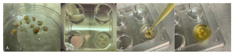
Figure 1: Flowchart of Consolidated Standards of Reporting Trials (CONSORT).
Conjunctival Epithelial Cell Culture
Human Primary Conjunctival Epithelial Cells (HPCEC) (ScienCell Catalog #6630 CA, US) were seeded from nitrogen on a poly-L-lysine (2 mg/ cm2) coated 24-well plates at 1.25 x 10³/cm2 and placed in 500 mL of Corneal Epithelial Cell Medium (ScienCell CEpiCM, Cat. #6511), 5 mL of corneal epithelial cell growth supplement (ScienCell CEpiCGS, Cat. No. 6552,) and 5 mL of penicillin/streptomycin solution (ScienCell P/S, Cat. No. 0503). Cells were then cultured in a 37oC, 5% CO2 incubator until cells were at least 70% confluent before the first passage. Media was changed every 2 to 3 days.
Cell Proliferation Assay
HPCECs were seeded on four 96-well plates at a density of 2 x 10³/cm2, and grown with Corneal Epithelial Cell Medium (ScienCell CEpiCM, Cat. #6511) in a 37°C, 5% CO2 incubator until at least 40% confluency was reached. Once confluent, the growth media was aspirated and replaced with SFM for 72 hours to deaccelerate cell growth. After 72 hours, the SFM media was removed and experimental conditions were added and left to incubate in a 37°C, 5% CO2 incubator for 5 minutes before new SFM media was added and placed back into the incubator overnight. Conditions included a) SFM only serving as control, b) SFM and aPRP membrane, c) SFM and orbital fat, and d) SFM and aPRP/Orbital fat membrane complex. CyQUANT NF Proliferation Assay Kit (Thermo Fisher Scientific) was used to determine cell proliferation every 48 hours for the following time points: 0, 48, 96, and 144 hours.
Cell Migration Assay
HPCECs were seeded on 24-well plates at a density of 5 x 10³/cm2, and grown with Corneal Epithelial Cell Medium (ScienCell CEpiCM, Cat. #6511) in a 37°C, 5% CO2 incubator until at least 70% confluency was reached. HPCECs were then incubated with SFM for 24 hours to deaccelerate cell growth. Next, a scratch (or wound) was made down the center of each well using SPLScarTM Scratcher 24-well (SPL lifescience), and images of the initial scratch were taken using a Keyence BZ-X800 fluorescence microscope. Experimental conditions were a) serum free media (SFM), b) SFM and aPRP membrane, c) SFM and orbital fat, and d) SFM and aPRP/Orbital fat membrane complex. Images were taken with a Keyence BZ-X800 fluorescence microscope every 48 hours for a total of 144 hours. Once a corresponding timepoint was imaged the tissues were removed to be fixed, while the cells were kept in the well with SFM until all timepoints were complete. Once all timepoints were imaged, the cells were immediately fixed with 100% cold methanol and stored in Phosphate Buffer Saline (PBS) in a 4°C refrigerator. The removal of the membrane at the end of the experiment simulates our clinical experience, as the aPRP membrane typically dissolves within 7 to 14 days in-vivo.
Cell Morphology Evaluation
Following aPRP/Orbital fat membrane complex removal, growth media was added to the HPCECs to assess cell morphology. Growth media was replenished every other day for a total of 10 days.
Cell/tissue Characterization
To evaluate for the morphology after removal of experimental conditions, phase contrast photographs in 4X magnification were taken at each timepoint. Additional staining for immunofluorescence was done with conjunctival cell marker CK13 and mesenchymal stem cell marker CD34. HPCECs and tissue removed from the migration assay were fixed with 100% cold methanol for 5 minutes at -20°C. Tissues were then washed PBS three times for 5 minutes, then permeabilized with 0.3% Triton X-100 (Sigma-Aldrich, Saint Louis, MO, USA) in PBS for 15 minutes at room temperature, and blocked with 0.2% BSA (Sigma-Aldrich, Saint Louis, MO, USA) in PBS for one hour at room temperature before the addition of primary antibodies for immunofluorescence staining overnight at 4°C. Primary antibodies used were: mouse monoclonal anti-cytokeratin 13 (Abcam, Cat# ab16112) and rabbit monoclonal recombinant anti-CD34 (Abcam; Cat # ab81289). Tissues were then washed three times with PBS for 5 minutes and then incubated with a 1:200 dilution of Alexa Fluor 488-goat anti-mouse or anti-rabbit or 594-goat anti-mouse or anti-rabbit conjugated secondary antibodies (Thermo Fisher Scientific) for one hour at room temperature. aPRP membranes, orbital fat, and aPRP/Orbital fat membrane complex were analyzed by immunofluorescence. Tissues were then imaged using the Keyence BZ-X800 analyzer fluorescence microscope and a Leica SP8 scanning laser confocal microscope.
Results
aPRP/Orbital Fat Complex Formation
Orbital fat was obtained from bilateral blepharoplasty surgeries from a total of 9 patients (n=9). Using the methods described above, the aPRP/Orbital fat complex was formed after combination of processed orbital fat and aPRP. As shown in Figure 1D, the cut orbital fat is noted to be incorporated into the PRGF with the subsequent formation of a membrane complex.
Cell Proliferation Assay
Cell proliferation fluorescence at different time points was recorded in Figure 2. Cells incubated with the aPRP/Orbital fat membrane had the highest readings for all timepoints but started to decrease at 144 hours. The cells incubated with the aPRP only condition was shown to have some growth of epithelial cells but to a lesser extent compared to aPRP/Orbital fat membrane. Cells incubated with the orbital fat only condition showed cell death.
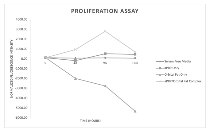
Figure 2: Proliferation assay. Cells incubated with serum free media served as control. Highest reading was seen with cells incubated with the aPRP/Orbital fat membrane, peaking at 96 hours.
Cell Migration Assay
To imitate the clinical condition of membrane dissolution around post-operative day 14, the experimental membranes were removed. At the time of membrane removal, some conjunctival cells were noted to lift from the plate in the aPRP only and aPRP/Orbital fat complex condition as they were partially attached to the aPRP and aPRP/Orbital fat complex membrane. As seen in Figure 3, at 96 hours the aPRP alone condition (Figure 3B) and the aPRP/Orbital fat membrane condition (Figure 3D) was noted to have complete wound closure when compared to the other conditions. HPCECs incubated with the aPRP at 96 hours appeared fibroblastic. HPCECs incubated with the aPRP/Orbital fat membrane at 96 hours appeared less fibroblastic than the cells at 48 hours. HPCECs at 96 hours incubated with the orbital fat only (Figure 3C) appear to be dying. Conjunctival cells in the aPRP/Fat complex initially appear fibroblastic. However, the conjunctival cells were shown to reach confluence and revert to normal morphology at day 10.
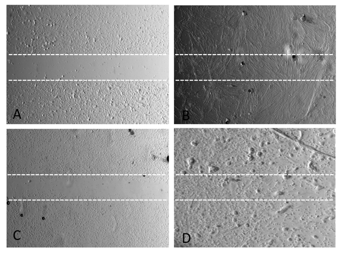
Figure 3: A) Control showed minimal number of cells over “wound” (within dotted lines) at 96 hours; B) aPRP condition shows growth of fibroblastic-appearing cells in the “wound”; C) In fat only condition, minimal migration of cells over the “wound” area was seen. D) in aPRP/Orbital fat complex, increased cell growth with migration of cells over “wound” area noted with cells appearing less fibroblastic.
Characterization via Immunofluorescence Staining
HPCECs from the migration assay were characterized via immunofluorescence staining. HPCECs incubated with aPRP only, orbital fat only, and aPRP/Orbital fat complex all showed positive immunofluorescence staining of the CK13 (Figure 4). HPCECs incubated with the orbital fat only condition and aPRP/Orbital fat membrane condition showed positive immunofluorescence staining for CD34, demonstrating that viable mesenchymal stem cells were present. No staining for CD34 was seen in the aPRP-only condition at 96 hours.
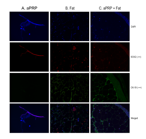
Figure 4: DAPI (4’, 6-diamidino-2-phenylindole) staining positive for all three conditions. CK13 (conjunctival cell marker) stained positively in all three experimental conditions. CD34 (mesenchymal cell marker) stained positively in condition B and condition C. It was negative in condition A. On the merged images, there is positive staining of both conjunctival and mesenchymal cell markers in condition C.
Discussion
Conjunctival fibrosis is a complex process that involves several pathways including inflammation, proliferation, and tissue remodeling. Key factors such as myofibroblasts, TGF-β, and fibroblast growth factor have all been shown to contribute to fibrosis [26-28]. Cicatrizing conjunctivitis with extensive fibrosis is particularly challenging to treat. Despite aggressive lubrication and immunosuppression, many patients require further surgical management due to symblepharon formation and fibrosis. Available options today include the excision of scar tissue with placement of amniotic or mucous membrane graft [4-6]. However, the absence of healthy conjunctival tissue in patients leads to unpredictable wound healing and surgical outcomes. In patients with mucous membrane involvement of systemic disease, donor site morbidity may be another concern for those requiring multiple mucous membrane graft.
Socket contracture is another condition where limited healthy conjunctiva is present. Treatment is typically surgical grafting to expand the anophthalmic socket. Oral mucosa such as labial or buccal mucous membrane graft has been widely used. However, these grafts involve donor site morbidity, and unpredictable post-operative contracture can still be seen [7, 29]. Amniotic membrane has been a popular adjunct given its unique anti-inflammatory and anti-fibrotic properties and its ability to promote epithelization [30, 31]. But with no viable epithelial cells, amniotic membranes require epithelialization by healthy proliferative conjunctival cells. As a result, their utility is limited in the setting of severe cicatrization [32, 33]. In addition, as amniotic grafts may only reduce the degree of fibrosis that occurs, studies have advocated for the concurrent application of antifibrotic medication such as mitomycin C in these cases [34]. Although several other grafting options have been proposed, recurrent contracture due to graft absorption, poor cell viability and donor site morbidity continues to be a dilemma for clinicians [16, 17].
In both cicatrizing conjunctivitis and orbital contracture, the ideal graft for surgical treatment would be one that is durable, contains anti-fibrotic and anti-inflammatory properties, can provide healthy substitute of conjunctiva tissue, and is easily obtainable without donor site morbidity. We propose here a combined orbital fat and aPRP scaffold as a treatment option for cicatrizing conjunctivitis and orbital contracture. The medial orbital fat contains OASCs that are neural crest in origin and have been shown to possess multi-lineage differentiation [19, 20]. Particularly, OASC have been demonstrated to differentiate into polygonal epithelial-like cells expressing epithelial-specific antigen after mix-culture with corneal epithelial cells [35]. In-vivo studies have shown that OASCs may have anti-inflammatory effects in the setting of acute corneal injuries via inhibition of macrophage infiltration and subsequent production of pro-inflammatory molecules [36]. Furthermore, transplanted OASCs have been shown to differentiate into corneal epithelial linage [36]. On the other hand, aPRP is integral in regeneration of tissue and bone, including the promotion of osteogenic induction via increasing the proliferation and migration of osteoblasts [37, 38]. Other studies have shown that autologous aPRP promotes healing and regeneration of the ocular surface, especially in cases of corneal epithelial defects [22]. The combination of adipose stem cells with aPRP may have synergistic effect in wound healing [25]. Platelet derived growth factors may function as important signaling ligand for adipose stem cells and to stimulate vascularization and wound repair [25].
In our study, we demonstrate a technique to create an aPRP/Orbital fat complex scaffold containing OASC and aPRP. Figure 1 demonstrates a membrane scaffold of aPRP/Orbital fat complex that can be easily harvested and processed intraoperatively. This membrane can be easily manipulated and can readily be sutured onto the ocular surface. In our proliferation assay (Figure 2), cells incubated with the aPRP/Orbital fat complex show the greatest growth. This is likely due to regenerative effects of the aPRP/Orbital fat complex. However, decrease in reading after 96 hours likely is due to cells dying after depleting growth factors from aPRP. In the clinical setting, patients would be provided with topical PRGF after surgery to support cells until the complex has been integrated into the patient’s vasculature. The condition with orbital fat only demonstrates that there is likely cell death, as the orbital fat initially contains many cells but does not have the necessary growth factors in the media for continued cell survival and growth. Without additional orbital fat stem cells, the condition of aPRP-only shows some growth of epithelial cell but comparatively less than the aPRP/Orbital fat complex. However, cell growth is observed which may reflect that aPRP contains important modulators for cell growth. It is important to note that the addition of orbital fat and aPRP/Orbital fat complex would inherently lead to an increase in cell number. However, when comparing the condition of aPRP/Orbital fat complex with orbital fat only condition, there was an increase of fluorescence intensity reading over time in the condition with aPRP/Orbital fat complex, showing that aPRP serves an important moderator in cell proliferation.
To mimic the clinical condition of scaffold dissolution in-vivo, we subsequently removed the experimental condition from each plate. The migration assay assessed the migratory response of cells in the “wound area” created by a scratch on the well plate. It was noted that some conjunctival cells were likely dislodged with the experimental condition of membrane removal, particularly with the aPRP and aPRP/Orbital fat complex removal. This could be due to the function of the aPRP and aPRP/Fat complex serving as a substrate for cell growth. Our result of the migration assay demonstrated that wound closure was achieved in the aPRP and aPRP/Orbital fat condition, and conjunctival cells in both were shown to undergo morphologic changes. While the HPCECs incubated with aPRP-only condition appeared fibroblastic at 96 hours, the HPCECs incubated with aPRP/Orbital fat membrane was less fibroblastic than they appeared at 48 hours. Finally, the conjunctival cells previously incubated with aPRP/Orbital fat complex demonstrated positivity for not only CK13 but also CD34, further implicating that OASC likely played an integral role in differentiation into conjunctival epithelial cells under the modulation of aPRP.
Conclusion
This study provides a description of the creation of an aPRP/Orbital fat complex that could potentially serve as a scaffold for conjunctival cell proliferation. Limitations of this study include that the ex-vivo condition might not adequately reflect the actual course of tissue reaction and degradation. Second, the experimental conditions were evaluated up to 96 hours, and the morphology description was followed up to day 10 and therefore it would be difficult to determine the final morphology and behavior of OASC and conjunctival cells in an in-vivo setting over an extended period. Furthermore, this scaffold is intended to be used in the treatment of cicatrizing conjunctivitis and socket contraction, and the lack of healthy conjunctival tissue may present a challenge for adequate cell proliferation and migration. However, the OASC from harvested orbital fat can potentially provide additional cells. This could also be mitigated by providing an alternative source of healthy conjunctival epithelial cells at the same time as creation of scaffold. Finally, the durability of the scaffold was not tested in-vitro. Given the known properties of aPRP fibrin membrane and OASC in cellular proliferation, migration, and reduction of fibrosis, we do believe that these initial results show promise in the potential for an easily obtainable aPRP/Orbital fat scaffold that could have benefit in the surgical treatments of cicatrizing conjunctivitis and socket contraction.
Acknowledgements
Funding and Support: None.
Financial Disclosures and Conflicts of Interest: The authors alone are responsible for the writing and content of this article. YC consults for Tarsus Inc. STW consults for Horizon Therapeutics, Immunovant, Lassen Therapeutics and Sling. ALS: Abbie, Brill Pharma, GlaxoSmithKline, PlasmaCord, TissueCor, LyoDrop, Department of Defense Air Force Research Laboratory FA864921P0155, Beauty of Sight Foundation. DP is an equity holder for EIR Biopharma.
References
- Elder MJ, et al. Progression of disease in ocular cicatricial pemphigoid. Br J Ophthalmol 80 (1996): 292-6.
- Bobba S, et al. Incidence, clinical features and diagnosis of cicatrising conjunctivitis in Australia and New Zealand. Eye (Lond) 32 (2018): 1636-1643.
- Vazirani J, et al. Limbal Stem Cell Deficiency-Demography and Underlying Causes. Am J Ophthalmol 188 (2018): 99-103.
- Kohanim S, et al. Acute and Chronic Ophthalmic Involvement in Stevens-Johnson Syndrome/Toxic Epidermal Necrolysis - A Comprehensive Review and Guide to Therapy. II. Ophthalmic Disease. Ocul Surf 14 (2016): 168-88.
- Iyer, G, et al, Mucous membrane grafting for lid margin keratinization in Stevens-Johnson syndrome: results. Cornea 29 (2010): 146-51.
- Fu Y, J Liu, and SC Tseng. Oral mucosal graft to correct lid margin pathologic features in cicatricial ocular surface diseases. Am J Ophthalmol 152 (2011): 600-608 e1.
- Tawfik HA, AO Raslan and N Talib. Surgical management of acquired socket contracture. Curr Opin Ophthalmol 20 (2009): 406-11.
- Bowen Jones EJ and E Nunes. The outcome of oral mucosal grafts to the orbit: a three-and-a-half-year study. Br J Plast Surg 55 (2002): 100-4.
- Foster JA, E Castro and FA Papay. Reconstruction of the irradiated contracted socket with an expanded superficial temporalis fascial flap. Am J Ophthalmol 127 (1999): 621-2.
- Karesh JW and AM Putterman. Reconstruction of the partially contracted ocular socket or fornix. Arch Ophthalmol 106 (1988): 552-6.
- Sullivan SA and RA Dailey. Graft contraction: a comparison of acellular dermis versus hard palate mucosa in lower eyelid surgery. Ophthalmic Plast Reconstr Surg 19 (2003): 14-24.
- Betharia SM, et al. Skin grafting in severely contracted socket with the use of 'Compo'. Indian J Ophthalmol 38 (1990): 88-91.
- Liu D, J Michon and J Choi. Galeal flap: clinical application in periorbital and orbital reconstruction. Ophthalmic Plast Reconstr Surg 12 (1996): 273-8.
- Przybyla VA, FG La Piana and DJ Bergin. Fitting of the dermis-fat grafted socket. Ophthalmology 88 (1981): 904-7.
- Levin PS and JJ Dutton. Polytef (polytetrafluoroethylene) alloplastic grafting as a substitute for mucous membrane. Arch Ophthalmol 108 (1990): 282-5.
- Choi CJ, AQ Tran and DT Tse. Hard palate-dermis fat composite graft for reconstruction of contracted anophthalmic socket. Orbit (2018): 1-6.
- Slentz DH and CC Nelson. Novel Use of Cryopreserved Ultra-Thick Human Amniotic Membrane for Management of Anophthalmic Socket Contracture. Ophthalmic Plast Reconstr Surg (2018).
- Fagien S and AM Putterman. Putterman’s Cosmetic Oculoplastic Surgery. 4th ed. 2008, Philadelphia: Saunders (2008).
- Johnston MC, et al. Origins of avian ocular and periocular tissues. Exp Eye Res 29 (1979): 27-43.
- Korn BS, DO Kikkawa and KC Hicok. Identification and characterization of adult stem cells from human orbital adipose tissue. Ophthalmic Plast Reconstr Surg 25 (2009): 27-32.
- Pal US, et al. Platelet-rich growth factor in oral and maxillofacial surgery. Natl J Maxillofac Surg 3 (2012): 118-23.
- Sabater AL, et al. Use of autologous plasma rich in growth factors fibrin membrane in the surgical management of ocular surface diseases. Int Ophthalmol 41 (2021): 2347-2358.
- Anitua, E, et al, Plasma rich in growth factors (PRGF) eye drops stimulates scarless regeneration compared to autologous serum in the ocular surface stromal fibroblasts. Exp Eye Res 135 (2015): 118-26.
- Lopez-Plandolit, S, et al, Efficacy of plasma rich in growth factors for the treatment of dry eye. Cornea 30 (2011): 1312-7.
- Blanton MW, et al. Adipose stromal cells and platelet-rich plasma therapies synergistically increase revascularization during wound healing. Plast Reconstr Surg 123 (2009): 56S-64S.
- Teller P and TK White The physiology of wound healing: injury through maturation. Surg Clin North Am 89 (2009): 599-610.
- Luttikhuizen DT, MC Harmsen and MJ Van Luyn. Cellular and molecular dynamics in the foreign body reaction. Tissue Eng 12 (2006): 1955-70.
- Khaw PT, et al. The control of conjunctival fibrosis as a paradigm for the prevention of ocular fibrosis-related blindness. "Fibrosis has many friends". Eye (Lond) 34 (2020): 2163-2174.
- Grixti A and R Malhotra. Oral mucosa grafting in periorbital reconstruction. Orbit 37 (2018): 411-428.
- Tseng SC, DQ Li and X Ma. Suppression of transforming growth factor-beta isoforms, TGF-beta receptor type II, and myofibroblast differentiation in cultured human corneal and limbal fibroblasts by amniotic membrane matrix. J Cell Physiol 179 (1999): 325-35.
- Shimmura S, et al. Antiinflammatory effects of amniotic membrane transplantation in ocular surface disorders. Cornea 20 (2001): 408-13.
- Kumar S, et al. Amniotic membrane transplantation versus mucous membrane grafting in anophthalmic contracted socket. Orbit 25 (2006): 195-203.
- Borrelli M, et al. Eye Socket Regeneration and Reconstruction. Curr Eye Res 45 (2020): 253-264.
- Mandour SS, et al. Mucous Membrane Grafting Augmented with Topical Mitomycin C Application in Contracted Socket Repair Surgeries. J Ocul Pharmacol Ther 32 (2016): 691-694.
- Ho JH, et al. Isolation and characterization of multi-potent stem cells from human orbital fat tissues. Tissue Eng Part A 17 (2011): 255-66.
- Lin KJ, et al. Topical administration of orbital fat-derived stem cells promotes corneal tissue regeneration. Stem Cell Res Ther 4 (2013): 72.
- Anitua E, et al. Plasma rich in growth factors promotes bone tissue regeneration by stimulating proliferation, migration, and autocrine secretion in primary human osteoblasts. J Periodontol 84 (2013): 1180-90.
- Paknejad M, et al. Evaluation of the Effect of Plasma Rich in Growth Factors (PRGF) on Bone Regeneration. J Dent (Tehran) 9 (2012): 59-67.