Bhavya Sharda1, Falguni Agarwal1, Shaina Gaikwad1, Antisha Tiwari1, Anand Kumar Maurya*,1, Debasis Biswas1, Shashank Purwar1, Jitendra Singh2, Sagar Khadanga3, Alkesh Kumar Khurana4
1Department of Microbiology, All India Institute of Medical Sciences, Bhopal, Madhya Pradesh, India
2Department of Translational Medicine, All India Institute of Medical Sciences, Bhopal, Madhya Pradesh, India
3Department of General Medicine, All India Institute of Medical Sciences, Bhopal, Madhya Pradesh, India
4Department of Pulmonary Medicine, All India Institute of Medical Sciences, Bhopal, Madhya Pradesh, India
*Corresponding author: Anand Kumar Maurya, Additional Professor, Department of Microbiology, All India Institute of Medical Sciences, Bhopal, Madhya Pradesh, India.
Received: 17 September 2026; Accepted: 21 September 2026; Published: 24 September 2026
Background: Non-tuberculous mycobacteria (NTM) can resemble tuberculosis (TB), particularly in TB-endemic settings, and species-level identification is important because antimicrobial susceptibility differs considerably among species.
Methods: We conducted a prospective diagnostic accuracy study comparing Matrix-Assisted Laser Desorption/Ionization-Time of Flight (MALDI-TOF) mass spectrometry with the GenoType Mycobacterium CM/AS line probe assay (LPA) for NTM identification at a tertiary care centre in Central India. Isolates with discordant results were further evaluated by 16S–23S internal transcribed spacer (ITS) PCR sequencing. Thirty culture-positive, NAAT-negative, MPT64Ag-negative isolates from patients with presumptive TB or suspected NTM infection were tested by both methods.
Results: LPA identified a single organism in 23/30 isolates (76.7%), while 7/30 (23.3%) showed co-detection of M. tuberculosis complex and/or another NTM species. MALDI-TOF most frequently identified M. fortuitum (11/30, 36.7%), followed by M. abscessus and M. intracellulare (8/30, 26.7% each). Among the 23 isolates with a single-organism LPA result, MALDI-TOF and LPA agreed at the species level in 17 (73.9%), with moderate-to-substantial agreement (kappa = 0.66, z = 5.41, p < 0.0001). ITS sequencing of the six discordant isolates supported the LPA result in five and the MALDI-TOF result in one. Using LPA as the reference standard in the single-organism subset, MALDI-TOF showed a sensitivity of 73.9% and specificity of 94.8%.
Conclusion: MALDI-TOF and LPA showed good agreement for most NTM isolates, although discrepancies were more evident among rapidly growing mycobacteria and M. simiae. When the methods disagreed, ITS sequencing more often supported the LPA identification.
Non-tuberculous mycobacteria; MALDI-TOF; Line probe assay; GenoType CM/AS; ITS sequencing; Diagnostic accuracy; Central India
Non-tuberculous mycobacteria articles; MALDI-TOF articles; Line probe assay articles; GenoType CM/AS articles; ITS sequencing articles; Diagnostic accuracy articles; Central India articles
Introduction
Non-tuberculous mycobacteria (NTM) comprise a diverse group of environmental acid-fast bacilli that are different from Mycobacterium tuberculosis and M. leprae. They are commonly referred to as atypical or environmental mycobacteria, and have also been described as mycobacteria other than tuberculosis (MOTT) [1]. NTM are widely distributed in soil, dust, and natural and municipal water sources, where they can persist within biofilms. In most situations, human infection is thought to result from environmental exposure rather than direct person-to-person transmission [1]. NTM disease is increasingly recognised as a public health concern, although its true burden is probably underestimated. The clinical and radiological features may closely resemble pulmonary or extrapulmonary tuberculosis, and NTM infection may consequently be mistaken for drug-resistant TB, with important implications for treatment [2]. NTM are commonly classified as rapidly growing mycobacteria (RGM), which form colonies within seven days on solid media, and slowly growing mycobacteria (SGM), which require seven days or longer [3]. Examples of RGM include Mycobacterium abscessus, Mycobacterium fortuitum and Mycobacterium chelonae, whereas the SGM group includes organisms such as the Mycobacterium avium complex, Mycobacterium kansasii and Mycobacterium intracellulare. The reported prevalence of NTM has increased in North America, Europe and Asia over the last two decades [4]. Indian studies have reported isolation prevalences ranging from 0.2% to 5.9% and disease prevalence of up to 0.8 per 100,000 population, with considerable geographic variation in the species recovered [5]. Studies from Central India have particularly reported M. fortuitum and M. abscessus among the commonly isolated species in both pulmonary and extrapulmonary specimens [6,23], consistent with observations from other TB-endemic regions [7,23].
Species-level identification is clinically important because treatment regimens differ substantially between NTM species. For example, M. avium complex, M. kansasii and M. abscessus are managed with different multidrug treatment approaches [8]. Accurate identification therefore plays an important role in clinical decision-making [9]. Traditional biochemical methods, including niacin accumulation, arylsulfatase activity, nitrate reduction and growth on MacConkey agar, are time-consuming and have largely been replaced by more rapid methods [9]. In many reference laboratories, the GenoType Mycobacterium CM/AS line probe assay (LPA; Hain Lifescience, Nehren, Germany) and MALDI-TOF mass spectrometry are now used for rapid identification. The LPA is a reverse-hybridisation assay targeting the 23S rRNA gene and can identify 37 species across the CM and AS kits [10]. MALDI-TOF identifies mycobacteria by analysing characteristic protein spectra following cell inactivation and protein extraction [11]. Both approaches offer a shorter turnaround time than sequencing, although 16S–23S ITS sequencing remains an important method for confirming isolates when results are discordant or ambiguous [12]. Although both methods are increasingly used for NTM identification, comparative data on their agreement remain limited in India, particularly from Central India. This is relevant because NTM are increasingly recognised among patients investigated for tuberculosis in the region [13]. We therefore conducted a prospective diagnostic accuracy study to compare MALDI-TOF and LPA for species-level identification of NTM and to use ITS sequencing to resolve discordant results.
Methods
Study design and setting
This was a prospective, hospital-based comparative diagnostic accuracy study that was conducted over 18 months, from February 2025 to August 2026, in the Department of Microbiology at the All India Institute of Medical Sciences (AIIMS), Bhopal, Madhya Pradesh, India. The study was carried out in collaboration with the Departments of General Medicine, Pulmonary Medicine and Translational Medicine.
Study population and case definitions
The study included patients with presumptive tuberculosis or suspected NTM infection whose clinical specimens were referred to the institutional TB laboratory for Ziehl–Neelsen (ZN) staining, mycobacterial culture and nucleic acid amplification testing (NAAT). Presumptive TB was considered in patients with symptoms such as cough or fever lasting two weeks or more, significant weight loss, haemoptysis, or an abnormal chest radiograph. NTM infection was considered in patients with persistent fever or respiratory symptoms, skin lesions not responding to conventional antibacterial treatment, or clinical syndromes known to be associated with NTM disease. Specimens were included when they were obtained from patients evaluated for presumptive TB or suspected NTM infection and were submitted for ZN staining, culture and NAAT, with subsequent culture positivity for Mycobacterium spp. Specimens were excluded when NAAT was positive for M. tuberculosis complex, when both ZN microscopy and culture were negative, when a culture-positive isolate was MPT64 antigen positive, or when the specimen met laboratory rejection criteria.
Specimen types and processing
Both pulmonary specimens, including sputum, bronchoalveolar lavage, and endotracheal/tracheal aspirate, and extrapulmonary specimens, including blood, urine, pus, peritoneal fluid, lymph node aspirate, pericardial fluid, ascitic fluid, cerebrospinal fluid and tissue biopsy, were eligible. Specimens were processed according to institutional biosafety procedures in a Class II A2 biosafety cabinet, BSL-3 Facility, Department of Microbiology, All India Institute of Medical Sciences (AIIMS), Bhopal, Madhya Pradesh, India. ZN microscopy and NAAT were performed before NALC–NaOH decontamination and inoculation into the MGIT 960 automated liquid culture system (Becton, Dickinson and Company), following the manufacturer's instructions.
MPT64 antigen testing
All culture-positive isolates were tested with MPT64 antigen, a rapid immunochromatographic assay used to differentiate M. tuberculosis complex from NTM. A pink band in the control (C) region indicated a valid test. An additional pink band in the test (T) region was interpreted as MPT64 positive and consistent with M. tuberculosis complex, whereas a control-only band was considered negative and suggestive of NTM [10].
Line Probe Assay (GenoType Mycobacterium CM/AS)
DNA was extracted from positive liquid cultures using the GenoLyse kit (Hain Lifescience, Nehren, Germany). Biotinylated primers supplied with the GenoType CM/AS assay were used to amplify the region required for species identification. The resulting amplicons were hybridised to species-specific oligonucleotide probes immobilised on nitrocellulose strips, and the banding pattern was interpreted according to the manufacturer's instructions. The CM kit identifies 23 species, while the AS kit covers a further 14 less commonly encountered mycobacterial species [14].
MALDI-TOF mass spectrometry
For MALDI-TOF analysis, mycobacterial aliquots were washed and inactivated for 30 minutes at room temperature using the manufacturer's reagents. The material was then treated with acetonitrile and 70% formic acid and centrifuged at 13,000 rpm for 2 minutes. 1 microlitre of the supernatant was spotted in triplicate onto the MALDI target plate. An Escherichia coli protein extract was used as the internal quality control. Spectra were acquired in linear positive-ion mode at a laser frequency of 60 Hz over an m/z range of 2,000–20,000 Da using a Microflex LT MALDI-TOF MS instrument (Bruker Daltonik GmbH, Bremen, Germany). A log score of ≥1.80 was considered highly probable, 1.60–1.79 probable, and <1.60 unassigned. An identification was accepted when at least one of the three replicate spectra produced a species- or complex-level match with a log score of ≥1.60 [15].
ITS sequencing (discordance resolution)
Isolates with discordant LPA and MALDI-TOF results, as well as isolates for which LPA detected more than one organism, were considered for confirmatory PCR sequencing of the 16S–23S internal transcribed spacer (ITS) region. Genus- and species-specific primers were used as previously described [12], and sequences were compared with curated reference databases. For isolates with a single-organism LPA result, the ITS sequence was used to adjudicate discordant MALDI-TOF and LPA identifications. Mixed LPA results could not be resolved by the sequencing method used in this study because Sanger sequencing of a mixed template produces overlapping chromatogram peaks at divergent positions. Such samples would require methods such as cloning or targeted deep sequencing for definitive resolution. This was therefore considered a methodological limitation rather than an unresolved pending result.
Statistical analysis
Cohen’s kappa and diagnostic accuracy were calculated based on the categorical results obtained by each method. Isolates in which LPA detected more than one organism were analysed separately and excluded from the primary concordance and diagnostic accuracy analyses. Agreement between LPA and MALDI-TOF was assessed among the remaining isolates with a single-organism LPA result using percentage agreement and Cohen's kappa, with a z-test used to determine whether the observed kappa differed from chance agreement. LPA was used as the reference standard for evaluating MALDI-TOF because it is a widely validated molecular method for mycobacterial speciation. For each NTM species, a 2×2 table was constructed by comparing that species with all other species, and sensitivity, specificity, positive predictive value (PPV) and negative predictive value (NPV) were calculated with 95% confidence intervals using the exact Clopper–Pearson method [22]. Confidence intervals were reported instead of p-values for diagnostic accuracy measures because these measures represent precision around a proportion rather than a comparison between independent groups.
Results
Study population
During the study period, 4,325 specimens from patients evaluated for presumptive tuberculosis or suspected NTM infection were screened by ZN microscopy, NAAT/CBNAAT and MGIT-960 liquid culture. Of these, 3,460 were culture-negative, and 865 were culture-positive. Among the culture-positive specimens, 835 grew M. tuberculosis complex, while 30 were culture-positive for Mycobacterium spp. and negative for MPT64 antigen. These 30 isolates met the inclusion criteria and were subjected to species identification by both LPA and MALDI-TOF, giving an NTM yield of 3.5% among culture-positive specimens (0.7% of all specimens screened). Twenty-four isolates (80.0%) were from pulmonary specimens and six (20.0%) from extrapulmonary specimens.
Species distribution by MALDI-TOF and Line Probe Assay
All 30 isolates underwent species identification by both LPA and MALDI-TOF. For MALDI-TOF, an identification was considered reliable when at least one of the three replicate spectra had a log score of ≥1.60. The mean log score across the 30 isolates was 1.79, with values ranging from 1.68 to 1.82. MALDI-TOF most frequently identified M. fortuitum (11/30, 36.7%), followed by M. abscessus and M. intracellulare (8/30, 26.7% each), while M. chelonae, M. simiae and M. avium were each identified in one isolate (3.3%).
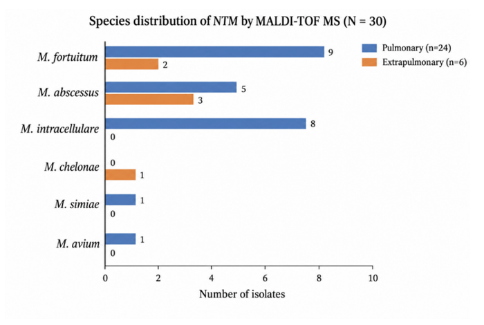
Figure 1: Species distribution by MALDI-TOF MS, pulmonary vs. extrapulmonary.
The distribution of species between pulmonary and extrapulmonary specimens did not show a statistically significant difference (Monte Carlo estimate of the Fisher–Freeman–Halton exact test, based on 100,000 replicates: χ² = 8.05, p = 0.16). Because more than half of the expected cell counts were below five, the exact simulation-based test was considered more appropriate than the asymptotic chi-square test. LPA also identified M. fortuitum most frequently (12/30, 40.0%, including its detection in all seven mixed results), followed by M. abscessus (7/30, 23.3%). In addition, LPA detected co-presence of M. tuberculosis complex with an NTM species in four isolates (13.3%), while three isolates (10.0%) showed two different NTM species without M. tuberculosis complex. Overall, 23/30 isolates (76.7%) had a single-organism NTM result. All seven mixed or dual detections were from pulmonary specimens. The four MTB–NTM co-infections involved M. fortuitum, with one also showing M. avium complex. The three dual-NTM detections consisted of M. fortuitum and M. chelonae.
Table 1: Mixed mycobacterial detections using the Line Probe Assay (N = 30)
|
Category |
No. of isolates |
Percentage |
|
MTB–NTM co-infection (NTM + M. tuberculosis complex) |
4 |
13.30% |
|
Dual/mixed NTM infection (two NTM species, no MTB) |
3 |
10.00% |
|
Single-organism NTM isolates |
23 |
76.70% |
Concordance between MALDI-TOF MS and Line Probe Assay
Among the 23 isolates for which LPA reported a single organism, the two methods produced the same species identification in 17 (73.9%), while 6 (26.1%) showed discordant results.
Table 2: Agreement between Line Probe Assay and MALDI-TOF MS, single-organism LPA subset (n = 23)
|
Category |
No. of isolates |
Percentage |
p-value |
|
Species-level concordance |
17 |
73.90% |
|
|
Discordant identification |
6 |
26.10% |
|
|
Cohen's kappa (κ) — agreement beyond chance |
0.66 (moderate–substantial) |
— |
<0.0001 |
For the 23-isolate single-organism subset, the isolate-level confusion matrix gave a Cohen's kappa of 0.655 (SE = 0.121). The corresponding z-test gave z = 5.41 with p < 0.0001, indicating that the agreement between LPA and MALDI-TOF was significantly greater than would be expected by chance.
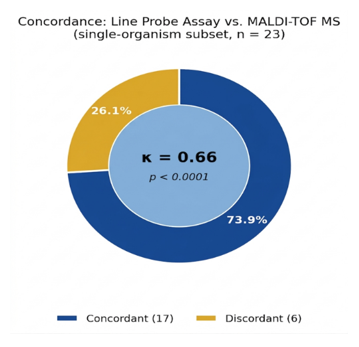
Figure 2: Concordance between Line Probe Assay and MALDI-TOF MS (single-organism subset, n = 23).
ITS sequencing resolution of discordant single-organism isolates
All six isolates with discordant single-organism results underwent confirmatory 16S–23S ITS PCR sequencing. Sequencing supported the LPA identification in five of the six isolates (83.3%) and the MALDI-TOF identification in one (16.7%). Of particular note, all three isolates identified as M. simiae by LPA were assigned different species by MALDI-TOF—M. intracellulare, M. abscessus and M. fortuitum, respectively. ITS sequencing confirmed M. simiae in all three cases.
Table 3: ITS sequencing resolution of discordant single-organism isolates (n = 6; all resolved)
|
Isolates |
LPA results |
MALDI-TOF results |
ITS (16S–23S) results |
Platform corroborated |
|
1 |
M. abscessus |
M. chelonae |
M. abscessus |
LPA |
|
2 |
M. chelonae |
M. abscessus |
M. abscessus |
MALDI-TOF |
|
3 |
M. abscessus |
M. intracellulare |
M. abscessus |
LPA |
|
4 |
M. simiae |
M. intracellulare |
M. simiae |
LPA |
|
5 |
M. simiae |
M. abscessus |
M. simiae |
LPA |
|
6 |
M. simiae |
M. fortuitum |
M. simiae |
LPA |
Overall, ITS sequencing supported the LPA result in five of the six discordant isolates (83.3%) and the MALDI-TOF result in one (16.7%). Thus, when the two methods gave different species assignments in this cohort, the LPA result was more frequently consistent with the sequence-confirmed identification.
MALDI-TOF concordance with multi-organism (mixed) LPA calls
Among the seven isolates in which LPA detected more than one organism, MALDI-TOF provided a single species identification and therefore could not directly indicate the presence of a mixed infection. Its result was compared with the organisms reported by LPA. In five of the seven isolates (71.4%), the MALDI-TOF identification corresponded to one of the organisms detected by LPA. In the remaining two isolates (28.6%), it did not match either LPA-reported organism. These seven mixed isolates could not be adjudicated by ITS sequencing because the mixed templates produced overlapping Sanger chromatograms that prevented reliable sequence interpretation.
Table 4: MALDI-TOF concordance with multi-organism LPA calls (n = 7; not resolvable by ITS sequencing)
|
Isolate |
MALDI-TOF results |
LPA results |
Concordance |
|
1 |
M. abscessus |
M. fortuitum + M. tuberculosis complex |
Full discordance |
|
2 |
M. simiae |
M. fortuitum + M. tuberculosis complex |
Full discordance |
|
3 |
M. fortuitum |
M. fortuitum + M. tuberculosis complex + M. avium complex |
Partial (matches 1 of 3) |
|
4 |
M. fortuitum |
M. tuberculosis complex + M. fortuitum |
Partial (matches 1 of 2) |
|
5 |
M. fortuitum |
M. chelonae + M. fortuitum |
Partial (matches 1 of 2) |
|
6 |
M. fortuitum |
M. fortuitum + M. chelonae |
Partial (matches 1 of 2) |
|
7 |
M. fortuitum |
M. fortuitum + M. chelonae |
Partial (matches 1 of 2) |
Diagnostic accuracy of MALDI-TOF MS against the Line Probe Assay
Diagnostic accuracy was assessed only in the 23 isolates with a single-organism LPA result, as specified in the statistical analysis.
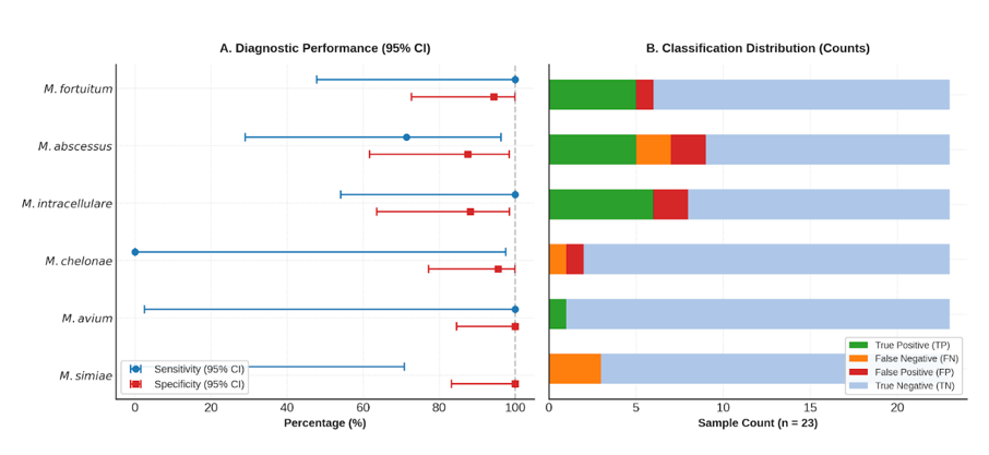
Figure 3: Species-wise diagnostic accuracy of MALDI-TOF MS with Line Probe Assay as reference standard (n = 23)
When species-level results from the 23 single-organism isolates were pooled, MALDI-TOF showed an overall sensitivity of 73.9% (95% CI 51.6–89.8%) and specificity of 94.8% (95% CI 89.0–98.1%) when LPA was used as the reference standard. Sensitivity was 100% for M. fortuitum, M. intracellulare and M. avium, although the estimate for M. avium was based on only one isolate. Sensitivity for M. abscessus was 71.4%, while it was 0% for M. chelonae and M. simiae. MALDI-TOF failed to correctly identify the single M. chelonae isolate, and all three M. simiae isolates were identified by LPA. The three M. simiae results were subsequently shown to agree with LPA by ITS sequencing, whereas the M. chelonae isolate was confirmed as M. abscessus, supporting the MALDI-TOF result. Specificity remained above 85% for all evaluated species, indicating that MALDI-TOF rarely assigned a species that was not identified by LPA. The seven isolates with mixed LPA results were not included in these calculations because they could not be reliably adjudicated by ITS sequencing.
Discussion
In this analysis, MALDI-TOF and the GenoType Mycobacterium CM/AS line probe assay agreed on species identification in just under three-quarters of NTM isolates for which LPA returned a single organism (17/23, 73.9%), with moderate-to-substantial overall agreement (κ = 0.66). This concordance rate is broadly consistent with prior head-to-head comparisons of MALDI-TOF against hybridisation-based assays, which have generally reported good but incomplete agreement, with discordance concentrated among closely related rapidly growing species [15] [16]. In our cohort, M. fortuitum, M. abscessus and M. chelonae — all members of the rapidly growing mycobacteria group that share overlapping proteomic spectra and hybridisation-probe cross-reactivity — accounted for the majority of isolates and a substantial share of discordant pairs, mirroring the difficulty other groups have reported in resolving this complex by either platform alone [17]. Notably, discordance was not confined to the RGM complex: all three isolates called M. simiae by LPA were assigned a different species by MALDI-TOF and ITS sequencing subsequently confirmed M. simiae in all three, suggesting the local MALDI-TOF spectral database may under-represent this slowly growing species in particular.
Recent literature published since the design of this study reinforces both the promise and the limits of MALDI-TOF for NTM speciation. A 2026 Indian study using a modified bead-based protein-extraction protocol reported considerably higher concordance between MALDI-TOF and LPA than observed here (92.7%, 108/110 isolates) [19], suggesting that extraction methodology may be an important, modifiable driver of platform agreement that could be explored in future work at this centre. A 2025 multi-marker Sanger-sequencing comparison against MALDI-TOF from Ecuador reported Cohen's kappa values ranging from 0.46 to 0.76 depending on the gene target used (16S, hsp65, rpoB, singly and concatenated) [20], situating our kappa of 0.66 well within the range typically observed across contemporary NTM identification studies. A 2025/2026 systematic review and meta-analysis of 55 studies estimated a pooled MALDI-TOF identification accuracy of 88% (95% CI 84–91%) against varied reference standards [21], underscoring that no single platform yet achieves uniform accuracy across the full diversity of NTM species, consistent with the species-specific discordance we observed for M. chelonae and M. simiae.
Now that ITS sequencing is complete for all six discordant single-organism isolates, a clear picture emerges: LPA was corroborated over MALDI-TOF in five of six discordant isolates (83.3%), and MALDI-TOF was corroborated over LPA in only one (16.7%). This asymmetry indicates that, at least for the rapidly growing mycobacteria and M. simiae discordant in this cohort, LPA more often reflected the sequence-confirmed species when the two platforms disagreed. This finding, together with the fact that all MALDI-TOF identifications in this cohort met at least the "probable" score threshold, suggests that score thresholds alone do not guarantee correct species assignment [18] and that sequence-based confirmation remains necessary for discordant results, particularly within the RGM complex and for M. simiae, as recommended by current diagnostic guidelines [8].
A separate and practically important finding concerns the 7/30 isolates (23.3%) for which LPA reported more than one organism. In five of these seven, MALDI-TOF's single call matched one of the organisms LPA detected, but in two it matched neither — meaning MALDI-TOF, which cannot itself flag mixed infection, may silently miss a clinically relevant second organism (including M. tuberculosis complex itself) in around a quarter of such mixed specimens. This has direct clinical relevance in TB-endemic settings, where an NTM-only MALDI-TOF call could mask underlying MTB co-infection detected by LPA. Unlike the single-organism discordants, these 7 mixed isolates cannot be adjudicated by ITS sequencing: Sanger sequencing of a mixed-template specimen yields overlapping chromatogram peaks that cannot be confidently base-called wherever the two organisms' ITS sequences diverge, so no reference identification is available for this subset with the methodology used here. This is a genuine limitation of sequence-based confirmation in mixed infections, not an outstanding result, and would require cloning or targeted deep sequencing to resolve. This analysis has important limitations. First, the diagnostic-accuracy estimates presented here, while based on completed ITS sequencing for the single-organism subset, are drawn from a small number of isolates per species (e.g., single isolates of M. avium and M. chelonae); confidence intervals around several species-wise estimates are correspondingly wide. Second, the 7 isolates with multi-organism LPA calls could not be included in the kappa or diagnostic-accuracy calculations, and could not be resolved by ITS sequencing for the methodological reasons described above, so the true concordance and accuracy of MALDI-TOF across the full 30-isolate cohort remains uncertain for this subset. Third, single-centre recruitment may limit generalisability to other regions of India with different NTM species distributions.
Despite these limitations, these findings support the use of MALDI-TOF as a rapid, comparatively low-cost first-line platform for NTM speciation in resource-limited, TB-endemic settings, while reinforcing that discordant calls — especially among rapidly growing mycobacteria and M. simiae — require sequence-based confirmation, and that mixed-organism LPA results warrant particular caution given both MALDI-TOF's inability to flag co-infection and the inherent limits of Sanger-based ITS sequencing in resolving mixed templates. A larger, multi-centre study incorporating cloning-based or next-generation sequencing approaches for mixed specimens would allow a definitive, algorithm-based diagnostic pathway for NTM identification to be defined for TB-endemic, resource-limited settings.
Conclusion
MALDI-TOF MS and the GenoType Mycobacterium CM/AS line probe assay showed moderate-to-substantial agreement (73.9% concordance, κ = 0.66) for NTM species identification among isolates with an unambiguous single-organism LPA result. ITS sequencing, now complete for all six discordant single-organism isolates, corroborated LPA in five and MALDI-TOF in only one — indicating that, for the species discordant in this cohort (principally rapidly growing mycobacteria and M. simiae), LPA more often reflected the sequence-confirmed identity. A further seven isolates showed multi-organism LPA calls, of which MALDI-TOF's single-species assignment partially matched in five and fully missed in two, including possible co-detection of M. tuberculosis complex; these mixed isolates could not be resolved by ITS sequencing, since a mixed-culture template precludes confident Sanger base-calling, and this is reported as a methodological limitation rather than a pending finding. MALDI-TOF remains an attractive rapid, low-cost first-line tool for NTM speciation in TB-endemic, resource-limited settings, provided discordant single-organism calls are routinely referred for ITS sequencing and mixed-organism LPA results are interpreted with the caution that neither platform, nor standard Sanger sequencing, can fully adjudicate them.
Ethical Approval and Consent to Participate
The study was approved by the Institutional Human Ethics Committee, All India Institute of Medical Sciences, Bhopal, Madhya Pradesh, India (Approval No. IHECR/AIIMSBPL/Jan25/120).
Informed Consent Statement
The requirement for individual written informed consent was waived by the Institutional Human Ethics Committee, All India Institute of Medical Sciences, Bhopal, Madhya Pradesh, India (Approval No. IHECR/AIIMSBPL/Jan25/120). All culture-positive isolates from patients evaluated for presumptive tuberculosis or suspected NTM infection during the study period were prospectively enrolled and tested by both methods using specimens already submitted for routine diagnostic workup; no additional sampling procedure was performed on patients, and all data were de-identified before analysis.
Clinical Trial Registration
Not applicable. This was an observational diagnostic accuracy study and was not registered as a clinical trial.
Data Availability Statement
The de-identified dataset generated and analysed during this study is available from the corresponding author on reasonable request.
Author Contributions
AKM, BS and DB conceived and designed the study. BS, SG and FA performed specimen processing, Line Probe Assay and MALDI-TOF testing, and collected the data. FA, AT, SP and JS contributed to ITS sequencing and its interpretation. SK and AKK contributed patients, clinical data and critical revision of the manuscript for clinical content. BS performed the statistical analysis and drafted the manuscript. AKM supervised the study and critically revised the manuscript. All authors read and approved the final manuscript.
Acknowledgements
The authors thank Dr Anand Kumar Maurya and all technical staff of the TB Lab in the Department of Microbiology, All India Institute of Medical Sciences, Bhopal, Madhya Pradesh, India for their assistance with specimen processing and laboratory testing.
Funding
This research received financial support under the MD/MS/DM/MCh/DNB/DrNB/MDS thesis program (2025 Batch) that comes under the Department of Health Research (DHR), HRD scheme, Indian Council of Medical Research, New Delhi, India.
Conflicts of Interest
The authors declare no conflicts of interest.
References
- About Nontuberculous Mycobacteria (NTM) Infections. Available at: https://www.cdc.gov/nontuberculous-mycobacteria/about/index.html [Accessed 18.08.2026].
- Sharma M, Malhotra B, Tiwari J, et al. Profile of Nontuberculous Mycobacteria in Patients Suspected of Tuberculosis and Drug-Resistant Tuberculosis. J Lab Physicians 12 (2020): 203-211.
- Khieu V, Ananta P, Kaewprasert O, et al. Whole-Genome Sequencing Analysis to Identify Infection with Multiple Species of Nontuberculous Mycobacteria. Pathogens 10 (2021): 879.
- Solanki P, Lipman M, McHugh TD, et al. Whole genome sequencing and prediction of antimicrobial susceptibilities in non-tuberculous mycobacteria. Front Microbiol 13 (2022): 1044515.
- Sharma SK, Upadhyay V. Epidemiology, diagnosis and treatment of non-tuberculous mycobacterial diseases. Indian J Med Res 152 (2020): 185.
- Dadheech M, Malhotra AG, Patel S, et al. Molecular Identification of Non-tuberculous Mycobacteria in Suspected Tuberculosis Cases in Central India. Cureus 15 (2023): e39992.
- Gopalaswamy R, Shanmugam S, Mondal R, et al. Of tuberculosis and non-tuberculous mycobacterial infections: a comparative analysis of epidemiology, diagnosis and treatment. J Biomed Sci 27 (2020): 74.
- Griffith DE, Aksamit T, Brown-Elliott BA, et al. An Official ATS/IDSA Statement: Diagnosis, Treatment, and Prevention of Nontuberculous Mycobacterial Diseases. Am J Respir Crit Care Med 175 (2007): 367-416.
- Daley CL, Iaccarino JM, Lange C, et al. Treatment of Nontuberculous Mycobacterial Pulmonary Disease: An Official ATS/ERS/ESCMID/IDSA Clinical Practice Guideline. Clin Infect Dis 71 (2020): e1-e36.
- Singh AK, Maurya AK, Umrao J, et al. Role of GenoType Mycobacterium Common Mycobacteria/Additional Species Assay for Rapid Differentiation Between Mycobacterium tuberculosis Complex and Different Species of Non-Tuberculous Mycobacteria. J Lab Physicians 5 (2013): 83-89.
- Pastrone L, Curtoni A, Criscione G, et al. Evaluation of Two Different Preparation Protocols for MALDI-TOF MS Nontuberculous Mycobacteria Identification from Liquid and Solid Media. Microorganisms 11 (2023): 120.
- Park H, Jang H, Kim C, et al. Detection and Identification of Mycobacteria by Amplification of the Internal Transcribed Spacer Regions with Genus- and Species-Specific PCR Primers. J Clin Microbiol 38 (2000): 4080-4085.
- Arora J, Kumar G, Verma AK, et al. Utility of MPT64 Antigen Detection for Rapid Confirmation of Mycobacterium tuberculosis J Glob Infect Dis 7 (2015): 66-69.
- Richter E, Rüsch-Gerdes S, Hillemann D. Evaluation of the GenoType Mycobacterium Assay for Identification of Mycobacterial Species from Cultures. J Clin Microbiol 44 (2006): 1769-1775.
- Costa-Alcalde JJ, Barbeito-Castiñeiras G, González-Alba JM, et al. Comparative evaluation of the identification of rapidly growing non-tuberculous mycobacteria by mass spectrometry (MALDI-TOF MS), GenoType Mycobacterium CM/AS assay and partial sequencing of the rpoβ gene with phylogenetic analysis as a reference method. Enferm Infecc Microbiol Clin 37 (2019): 160-166.
- O'Connor JA, O'Reilly B, Corcoran GD, et al. A comparison of the HAIN Genotype CM reverse hybridisation assay with the Bruker MicroFlex LT MALDI-TOF mass spectrometer for identification of clinically relevant mycobacterial species. Br J Biomed Sci 77 (2020): 152-155.
- Kehrmann J, Schoerding AK, Murali R, et al. Performance of Vitek MS in identifying nontuberculous mycobacteria from MGIT liquid medium and Lowenstein-Jensen solid medium. Diagn Microbiol Infect Dis 84 (2016): 43-47.
- Rychert J. Benefits and Limitations of MALDI-TOF Mass Spectrometry for the Identification of Microorganisms. J Infect 2 (2019): 1-5.
- Salvi A, Basu S, Shetty A, et al. Rapid and accurate identification of nontuberculous mycobacteria (NTM) by MALDI-TOF MS using a modified extraction protocol. Indian J Med Microbiol 63 (2026): 101202.
- Rodriguez-Pazmiño AS, Carvajal E, Paredes-Núñez D, et al. Comparative evaluation of MALDI-ToF mass spectrometry and Sanger sequencing of the 16S, hsp65, and rpoB genes for non-tuberculous mycobacteria species identification. Front Cell Infect Microbiol 15 (2025): 1612459.
- Wang X, Zhao L, Zhao H, et al. Diagnostic performance of MALDI-TOF MS for non-tuberculous mycobacteria: a systematic review and meta-analysis. Eur J Clin Microbiol Infect Dis 45 (2026): 315-329.
- Clopper CJ, Pearson ES. The use of confidence or fiducial limits illustrated in the case of the binomial. Biometrika 26 (1934): 404-413.
- Sharda B, Agarwal F, Gaikwad S, et al. Beyond tuberculosis: A seven-case series of Central Indian pulmonary and extrapulmonary nontuberculous mycobacterial infections. Indian J Med Microbiol 64 (2026): 101249.