Raed Khaled ALkhub MD, Mohammad Juma Al Hawawsheh MD, Mahdi Saleh Jaradat MD, Mohammad Adel Alsaaideh MD, Mohammad AlHawmdeh MD, Mohammad Ali Al-Dweeri MD, Naser Fuad Shari MD, Laith Mohammad ALHseinat MD*
Department of Orthopedics, Jordanian Royal Medical Services, King Abdullah II St 230, Amman, 11733, Jordan
*Corresponding Author: Laith Mohammad ALHseinat, MD, Department of Orthopedics, Jordanian Royal Medical Services, King Abdullah II St 230, Amman, 11733, Jordan.
Received: 28 August 2026; Accepted: 04 September 2026; Published: 11 September 2026
Background: Chevron osteotomy is a widely accepted surgery for treating mild to moderate hallux valgus. Despite its effectiveness, some patients experience lingering misalignment or additional issues with the big toe’s phalangeal bone, which can limit the overall correction achieved by Chevron osteotomy alone. In these situations, adding an Akin osteotomy to the procedure might provide better alignment and improved patient outcomes.
Objective: This study aimed to evaluate and compare the effectiveness, functional outcomes, and complication rates of Chevron osteotomy alone versus Chevron osteotomy combined with an Akin osteotomy for patients suffering from mild to moderate hallux valgus.
Methods: A retrospective comparative study was conducted involving 52 patients operated in Queen Alia Military Hospital (a total of 60 feet), in the period between June 2022 to June 2024 diagnosed with mild to moderate hallux valgus. Participants were divided into two divisions: one group (Group A) underwent only the Chevron osteotomy, while the other group (Group B) received both Chevron and Akin osteotomies. The results were assessed using x-ray measurements—specifically the hallux valgus angle (HVA) and intermetatarsal angle (IMA). Additionally, gross outcomes were evaluated with the American Orthopaedic Foot and Ankle Society (AOFAS) hallux score, a visual analog scale (VAS) for pain, and overall patient satisfaction. All patients were followed for at least 12 months after surgery.
Results: Both surgical approaches led to significant improvements in x-ray and gross outcomes. However, the group that received both procedures (Group B) showed greater correction of the hallux valgus angle and reported higher functional scores and less postoperative pain compared to the Chevron-only group. Satisfaction was also notably higher in the combined group, with 93% of patients rating their results as excellent or good, versus 83% in the Chevron group. The rate of complications was low and similar between the two divisions.
Conclusion: Chevron osteotomy remains a dependable option for correcting mild to moderate hallux valgus. For patients with additional phalangeal deformity, supplementing Chevron osteotomy with an Akin procedure offers greater correction and better gross outcomes, without raising the risk of complications.
Hallux valgus; Chevron osteotomy; Akin osteotomy; Hallux valgus angle; Intermetatarsal angle
Hallux valgus articles; Chevron osteotomy articles; Akin osteotomy articles; Hallux valgus angle articles; Intermetatarsal angle articles
1. Introduction
Hallux valgus is one of the most common forefoot deformities and is characterized by progressive lateral deviation of the hallux in association with medial deviation of the first metatarsal and varying degrees of rotational and soft-tissue imbalance. The deformity may progressively alter the biomechanics of the first ray and forefoot, leading to pain, difficulty with footwear, cosmetic concerns, reduced walking tolerance, and limitations in daily activities. Although the clinical presentation and severity vary considerably among patients, symptomatic hallux valgus can substantially affect physical function and quality of life [1-3].
Initial management of hallux valgus is generally conservative and may include footwear modification, orthotic devices, activity modification, and symptomatic pain management. However, conservative measures primarily address symptoms and do not correct the underlying structural deformity. Surgical intervention is therefore considered in patients with persistent pain, progressive deformity, functional impairment, or difficulty with footwear despite appropriate nonoperative treatment. More than one hundred surgical techniques and modifications have been described, reflecting the heterogeneous nature of hallux valgus and the absence of a single procedure suitable for all patients. Selection of the surgical technique depends on several factors, including the severity of the deformity, radiographic parameters, first metatarsal anatomy, joint congruency, associated phalangeal deformity, and the presence of additional forefoot pathology [1-4].
Distal first-metatarsal osteotomies remain widely used for the treatment of mild to moderate hallux valgus. Among these procedures, the Chevron osteotomy has become one of the most commonly performed techniques because of its relative technical simplicity, inherent stability, reproducibility, and ability to achieve satisfactory correction of the hallux valgus angle (HVA) and intermetatarsal angle (IMA). Contemporary comparative evidence continues to support Chevron osteotomy as an effective procedure with favorable radiographic and functional outcomes and acceptable complication rates in appropriately selected patients [5].
Despite the established effectiveness of Chevron osteotomy, correction of the first metatarsal alone may not fully address all components of hallux deformity. In particular, an associated deformity of the proximal phalanx, commonly described as hallux valgus interphalangeus, may contribute to persistent valgus alignment or residual deformity despite adequate correction of the metatarsal component. In these patients, correction of the first metatarsal may improve the overall alignment of the first ray while leaving a clinically relevant phalangeal deformity uncorrected. Residual phalangeal valgus may therefore influence the final hallux position and potentially contribute to loss of correction or recurrence over time (Figure 1) [4-6,8].
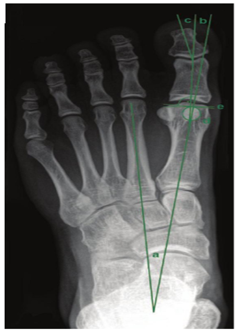
Figure 1: Angles definitions: a) intermetatarsal angle (IMA); b) hallux valgus angle (HVA); c) hallux valgus interphalangeus angle (HVI); d) position of the medial sesamoid in relation to the axis of the metatarsal head; e) distal metatarsal articular angle (DMAA).
The Akin osteotomy is a medial closing-wedge osteotomy of the proximal phalanx designed to correct residual hallux valgus interphalangeus and improve alignment of the hallux. It is frequently performed as an adjunct to first-metatarsal osteotomy when additional correction is required. Modern literature has demonstrated generally favorable union rates and acceptable complication profiles for Akin osteotomy, regardless of the fixation method used. However, an additional osteotomy also increases surgical intervention and fixation requirements, making it important to establish when the procedure provides meaningful additional benefit rather than being performed routinely [5,7].
The decision to add an Akin osteotomy to Chevron correction remains an area of ongoing clinical interest. Previous comparative studies have suggested that the combined Chevron-Akin procedure may improve maintenance of hallux alignment and reduce postoperative loss of correction in selected patients. In a large retrospective comparative series, a preoperative proximal phalanx deformity measurement exceeding 8° was associated with greater loss of correction following Chevron osteotomy alone, suggesting that preoperative assessment of the phalangeal component may help identify patients who would benefit from a concomitant Akin osteotomy. Similarly, another retrospective comparative study reported better maintenance of HVA and interphalangeal alignment during longer-term follow-up in patients undergoing combined Chevron-Akin osteotomy, although differences in some other radiographic parameters were limited. These findings suggest that the benefit of the Akin osteotomy may depend on appropriate patient selection and the specific morphological characteristics of the deformity.
Nevertheless, important questions remain regarding the incremental clinical value of adding an Akin osteotomy to Chevron correction. While previous studies have primarily focused on radiographic parameters and maintenance of correction, fewer comparative investigations have simultaneously evaluated radiographic correction together with patient-centered outcomes such as postoperative pain, functional recovery, satisfaction, and complications. Because radiographic correction does not necessarily correspond directly to patient satisfaction or functional improvement, a comprehensive comparison incorporating both objective and clinical outcomes is clinically relevant [5,7].
Therefore, the present study aimed to compare the outcomes of Chevron osteotomy alone with those of combined Chevron and Akin osteotomies in patients with mild to moderate hallux valgus. The primary objective was to evaluate and compare radiographic correction using the HVA and IMA. Secondary objectives included assessment of functional outcomes using the American Orthopaedic Foot and Ankle Society (AOFAS) hallux score, postoperative pain using the visual analog scale (VAS), patient satisfaction, and postoperative complications. We hypothesized that the addition of an Akin osteotomy would provide improved correction of hallux alignment and superior clinical outcomes in appropriately selected patients without significantly increasing the risk of postoperative complications [5,6].
2. Materials and Methods
2.1 Study Design
This retrospective comparative study included 52 patients (60 feet) with mild to moderate hallux valgus who underwent surgical correction at Queen Alia Military Hospital between June 2022 and June 2024. Patients were divided into two groups according to the surgical procedure performed: Group A underwent Chevron osteotomy alone, whereas Group B underwent Chevron osteotomy combined with an Akin osteotomy.
Outcomes were evaluated using radiographic and clinical parameters. Radiographic assessment included measurement of the hallux valgus angle (HVA) and intermetatarsal angle (IMA). Clinical outcomes were assessed using the American Orthopaedic Foot & Ankle Society (AOFAS) Hallux Metatarsophalangeal-Interphalangeal score, the visual analog scale (VAS) for pain, and patient satisfaction. All patients were followed for a minimum of 12 months after surgery.
The study was conducted following approval from the relevant Institutional Review Board (IRB). Appropriate informed consent was obtained from all participants in accordance with institutional requirements.
2.2 Patient Selection
A total of 52 patients involving 60 feet with mild to moderate hallux valgus were included in the study. Patients were eligible for inclusion if they had a preoperative HVA between 20° and 40° and an IMA of less than 15°.
Patients were excluded if they had severe hallux valgus, defined as an HVA greater than 40°, rheumatic joint disease, previous surgery involving the first ray, or upper motor neuron disorders. All included patients were followed for at least 12 months after surgical correction.
2.3 Group Allocation
The study population was divided into two groups according to the surgical procedure performed. Group A included 30 feet treated with Chevron osteotomy alone, whereas Group B included 30 feet treated with combined Chevron and Akin osteotomies.
The decision to perform an additional Akin osteotomy was based on the presence of hallux valgus interphalangeus identified during clinical and/or radiographic evaluation, or when residual phalangeal deformity remained after correction of the metatarsal component with Chevron osteotomy.
2.4 Surgical Technique
All procedures were performed by experienced orthopaedic foot and ankle surgeons using a standardized medial surgical approach. A Chevron osteotomy of the first metatarsal was performed in all cases and stabilized using a single screw (Figure 2).
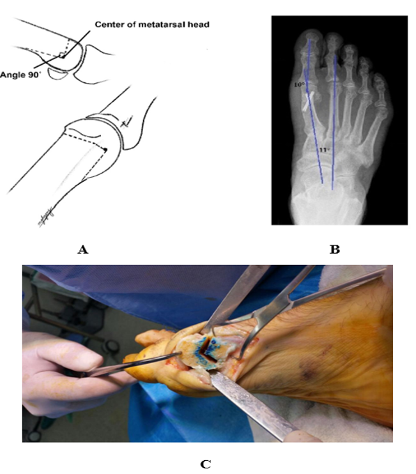
Figure 2: Chevron osteotomy technique and fixation. (A) Surgical illustration demonstrating the Chevron osteotomy. (B) Postoperative anteroposterior radiograph showing the Chevron osteotomy stabilized with a headless compression screw. (C) Intraoperative photograph demonstrating the marking of the osteotomy level at the first metatarsal head.
In patients included in Group B, an additional Akin osteotomy was performed to address associated hallux valgus interphalangeus or residual phalangeal deformity following correction of the metatarsal component. The Akin procedure consisted of a medial closing-wedge osteotomy of the proximal phalanx and was fixed using either a staple or a screw, depending on the intraoperative assessment and surgeon preference (Figure 3) [6,7].
The combined Chevron and Akin procedures provided correction of both the metatarsal and phalangeal components of the deformity, respectively (Figure 4).
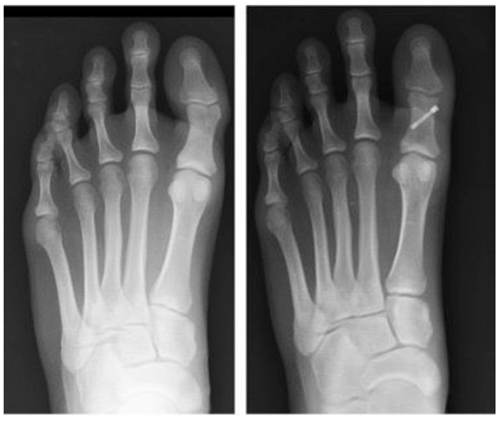
Figure 3: Preoperative and postoperative radiographs demonstrating the level of the Akin osteotomy and fixation with a Herbert screw.
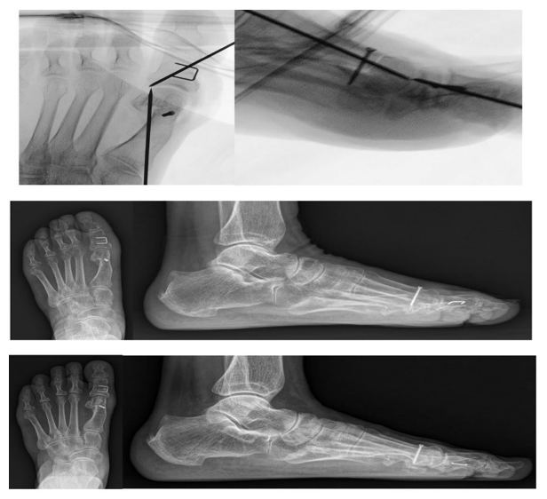
Figure 4: Intraoperative and postoperative radiographs demonstrating the combined Chevron and Akin osteotomies used for correction of hallux valgus.
2.5 Postoperative Protocol
Following surgery, all patients were permitted partial weight bearing using a postoperative medical orthosis for six weeks. After radiographic evidence of satisfactory bone healing, patients were gradually progressed to full weight bearing, and a physiotherapy program was initiated to restore foot and ankle function and facilitate return to normal activities.
2.6 Outcome Measures
The study evaluated radiographic, clinical, and postoperative outcomes. Radiographic assessment included measurement of the hallux valgus angle (HVA) and intermetatarsal angle (IMA). Clinical outcomes were assessed using the American Orthopaedic Foot & Ankle Society (AOFAS) Hallux Metatarsophalangeal-Interphalangeal score, the Visual Analog Scale (VAS) for pain, and patient satisfaction. Postoperative complications, including infection, recurrence, joint stiffness, nonunion, and hardware-related complications, were also recorded.
2.7 Statistical Analysis
Statistical analyses were performed using IBM SPSS Statistics version 25. Continuous variables were expressed as appropriate and analyzed using paired t-tests for within-group comparisons between preoperative and postoperative outcomes. Independent-samples t-tests were used to compare continuous variables between the two groups. Categorical variables were analyzed using the chi-square test. A p-value of <0.05 was considered statistically significant.
3. Results
3.1 Baseline Characteristics
A total of 60 feet from 52 patients were included in the study, with 30 feet in each group. Group A underwent Chevron osteotomy alone, whereas Group B underwent combined Chevron and Akin osteotomies.
|
Parameter |
Group A (Chevron) |
Group B (Chevron + Akin) |
p-value |
|
Number of feet |
30 |
30 |
— |
|
Mean age (years) |
48.2 ± 9.1 |
49.0 ± 8.7 |
0.74 |
|
Female : Male |
0.209028 |
0.209028 |
— |
|
Preop HVA (°) |
31.4 ± 4.1 |
32.1 ± 4.4 |
0.56 |
|
Preop IMA (°) |
13.2 ± 2.0 |
13.4 ± 2.3 |
0.68 |
Table 1: Baseline demographic and preoperative characteristics.
The baseline demographic and preoperative characteristics were comparable between the two groups. The mean age was 48.2 ± 9.1 years in Group A and 49.0 ± 8.7 years in Group B (p = 0.74). Female patients predominated in both groups, with a female-to-male ratio of 5:1. The mean preoperative HVA was 31.4 ± 4.1° in Group A and 32.1 ± 4.4° in Group B (p = 0.56), while the mean preoperative IMA was 13.2 ± 2.0° and 13.4 ± 2.3°, respectively (p = 0.68). No statistically significant differences were observed between the groups in baseline demographic or preoperative radiographic parameters (Table 1) [8,9].
3.2 Radiographic Outcomes
Both groups demonstrated substantial improvement in radiographic alignment following surgery. The mean postoperative HVA decreased to 14.8 ± 3.9° in Group A and 10.6 ± 3.2° in Group B, with significantly greater correction observed in the combined Chevron-Akin group (p = 0.02). The mean HVA correction was 16.6° in Group A and 21.5° in Group B.
The postoperative IMA was 8.6 ± 1.8° in Group A and 8.2 ± 1.7° in Group B, with no statistically significant difference between the groups (p = 0.21). The mean IMA correction was 4.6° in the Chevron-only group and 5.2° in the combined procedure group (Table 2) [10,11].
|
Parameter |
Group A |
Group B |
Intergroup p-value |
|
Postop HVA (°) |
14.8 ± 3.9 |
10.6 ± 3.2 |
0.02 |
|
HVA correction (°) |
16.6 |
21.5 |
— |
|
Postop IMA (°) |
8.6 ± 1.8 |
8.2 ± 1.7 |
0.21 |
|
IMA correction (°) |
4.6 |
5.2 |
— |
Table 2: X-ray outcomes.
3.3 Clinical Outcomes
Both groups demonstrated improvement in functional outcomes and pain scores following surgery. The mean preoperative AOFAS score was comparable between Group A and Group B (56.8 ± 8.2 vs. 55.9 ± 7.9, respectively; p = 0.81). Postoperatively, the mean AOFAS score improved to 84.6 ± 7.5 in Group A and 90.2 ± 6.4 in Group B, with the combined Chevron-Akin group demonstrating significantly higher postoperative functional scores (p = 0.03).
Similarly, the mean preoperative VAS pain score was comparable between the groups (6.7 ± 1.4 in Group A vs. 6.9 ± 1.5 in Group B; p = 0.64). Postoperatively, the VAS score decreased to 2.1 ± 1.0 in Group A and 1.2 ± 0.9 in Group B, with significantly lower postoperative pain scores observed in Group B (p = 0.04).
Patient satisfaction was also higher in the combined procedure group, with 93% of patients reporting an excellent or good outcome compared with 83% in the Chevron-only group (Table 3) [12,13].
|
Outcome |
Group A |
Group B |
p-value |
|
Preop AOFAS |
56.8 ± 8.2 |
55.9 ± 7.9 |
0.81 |
|
Postop AOFAS |
84.6 ± 7.5 |
90.2 ± 6.4 |
0.03 |
|
Preop VAS |
6.7 ± 1.4 |
6.9 ± 1.5 |
0.64 |
|
Postop VAS |
2.1 ± 1.0 |
1.2 ± 0.9 |
0.04 |
|
Satisfaction (Excellent/Good) |
0.83 |
0.93 |
— |
Table 3: Gross Outcomes.
3.4 Complications
The overall complication rate was low in both groups. Superficial infection occurred in two feet in each group, while recurrence was observed in one foot in each group. No cases of nonunion or hardware failure were recorded.
The total reported complication rate was 10% in Group A and 10% in Group B. Overall, no major differences in the pattern of postoperative complications were observed between the two groups (Table 4) [14].
|
Complication |
Group A |
Group B |
|
Superficial infection |
2 |
2 |
|
Recurrence |
1 |
1 |
|
Nonunion |
0 |
0 |
|
Hardware failure |
0 |
0 |
|
Total complication rate |
0.1 |
0.1 |
Table 4: Complications.
4. Discussion
This retrospective comparative study evaluated the additional value of concomitant Akin osteotomy in patients undergoing Chevron osteotomy for mild to moderate hallux valgus. The two groups were comparable at baseline with respect to age, sex distribution, and preoperative radiographic parameters, supporting comparison of the postoperative outcomes between the two surgical strategies [9]. Both procedures resulted in substantial improvements in deformity correction and clinical outcomes; however, the addition of an Akin osteotomy was associated with greater correction of the hallux valgus deformity and superior postoperative functional outcomes.
The principal radiographic finding of the present study was the significantly lower postoperative HVA in the combined Chevron-Akin group compared with the Chevron-only group (10.6° vs. 14.8°, respectively; p = 0.02). Similarly, the magnitude of HVA correction was greater in patients undergoing the combined procedure (21.5° vs. 16.6°). These findings suggest that correction of the metatarsal component alone may be insufficient in selected patients with an associated phalangeal deformity or residual hallux malalignment. The Akin osteotomy provides additional correction at the level of the proximal phalanx and may therefore improve the overall alignment achieved following metatarsal realignment [10-13].
Our findings are consistent with previous comparative studies. Kaufmann et al. [5] evaluated 859 feet treated with either Chevron osteotomy alone or combined Chevron-Akin osteotomy and reported superior correction and better maintenance of hallux alignment in the combined group. They further demonstrated that preoperative phalangeal alignment may help identify patients who are more likely to benefit from an additional Akin osteotomy [13]. Similarly, a prospective matched-group analysis comparing isolated Chevron osteotomy with Chevron-Akin double osteotomy demonstrated greater HVA correction with the combined procedure, although the superiority of the combined technique appeared to be most relevant in selected clinical settings rather than universally applicable to all patients [18]. These findings support the selective use of an Akin osteotomy when an associated phalangeal deformity or residual hallux malalignment is present.
Although the combined procedure resulted in significantly greater HVA correction in our study, postoperative IMA values were similar between the two groups. This finding is consistent with the different biomechanical roles of the two procedures. Chevron osteotomy primarily corrects the metatarsal component of the deformity and reduces the IMA, whereas the Akin osteotomy addresses deformity at the level of the proximal phalanx. Therefore, the additional Akin procedure would be expected to improve hallux alignment without substantially affecting the intermetatarsal relationship [10,13]. The similar postoperative IMA values observed in both groups support the effectiveness of Chevron osteotomy in correcting first-metatarsal alignment, while the greater improvement in HVA observed in the combined group likely reflects correction of the additional phalangeal component.
The clinical findings further support the radiographic results. Both groups demonstrated substantial improvement in AOFAS scores and VAS pain scores following surgery; however, patients treated with the combined procedure achieved significantly higher postoperative AOFAS scores and lower postoperative pain scores. Patient satisfaction also favored the combined procedure, with 93% of patients reporting excellent or good outcomes compared with 83% in the Chevron-only group. Although radiographic correction does not always directly correlate with clinical improvement, more complete correction of the deformity may contribute to improved comfort, function, and patient satisfaction [11,15].
Importantly, the improved radiographic and clinical outcomes observed with the combined procedure were not accompanied by an apparent increase in major postoperative complications. Superficial infections occurred in two feet in each group, and one recurrence was observed in each group, with no cases of nonunion or hardware failure [15-17]. These findings are consistent with previous studies suggesting that the addition of an Akin osteotomy does not appear to substantially increase the risk of major complications when appropriately performed [12,14]. However, the complication data should be interpreted cautiously given the relatively small sample size of the present study.
Kuliński et al. [6] similarly reported that the addition of an Akin osteotomy may help maintain lower HVA and interphalangeal alignment during longer-term follow-up without adversely affecting other radiographic parameters [14,16]. Their findings, together with those of the present study, suggest that the potential benefit of the Akin procedure may extend beyond immediate deformity correction by improving the overall alignment and potentially reducing loss of correction in appropriately selected patients.
The decision to perform an additional Akin osteotomy should not, however, be interpreted as a recommendation for its routine use in all patients undergoing Chevron osteotomy. In the present study, the Akin procedure was selectively performed in the presence of clinically or radiographically evident hallux valgus interphalangeus or residual phalangeal deformity after correction of the metatarsal component. Previous evidence similarly supports an individualized approach, with the indication for concomitant Akin osteotomy based on the characteristics of the phalangeal deformity and the degree of residual hallux malalignment rather than its routine application in every case [13,15,17].
Overall, the findings of the present study suggest that Chevron osteotomy alone remains an effective treatment option for mild to moderate hallux valgus, particularly when the deformity is predominantly related to the metatarsal component. However, in patients with an associated phalangeal deformity or persistent hallux malalignment following metatarsal correction, the addition of an Akin osteotomy may provide greater overall correction and improved functional outcomes without an apparent increase in major complications. These findings support an individualized surgical approach in which correction is tailored to the specific anatomical components of the deformity [9,12,18,19].
5. Limitations
This study has several limitations that should be considered when interpreting the findings. First, its retrospective and non-randomized design may have introduced selection bias, particularly because the decision to perform an additional Akin osteotomy was based on clinical, radiographic, and intraoperative findings. Second, the relatively small sample size and single-center design may limit the generalizability of the results to other populations and surgical settings. Third, although all patients were followed for at least 12 months, longer-term follow-up is required to evaluate the durability of correction, recurrence rates, and long-term functional outcomes. Finally, the study did not include direct measurements of the hallux interphalangeal angle, which could have provided a more detailed assessment of phalangeal deformity and the specific contribution of the Akin osteotomy to its correction. Future prospective, multicenter studies with larger sample sizes and longer follow-up periods are warranted to confirm these findings.
6. Conclusion
Chevron osteotomy remains an effective surgical option for the treatment of mild to moderate hallux valgus. In appropriately selected patients with an associated phalangeal deformity or residual hallux malalignment, the addition of an Akin osteotomy may provide greater correction of the hallux valgus angle and improved postoperative functional outcomes. Importantly, the combined procedure was not associated with an apparent increase in major postoperative complications in the present study. These findings support an individualized surgical approach in which the correction strategy is tailored to the specific anatomical components of the deformity.
7. Declarations
Ethics approval and consent to participate
Ethical approval was obtained from the Jordanian Royal Medical Services Institutional Review Board, and the study adhered to international standards for research involving human subjects, under the IRB #: 23/17/2025.
Patient confidentiality was strictly maintained, and identifying information was anonymized. Where possible, informed consent was obtained from patients or their legal guardians.
Consent for publication
Written informed consent was obtained from all participants prior to inclusion in this study. All individuals provided authorization for the use of their gross data for research purposes, including all images, analysis and publication. No identifying information was collected or reported, and confidentiality was strictly maintained throughout the study.
Availability of data and material
Available upon request.
Competing interests
The authors declare no competing interests.
Funding
This research did not receive any funds.
Acknowledgements
The authors would like to thank the medical and administrative staff of the Military Medical Services for their support during data collection and record retrieval. We also extend our appreciation to the trainees who participated in this study for their cooperation and contribution to the research.
References
- Coughlin MJ, Jones CP. Hallux Valgus: Demographics, Etiology, and Radiographic Assessment. Foot & Ankle International 28 (2007): 759-777.
- Austin DW, Leventen EO. A new osteotomy for hallux valgus: a horizontally directed "V" displacement osteotomy of the metatarsal head for hallux valgus and primus varus. Clinical Orthopaedics and Related Research 157 (1981): 25-30.
- Robinson AH, Limbers JP. Modern concepts in the treatment of hallux valgus. Journal of Bone and Joint Surgery British 87 (2005): 1038-1045.
- Steinböck G. Chevron-osteotomy for the treatment of Hallux valgus. Foot and Ankle Surgery 9 (2003): 95-102.
- Kaufmann G, Dammerer D, Heyenbrock F, et al. Need for concomitant Akin osteotomy in patients undergoing Chevron osteotomy can be determined preoperatively: a retrospective comparative study of 859 cases. Journal of Orthopaedic Surgery and Research 14 (2019).
- Kuliński P, Rutkowski M, Tomczyk Ł, et al. Outcomes After Chevron Osteotomy with and Without Additional Akin Osteotomy: A Retrospective Comparative Study. Indian Journal of Orthopaedics 57 (2023): 907-916.
- Eldessouky AH, Khattak MU, Srour AM. Akin Osteotomy: A Review of Modern Fixation Techniques. Cureus 16 (2024): e57026.
- Peng YN, Peng YH, Chen CPC, et al. Chevron osteotomy and scarf osteotomy for hallux valgus angle and intermetatarsal angle correction: a systematic review and meta-analysis of randomized controlled trials. Journal of Orthopaedic Surgery and Research 19 (2024): 566.
- Trnka HJ. Osteotomies for hallux valgus correction. Foot and Ankle Clinics 10 (2005): 15-33.
- Donahue WE. The Proximal Phalangeal Osteotomy: A Technically Advanced Approach. Clinics in Podiatry 2 (1985): 449-455.
- Okuda R, Kinoshita M, Yasuda T, et al. Proximal Metatarsal Osteotomy for Hallux Valgus: Comparison of Outcome for Moderate and Severe Deformities. Foot & Ankle International 29 (2008): 664-670.
- Liu J, Shi X, Zhang C, et al. Outcomes following modified chevron osteotomy combined with akin osteotomy for severe hallux valgus: A prospective study. The Journal of Foot and Ankle Surgery 64 (2025): 387-391.
- Genuth G, Moses N, Hramov K, et al. Combined Minimally Invasive Chevron-Akin and Distal Minimally Invasive Metatarsal Osteotomy for Hallux Valgus and Metatarsalgia: 12-Month Outcomes and High Radiographic Recurrence in a Dual-Center Study. Foot & Ankle International 47 (2026): 10-18.
- Wanivenhaus A. Decision-making in Hallux Valgus Correction. Foot and Ankle Clinics 30 (2025): 239-250.
- Ezzatvar Y, López-Bueno L, Fuentes-Aparicio L, et al. Prevalence and Predisposing Factors for Recurrence after Hallux Valgus Surgery: A Systematic Review and Meta-Analysis. Journal of Clinical Medicine 10 (2021): 5753.
- Haskell A, Coughlin MJ. Coughlin and Mann’s Surgery of the Foot and Ankle. 10th Edition (2023).
- Nilsdotter A-K, Cöster ME, Bremander A, et al. Patient-reported outcome after hallux valgus surgery — a two year follow up. Foot and Ankle Surgery 25 (2019): 478-481.
- Nguyen JK, Sullivan M, Alpuerto BB, et al. A radiographic analysis of the abnormal hallux interphalangeus angle range: Considerations for surgeons performing Akin osteotomies. Journal of Orthopaedic Surgery 27 (2019).
- Reeves CL, Shane AM. Complications of Hallux Valgus Surgery. In: Lee M, Grossman J, eds. Complications in Foot and Ankle Surgery. Springer, Cham (2017).
- Perera AM, Mason L, Stephens MM. The pathogenesis of hallux valgus. Journal of Bone and Joint Surgery American 93 (2011): 1650-1661.