Sonali D Modi1*, Dharita S Shah2, Krati S Mundhra3, Bhargav Gandhi1, Rahul Shah1, Vikas Kagathara1, Madhav Kiritbhai Dharaiya4, Dhruv Modi5
1Department of Radio-diagnosis, SVP Hospital, Smt. NHL Municipal Medical College, Ahmedabad, India
2Professor and Head of the Department, Department of Radio-diagnosis, SVP Hospital, Smt. NHL Municipal Medical College, Ahmedabad, India
3Associate professor, Department of Radio-diagnosis, SVP Hospital, Smt. NHL Municipal Medical College, Ahmedabad, India
4MBBS student and Data Analyst, Smt. NHL Municipal Medical college, Ahmedabad, India
5Data Scientist, B.Tech. Computer Science and Engineering, Nirma University, Ahmedabad, India
*Corresponding Author: Sonali D Modi, Department of Radio-diagnosis, SVP Hospital, Smt. NHL Municipal Medical College, Ahmedabad, India
Received: 03 August 2021; Accepted: 25 August 2021; Published: 16 September 2021
SARS-CoV-2 virus typically causes lung infiltration causing acute respiratory distress syndrome (ARDS) and in later stages pulmonary fibrosis. Vaccines provide immunity against desired pathogen. Vaccines available in India against this virus are Covishield and Covaxin (majority of the patients are vaccinated with Covishield). COVID-19 causes lung parenchymal infiltration which leads to pneumonitis. The best modality available to study this is the HRCT. Analysis of the extent of lung parenchyma invasion of patients despite taking one or both the doses was done by us. For which , we did a single centered retrospective observational study in laboratory confirmed COVID-19 positive patients with sample size (n=274) being duration dependent (2 months). We found a statistically significant correlation between vaccination status and lung involvement based on HRCT Thorax. In conclusion the presence of vaccination reduces the severity of the CT severity score and improves the outcome in terms of survival of the patient.
Covid 19; Vaccine
1. Introduction
COVID 19/ SARS CoV-2 has spread rapidly throughout the world. WHO has declared COVID 19 as a pandemic in March 2020. Global health system, economy and social progress is severely affected by this disease. To combat COVID many countries have developed vaccines for COVID. Vaccine against COVID can boost immunity and prevent the spread of disease. Vaccine does not eliminate reinfection or infection but surely reduces the severity of symptoms and infectivity and improves the survival of the patients.
The two vaccines developed in INDIA till now are COVISHIELD and COVAXIN. COVID-19 pneumonia manifests with chest CT imaging abnormalities, even in asymptomatic patients, with rapid evolution from focal unilateral to diffuse bilateral ground-glass opacities that progressed to or co-existed with consolidations within 1–3 weeks [1]. The CT value of the patient gives us a comparable numeric modulus towards the extent of damage that has taken place. Our hypothesis in this study is to prove that in comparison to non-vaccinated controls, vaccinated cases show reduced lung involvement in terms of low CT severity index. This study aims to understand the extent to which the post vaccination infection affects the body, primarily in the lungs.
1.1 Pathogenesis
Covid-19 causes abnormality in the immune system response which causes increased release of Tumor Necrosis Factor – alpha [TNF- α] an interleukin-6 [IL-6], which is referred to as cytokine storm. This cytokine storm causes destruction of alveolar epithelial cells[2]. Cytokine storm is caused by an abnormal immune mechanism that may lead to initiation and promotion of pulmonary fibrosis. Epithelial and endothelial injury occurs in the inflammatory phase of ARDS due to dysregulated release of matrix metalloproteinases. VEGF and cytokines such as IL-6 and TNF-α are also involved in the process of fibrosis. The 2 vaccines in study here work on the principles inducing immunity by introduction of 1) inactivated virus and 2) spike proteins by the Covaxin and Covishield vaccines respectively.
2. Method
MDCT was performed on 128 slice PHILIPS CT Scanner machines on COVID +ve patients from 1st April 2021 to 31st May 2021 in our SVP Hospital, NHLMMC, Ahmedabad. Comorbidities are not taken into consideration. Three readers independently and blindly reviewed all HRCT thorax, rating the pulmonary parenchymal involvement.
2.1 Inclusion criteria
- All hospitalised patients with positive RT-PCR result forSARS-CoV-2/Suspected for COVID during the specified period with mean age group being 20-75 years.
- Vaccinated and non-vaccinated patients
- Patients with altered Lab values
- Patients not maintaining spO2 levels
2.2 Exclusion criteria
- Pregnant females
- Patients < 20 years of age
- Patients with pre-existing ILD as GGO of ILD and Covid-19 are overlapping so actual lung parenchyma involvement cannot be predicted.
2.3 Imaging parameters and interpretation
All scans were performed on 128 slice PHILIPS CT Scanner.
Scan Parameters:
- Slice Thickness: 1.00 mm
- Collimation: 128 x 1.00
- Pitch: 0.95
- mAS: 160
- Kvp: 120
- Volumetric data was reconstructed in the multiple planes.
- A semi-quantitative CT score was calculated based on the extent of each lobar involvement.
- <5% lung parenchyma involved =1
- 5-25% lung parenchyma involved =2
- 25-50% lung parenchyma involved =3
- 50-75% lung parenchyma involved =4
- >75% lung parenchyma involved =5
The total CT score would be the sum of the individual lobar scores and can range from 0(No involvement) to 25(Maximum involvement), when all the five lobes show more than 75% involvement.
- CT Severity index <10 is considered to be MILD involvement.
- CT Severity index 10-16 is considered to be MODERATE involvement.
- CT Severity index >16 is considered to be SEVERE involvement.
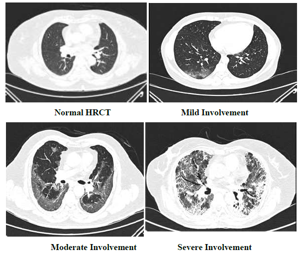
Figure 1: CT images of COVID +ve patients.1st image shows normal HRCT.2nd image shows subpleural areas off ground glass opacities in right lower lobe (mild involvement). 3rd image shows subpleural areas and peribronchovascular areas of ground glass opacities in bilateral lower lobes (moderate involvement). 4th image shows diffuse areas of ground glass opacities with interstitial septal thickening and tractional bronchiectasis (severe involvement).
2.4 Statistics
The Shapiro Wilk Test is performed to check the normality of CT score under each group.
Shapiro Wilk Test:
Null hypothesis: Sample is normal
Alpha: 0.05
|
Vaccine doses |
p-value |
is_normal |
|
0 |
0.353 |
normal |
|
1 |
0.004 |
Non normal |
|
2 |
0.00017 |
Non normal |
Table 1: Shapiro Wilk Test’s p-value for each vaccine dose group.
This shows our vaccine data in two categories is non normal, hence we can’t use one way Anova test, as it doesn’t satisfy this test’s normality assumption. Instead, a non parametric test, named Kruskal wallis test can be used, when normality assumptions of one way Anova is not met.
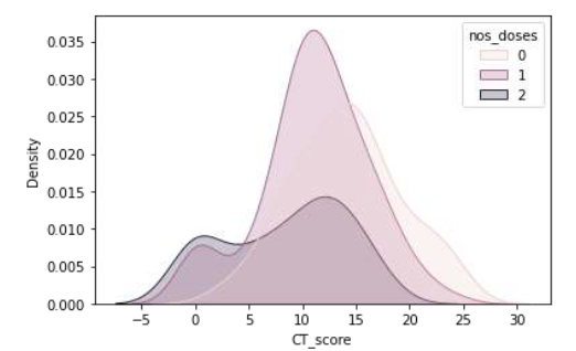
Figure 2: Distribution of CT score for each vaccine dose group.
2.4.1 Data Statistics
|
Vaccine doses |
Cases |
Median |
IQR |
Median ± Quartile Deviation |
|
0 |
98 |
14 |
7 |
14 ± 3.5 |
|
1 |
116 |
11 |
6 |
11 ± 3 |
|
2 |
60 |
10 |
9.5 |
10 ± 4.75 |
Table 2: Showing numerical spread of CT score for each vaccine dose group.
Kruskal Wallis test:
Null hypothesis: Median of all samples are same
Alpha: 0.05
Independent: number_of_doses
Dependent: ct_score
P-value: 7.09e-08
As our p-value < 0.05, we reject our null hypothesis and consider alternative hypothesis i.e. At Least one group is showing a significant difference. To know which group(s) is/are different, let’s perform Dunn’s post-hoc test.
Dunn’s post-hoc test:
Null hypothesis: Two samples under study are the same.
Alpha: 0.05
Note: 0, 1, 2 in the below tables means number of vaccine doses taken and cells represent p-value.
|
p-val |
0 |
1 |
2 |
|
0 |
1 |
0.000476 |
7.96E-08 |
|
1 |
4.75E-04 |
1 |
3.99E-02 |
|
2 |
7.96E-08 |
0.03994 |
1 |
Table 3: Dunn’s Post Hoc’s p-value among two vaccine dose groups.
If p-value < 0.05, then the test is statistically significant i.e. the groups are different
This shows, the groups which are different are:
- 0 and 1
- 0 and 2
- 1 and 2
To conclude, all vaccine groups are different. Median value of CT severity score in non-vaccinated patients is 14, in single dose vaccinated patients is 11 and in two dose vaccinated patients is 10. Hence this study statistically proves that vaccination reduced lung parenchymal involvement in covid 19 patients in terms of CT severity index.
3. Result
We analysed the data of 274 patients (pie chart). Mean age group is 22-75. Study includes 174 male and 100 females. We further divided patients into the patients having co morbidity and not having comorbidity. Vaccinated patients with comorbidity show lower CT severity scores in comparison to non vaccinated patients with comorbidity.
Death
Non vaccinated patients=10
1 Dose vaccinated patients =2
2 dose patient= 1
Vaccination reduces mortality and improves survival in vaccinated patients.
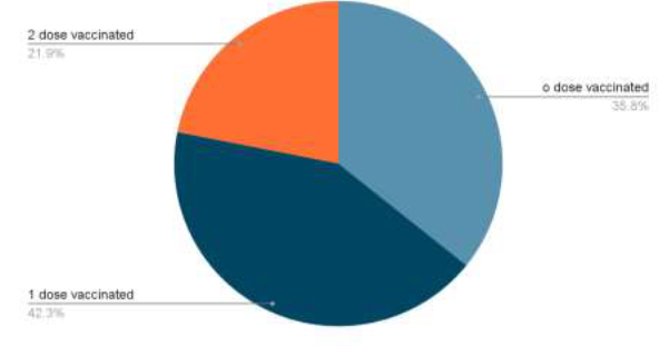
Figure 3: Distribution of patients with respect to their vaccination status.
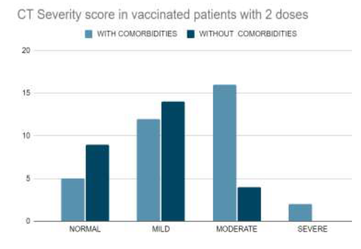
Figure 4: CT severity score in vaccinated patients with 2 doses.
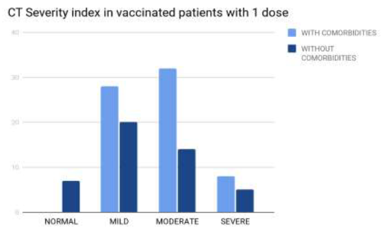
Figure 5: CT severity score in vaccinated patients with 1 dose.
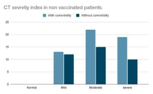
Figure 6: CT severity score in non vaccinated patients.
4. Discussion
The study shows statistical significance between the presence of vaccination and lung parenchymal involvement in terms of CT severity index. Non vaccinated patients with comorbidity have higher CT severity scores in comparison to the vaccinated patients with comorbidity. Based on data available we found that the vaccinated patients have better outcomes in terms of survival than non vaccinated patients.
4.1 Limitations
This study has several limitations. First, owing to limited medical resources, only patients with relatively severe COVID-19 pneumonia were hospitalized during this period. Second, this study was conducted at a single-center hospital with limited sample size. As such, this study may have included disproportionately more patients with poor outcomes. There may also be selection bias because not all vaccinated individuals were hospitalised and not all the hospitalised patients undergo CT imaging as selection criteria in our institute for CT imaging is related to their lab markers and clinical findings.
5. Conclusion
From comparison between vaccinated and non vaccinated COVID patients in our hospital, the study concluded that non vaccinated patients have higher CT severity scores and low survival rate compared to vaccinated patients.
Declarations
Funding
None
Potential conflicts of interest
The authors declare that they have no conflicts of interest.
Author’s contribution
All authors have made substantial contributions to the interpretation and analysis of data. The corresponding author had full access to the data and responsibility for the decision to submit the manuscript for publication.
References
- Heshui Shi, Xiaoyu Han, Nanchuan Jiang, et al. Radiological findings from 81 patients with COVID-19 pneumonia in Wuhan, China: a descriptive study. Lancet (2020).
- Xue-Yan Chen, Bing-Xi Yan, Xiao-Yong Man. TNFα inhibitor may be effective for severe COVID-19: learning from toxic epidermal necrolysis. Ther Adv Respir Dis (2020).