Comparison of Serum Vitamin D level in Women with and without Uterine Fibroid
Dr. Sabiha Islam1, Prof. Dr. Tripti Rani Das2, Farah Noor3, Dr. Jinat Fatema3, Dr. Iffat Rahman4, Dr. Shah Noor Sharmin4, Dr. Bidisha Chakma1, Dr. Tanzina Iveen Chowdhury*,3
1Associate Professor, Department of Obstetrics & Gynecology, Bangladesh Medical University, Dhaka, Bangladesh.
2Professor, Department of Obstetrics & Gynecology, Bangladesh Medical University, Dhaka, Bangladesh
3Assistant Professor, Department of Obstetrics & Gynecology, Bangladesh Medical University, Dhaka, Bangladesh.
4Medical officer, Department of Obstetrics & Gynecology, Aliahat Hospital, Bogra, Bangladesh
*Corresponding author: Tanzina Iveen Chowdhury, Assistant Professor, Department of Obstetrics & Gynecology, Bangladesh Medical University, Dhaka, Bangladesh.
Received: 17 June 2025; Accepted: 23 June 2025; Published: 07 July 2025
Article Information
Citation: Dr. Sabiha Islam, Prof. Dr. Tripti Rani Das, Farah Noor, Dr. Jinat Fatema, Dr. Iffat Rahman, Dr. Shah Noor Sharmin, Dr. Bidisha Chakma, Dr. Tanzina Iveen Chowdhury. Comparison of Serum Vitamin D level in Women with and without Uterine Fibroid. Fortune Journal of Health Sciences, 8 (2025): 632-636.
Share at FacebookAbstract
Background: Uterine fibroids are common, affecting 70-80% of reproductive-aged women universal. They are more regular in African and South Asian women (Stewart et al, 2017). Vitamin D deficiency is greatly prevalent 60-90% in Bangladesh and the Indian subcontinent (Siddiqi et al, 2021; Chowdhury et al, 2018).
Aim: To find out the association of serum vitamin D levels among women with and without uterine fibroid.
Methods: A case-control study was conducted at the Bangladesh Medical University (BMU), Dhaka, from September 2021 to August 2022, involving 128 women aged 18-49 years. Among them 64 women with ultrasonographically proven uterine fibroids were associated to 64 age-matched controls without fibroids. Serum vitamin D levels were considered, and significant socio-demographic and lifestyle data were gathered. Statistical analysis was performed using Chi-square and independent t-tests with a significant level of p<0.05 OR indicates the association between the case and control group.
Results: Among the participants, 84.4% were identified with uterine fibroids. A significant proportionality 88.3% had inadequate vitamin D levels (20–30 ng/mL), with only 11.7% proving sufficient levels (>30 ng/mL). The prevalence of vitamin D deficiency was significantly greater in women with fibroids associated to individuals without (p=0.004), with an odds ratio (OR) of 1.886. In addition, the mean vitamin D level varied significantly among the fibroid and non-fibroid groups (p=0.043).
Conclusion: Women with uterine fibroids had significantly reduce serum vitamin D levels associated to persons devoid of fibroids. This advises that vitamin D deficiency may be related with fibroid growth, emphasizing the significance of vitamin D evaluation in clinical observe.
Keywords
Case-control study, Uterine fibroid, Vitamin D, Reproductive-aged women, Uterine Fibroid
Article Details
Introduction
A study in Bangladesh by Islam et al. (2022) examined the effect of vitamin D supplement on the size of uterine fibroids in women with fibroids insignificant than 5 cm. The decision implied that vitamin D supplement over three months guided to a decrease in the mean size of fibroids, with a more proclaimed impact examined in minor fibroids. This recommends a prospective therapeutic role for vitamin D in controlling uterine fibroids, particularly in lowering their development (Islam et al, 2022). Another study acted in eastern India by Singh et al. (2019) noticed a considerably decrease mean serum vitamin D3 intensity in women with uterine fibroids evaluated to the control group. A strong comparison of women with fibroids shown rigorous vitamin D3 deficiency. The study assumed that serum vitamin D3 levels were in reverse associated with the incidence of uterine fibroids, indicating that vitamin D shortage might be a danger factor for their improvement in the Indian population (Singh et al, 2019). Likewise, a case-control study in India by Kapoor and Maheshwari (2023) also narrated an excessive prevalence of vitamin D shortage among women with uterine fibroids and observed an adverse association between vitamin D levels and fibroid volume.
Nonetheless, not all the latest studies in the area have disclosed a direct correlation. A modern case-control study containing 366 women in the Indian subcontinent did not observe a statistically significant variation in serum vitamin D levels between women with and without uterine fibroids (ResearchGate, 2025). Allowing the enhancing interest in the non-skeletal results of vitamin D, researchers have started to examine its prospective role in uterine fibroid growth. Several studies have advocated an opposite correlation between serum vitamin D levels and the prevalence or size of uterine fibroids (Ciavattini et al, 2016; Halder et al, 2021). It has been hypothesized that vitamin D may apply its impacts on fibroids via different methods, involving preventing cell proliferation, advocating apoptosis, and modulating inflammatory pathways (Fauser et al, 2021). Current studies in Bangladesh observed this link. Islam et al. (2022) studied vitamin D and fibroid size. Supplement appeared to decrease fibroid size. This was more apparent in reduced fibroids. Vitamin D might assist in accomplishing fibroid growth. Singh et al. (2019) studied women in eastern India. They realized reduce vitamin D in women with fibroids. Several had serious vitamin D deficiency. Low vitamin D might be a risk cause in these inhabitants. Kapoor and Maheshwari (2023) in India also found low vitamin D. They stated a link between low vitamin D and bigger fibroids.
Uterine fibroids are widespread, non-malignant tumors in the uterus. They influence many women throughout their reproductive years (Stewart et al, 2017). Okoro et al. (2022) examined the association between serum vitamin D significance and uterine leiomyomas. Their results are circulated in Obstetrics & Gynecology Science. Kumari et al. (2022) investigated the association between serum vitamin D levels and uterine fibroids in premenopausal Indian women. This research happened in Drug Inventions& Therapeutics. Chowdhury et al. (2018) evaluated the vitamin D status of adult outpatients in Bangladesh. Their study was available in the Journal of Dhaka Medical College. Siddiqee et al. (2021) guided a systematic review and meta-analysis on the high prevalence of vitamin D deficiency among South Asian pregnant women. Siddiqi et al. (2021) also executed a systematic review and meta-analysis, focusing on the high prevalence of vitamin D shortage among South Asian adults. This study was announced in BMC Public Health Journal.
Methodology
A case-control study was conducted at the Department of Obstetrics and Gynecology, BMU, Dhaka, from September 2021 to August 2022, to find out the association between serum vitamin D levels and uterine fibroids status among women. A total of 128 women aged 18-49 years were nominated with a convenient sampling method, among them 64 cases (with fibroids) and 64 controls (without fibroids), confirmed by abdominal ultrasonography. Women were excluded if they were postmenopausal, pregnant, lactating, on vitamin D supplements, hormonal therapy, or had chronic setting. Data on sociodemographic, menstrual history, parity, sun display, diet, and physical action were collected. Serum vitamin D levels were considered and categorized as sufficient (>30 ng/mL) or insufficient (20–30 ng/mL). Ethical consent was taken from the BMU IRB. Written informed approval was taken from all contributors. Statistical analysis was accomplished employing Chi-square and independent t-tests, with p<0.05 deemed significant.
Results
A case-control study was conducted at the BMU, Dhaka, involving 128 (64 case and 64 control) women aged 18-49 years.
Table 1: Distribution of respondents by socio-demographic factors
|
Variables |
Frequency |
Percent |
|
Marital status |
||
|
Married |
62 |
48.4 |
|
Unmarried |
32 |
25 |
|
Divorced |
18 |
14.1 |
|
Widowed |
16 |
12.5 |
|
Occupation |
||
|
Housewife |
58 |
45.3 |
|
Service holder |
20 |
15.6 |
|
Business |
15 |
11.7 |
|
Others |
35 |
27.3 |
|
Monthly family income (BDT) |
||
|
<30,000 |
32 |
25 |
|
30,000–50,000 |
64 |
50 |
|
>50,000 |
32 |
25 |
|
Total |
128 |
100 |
Table 1 shows that among the 128 participants, 48.4% were married, 25.0% unmarried, 14.1% divorced, and 12.5% widowed. Occupationally, 45.3% were housewives, 15.6% service holders, 11.7% engaged in business, and 27.3% reported other occupations. Half of the respondents had a monthly family income of BDT 30,000–50,000, while 25.0% earned below and 25.0% above this range.
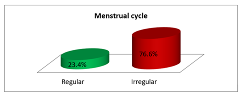
Figure 1 shown majority of participants 76.6% reported having irregular menstrual cycles, while only 23.4% had regular cycles.
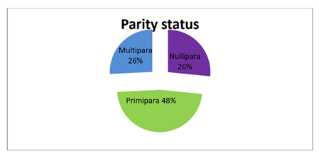
Figure 2 displays, 48% were primipara, while 26% were nullipara and 26% multipara.
Table 2: Uterine fibroids related information
|
Variables |
Frequency |
Percent |
|
Diagnosis of uterine fibroids |
||
|
Yes |
108 |
84.4 |
|
No |
20 |
15.6 |
|
Diagnosis technique |
||
|
Ultrasound |
96 |
75 |
|
MRI |
6 |
4.7 |
|
Clinical examination |
16 |
12.5 |
|
Other |
10 |
7.8 |
|
Duration of diagnosis |
||
|
Less than 1 year |
109 |
85.2 |
|
1–3 years |
10 |
7.8 |
|
More than 3 years |
9 |
7 |
|
Total |
128 |
100 |
Table 2 indicates that Uterine fibroids were diagnosed with 84.4% of participants. Most diagnoses were made via ultrasound 75.0%, followed by clinical examination 12.5%, MRI 4.7%, and other methods 7.8%. In 85.2% of cases, fibroids had been diagnosed within the past year, while 7.8% had been diagnosed 1-3 years ago and 7.0% over 3 years ago.
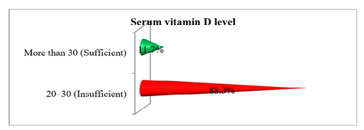
Figure 3 establishes most participants 88.3% had insufficient serum vitamin D levels (20-30 ng/mL), while only 11.7% had sufficient levels >30 ng/mL.
Table 3: Association between Uterine fibroids and vitamin D status
|
Uterine fibroids |
Category of Vit D |
p-value |
OR |
95% Confidence Interval (CI) |
||
|
Lower |
Upper |
|||||
|
20-30 |
>30 |
0.004 |
1.886 |
0.645 |
5.512 |
|
|
Yes |
88 |
20 |
||||
|
No |
14 |
6 |
||||
|
Total |
102 |
26 |
128 |
|||
Table 3 shows that there was a highly significant association between uterine fibroids and vitamin D status p=0.004 at 95% CI and OR=1.886 indicates that uterine fibroids may have hada 1.886 times higher risk to lower the Vit D status.
Table 4: Descriptive statistics of category of Vit. D by grouping variable (uterine fibroids)
|
Vit D |
Uterine fibroids |
N |
Mean |
SD |
F |
Sig. |
T |
|
Yes |
108 |
1.19 |
0.39 |
4.166 |
0.043 |
-1.169 |
|
|
No |
20 |
1.3 |
0.47 |
*Independent Samples t-Test
Table 4 explores that the vit.D status among uterine fibroids yes and no in the population where samples were drawn in different (p=0.043)
Table 5: Distribution of the respondents by lifestyle related factors
|
Daily sun exposure |
Frequency |
Percent |
|
Less than 15 minutes |
64 |
50 |
|
15-30 minutes |
42 |
32.8 |
|
More than 30 minutes |
22 |
17.2 |
|
Vitamin D-rich food intake |
||
|
Regular |
93 |
72.7 |
|
Occasionally |
24 |
18.8 |
|
Rarely/never |
11 |
8.6 |
|
Physical activity |
||
|
Sedentary |
17 |
13.3 |
|
Moderate |
38 |
29.7 |
|
Active |
73 |
57 |
|
Total |
128 |
100 |
Table 5 explains that half of the participants, 50.0% had less than 15 minutes of daily sun exposure, while 32.8% had 15-30 minutes, and 17.2% had over 30 minutes. Regular intake of vitamin D-rich foods was reported by 72.7%, with 18.8% consuming them occasionally and 8.6% rarely or never. In terms of physical activity, 57.0% were physically active, 29.7% moderately active, and 13.3% led a sedentary lifestyle.
Discussion
This case-control study confirmed a significant association between low serum vitamin D levels and the existence of uterine fibroids in reproductive-aged women, consistent with previous research across various populations. The majority 88.3% of participants exposed insufficient vitamin D levels (20–30 ng/mL), and vitamin D insufficiency was closely 1.9 times more probable among women with fibroids, highlighting a viable etiological role. These results associated with those of Kapoor and Maheshwari (2023), who perceived significantly lower serum vitamin D levels among Indian women with uterine fibroids associated to controls. Relatedly, Singh et al. (2019) stated that women with fibroids in Eastern India had significantly lowered vitamin D3 levels, indicating regional dietary, lifestyle, and sun experience examples may impact serum levels. The biological credibility of this correlation is reinforced by the existence of vitamin D receptors in uterine fibroid tissues, where vitamin D exerts antiproliferative effects. Ciavattini et al. (2016), in a systematic review and meta-analysis, emphasized vitamin D shortage as a modifiable risk factor for fibroid growth, suggesting that vitamin D may control fibroid development via anti-inflammatory and antiproliferative processes.
Our study also observed that women with <15 minutes of daily sun exposure and irregular intake of vitamin D-rich foods were more prospective to have insufficient vitamin D. These lifestyle forms are usual in South Asian territories, where traditional clothing and restricted outdoor occupation decrease sunlight coverage (Siddiqi et al, 2021; Chowdhury et al, 2018). Islam et al. (2022) further confirmed the therapeutic potential of vitamin D supplementation in reduction fibroid size, proving the reason for protective and therapeutic strategies pointing hypovitaminosis D. A study by Halder et al. (2021) showed in Bangladesh also informed a significant association between vitamin D deficiency and uterine fibroids, supporting the reliability of results in similar demographic circumstances. The present study supplements to this developing body of proof by importance the prevalence of insufficiency in Bangladeshi women and strengthening the essential for vitamin D screening in gynecological evaluations. From a wider public health scene, the high prevalence of vitamin D insufficiency among South Asian women incorporating reproductive-aged population has been well-familiar in meta-analyses by Siddiqee et al. (2021) and Siddiqi et al. (2021). Given this backdrop, identifying uterine fibroids as a possible significance of long-term deficiency adds insistence to protective interventions.
Remarkably, ultrasound was the elementary mode of diagnosis in our study, which may exhibit diagnostic convenience and the clinical reliance on imaging. Most fibroid cases were diagnosed within the past year, advising an expanding awareness or probably a growth occurrence, though longitudinal studies are required to prove developments. While the results are consistent with universal literature (Stewart et al, 2017; Okoro et al, 2024; Kumari et al, 2022), this study is restricted by its minor sample size and the use of accessible sampling, which may restrict generalizability. Furthermore, being a single-center study, external validity may be controlled. However, the significant association and reliability with regional and global studies gives weight to its results.
Conclusion
This study shown a significant association between low serum vitamin D levels and the presence of uterine fibroids among reproductive-aged women. The high prevalence of vitamin D insufficiency in both groups, particularly among those with fibroids, suggests a potential role of vitamin D deficiency in fibroid growth. These discoveries proof the necessity for regular screening and promising supplementation of vitamin D in women at danger of uterine fibroids. Other longitudinal and interventional studies are necessary to confirm causality and evaluate the therapeutic prospective of vitamin D in fibroid prevention and management.
Declaration of Interest: The authors affirm that they have no known disagreements of interest that could have persuaded the conduct or writing of this study.
Conflict of Interest: None to proclaim.
Authors Contributions: Prof. Dr. Tripti Rani Das and Dr. Sabiha Islam intellectualized the study and invented the methodology. Dr. Dipika Majumder and Dr. Iffat Rahman contributed data management and statistical evaluation. Dr. Shah Noor Sharmin, Dr. Jinat Fatema and Dr. Bidisha Chakma helped with manuscript writing and critical corrections. Prof. Dr. Tripti Rani Das and Dr. Tanzina Iveen Chowdhury supervised the research and afforded final manuscript endorsement. All authors examined and accepted the final version
References
- Islam MN, Akter S, Hossain MS, et al. Impact of vitamin D supplementation among the women with uterine fibroid in different age groups. Scholars Journal of Applied Medical Sciences 10 (2022): 2052-2056.
- Kapoor S & Maheshwari S. A case control study of association of vitamin D levels with uterine fibroids. The New Indian Journal of 1 OBGYN 10 (2023): 362-367.
- Does vitamin D play a role in uterine fibroids? A case control study (2025).
- Singh V, Barik A & Imam N. Vitamin D 3 Level in Women with Uterine Fibroid: An Observational Study in Eastern Indian Population. Journal of Obstetrics and Gynaecology of India 69 (2019): 161-165.
- Ciavattini A, Rossi S, Delli Carpini G, et al. Vitamin D deficiency and uterine myomas: a systematic review and meta-analysis. Nutrients 8 (2016):
- Halder A, Hossain MS, Akter S, et al. Association of vitamin D deficiency with uterine fibroids: a case-control study in Bangladeshi women. BMC Women's Health 21 (2021): 1-8.
- Fauser BCJM, Tarlatzis BC, Rebar RW, et al. Revised 2003 consensus on diagnostic criteria and long-term health risks related to polycystic ovary syndrome (PCOS). Human Reproduction 36 (2021): 1-7.
- Islam MN, Akter S, Hossain MS, et al. Impact of vitamin D supplementation among the women with uterine fibroid in different age groups. Scholars Journal of Applied Medical Sciences 10 (2022): 2052-2056.
- Singh V, Barik A & Imam N. Vitamin D 3 Level in Women with Uterine Fibroid: An Observational Study in Eastern Indian Population. Journal of Obstetrics and Gynaecology of India 69 (2019): 161-165.
- Kapoor S & Maheshwari S. A case control study of association of vitamin D levels with uterine fibroids. The New Indian Journal of 1 OBGYN 10 (2023): 362-367.
- Islam MN, Akter S, Hossain MS, et al. Impact of vitamin D supplementation among the women with uterine fibroid in different age groups. Scholars Journal of Applied Medical Sciences 10 (2022): 2052-2056.
- Kapoor S & Maheshwari S. A case control study of association of vitamin D levels with uterine fibroids. The New Indian Journal of 1 OBGYN 10 (2023): 362-367.
- Does vitamin D play a role in uterine fibroids? A case control study (2025).
- Stewart EA, Laughlin-Tommaso SK, Catherino WH, et al. Uterine fibroids. The Lancet 389 (2017): 1603-1612.
- Okoro CC, Ikpeze OC, Eleje GU, et al. Association between serum vitamin D status and uterine leiomyomas: a case-control study. Obstetrics & gynecology science 67 (2024): 101–111.
- Kumari R, Nath B, Kashika Gaikwad HS & et al. Association between serum vitamin D level and uterine fibroid in premenopausal women in Indian population. Drug Discoveries & Therapeutics 16 (2022): 8–13.
- Chowdhury N, Hossain MZ, Mia M, et al. Vitamin D Status of Adults in the Outpatient Department in Bangladesh. Journal of Dhaka Medical College 27 (2018): 94–97.
- Siddiqee MH, Bhattacharjee B, Siddiqi UR, et al. High prevalence of vitamin D insufficiency among South Asian pregnant women: a systematic review and meta-analysis. British Journal of Nutrition 126 (2021): 742–749.
- Siddiqi UR, Rahman MM & Rahman MM. High prevalence of vitamin D deficiency among the South Asian adults: a systematic review and meta-analysis. BMC Public Health 21 (2021):


