Tanushree Paul*,1, Sushanta Barua2,Rita Rani Barua3, Rubeyatul Jannat4,Rajib Kumar Dhar5
1Assistant Professor, Department of Pathology MH Samorita Hospital & Medical College, Dhaka, Bangladesh.
2Assistant Professor, Interventional Cardiology, National Institute of Cardiovascular Diseases &Hospital, Dhaka, Bangladesh.
3Professor& Head of Department of Pathology MH, Samorita Hospital & Medical College, Dhaka, Bangladesh.
4 Lecturer, School of Medicine IMU University, Kualalampur, Malaysia.
5Junior Consultant Medicine, Upazila Health Complex, Kaptai, Rangamati, Bangladesh
*Corresponding author: Tanushree Paul, Assistant Professor, Pathology, Department of Pathology, MH Samorita Hospital & Medical College, Dhaka, Bangladesh.
Received: 20 September 2026; Accepted: 28 September 2026; Published: 23 October 2026
Background: Breast cancer remains a leading cause of mortality and morbidity among women globally. The tumor microenvironment plays a critical role in tumor progression and invasion, with mast cells being key modulators within the tumor stroma. Intratumoral mast cell density (I-MCD) has been implicated in higher histologic grades, molecular subtypes, and lymph angiogenesis in invasive breast carcinoma.
Objective: To determine the correlation between lymphovascular proliferation and mast cell density across different histologic grades and molecular subtypes of invasive breast carcinoma.
Methods: This cross-sectional observational study was conducted at the Department of Pathology, Bangabandhu Sheikh Mujib Medical University, from March 2021 to January 2023. A total of 51 histopathologically diagnosed female cases of invasive breast carcinoma were included after obtaining informed written consent. Mast cells were identified using Toluidine blue stain, and lymph vessels were detected using podoplanin (D2-40) immunostaining.
Results: Two-thirds of breast carcinomas were located on the left side. Infiltrating duct carcinoma was the most frequent type, with histologic grade 2 being the most common. A significant correlation was observed between I-MCD and lymph vessel density (LVD). I-MCD was significantly associated with the luminal molecular subtype (p < 0.05) and with increased histologic grades (p < 0.05). Furthermore, peritumoral lymph vessel density was significantly associated with lymph node metastasis (p < 0.05).
Conclusion: Intratumoral mast cell density is directly proportional to higher histologic grades and positively associated with ER/PR receptor positivity in breast carcinoma. I-MCD correlates with increased lymph vessel density, which is further linked to lymph node metastasis. These findings suggest that mast cell density is associated with both poor prognostic indicators and potentially favorable treatment outcomes in hormone receptor-positive tumors.
Mast cell density, invasive breast carcinoma, lymphovascular density, tumor microenvironment, molecular subtypes, podoplanin (D2-40), Toluidine blue
Mast cell density articles, invasive breast carcinoma articles, lymphovascular density articles, tumor microenvironment articles, molecular subtypes articles, podoplanin (D2-40) articles, Toluidine blue articles
Introduction
Breast cancer accounts for approximately 685,000 deaths annually worldwide (1). (WHO, 20201. Holds the highest prevalence rate among Bangladeshi women (35.19 per 100,000), with an incidence rate of 8.5 per 100,000(2). It accounts for one-third of all female cancers. Etiology is driven by long-standing reproductive/hormonal factors (early menarche, late menopause, late childbirth, lower parity, reduced breastfeeding, exogenous hormone use) and lifestyle factors (obesity, alcohol, physical inactivity), paired with increased detection through screening (3). Invasive breast carcinoma of No Special Type (NST) is the most common form (~83% of cases). Scored I-III using the Nottingham modified Scarff-Bloom-Richardson system (evaluating tubule formation, nuclear pleomorphism, and mitotic rate). Defined by key protein markers Estrogen receptor positive (ER+), HER2 negative (HER2−). HER2 over expressed (HER2+), estrogen receptor negative (ER−). Negative for ER, PR, and HER2 (ER−/PR−/HER2−) (4). Tumors recruit inflammatory cells—including mast cells, macrophages, lymphocytes, and plasma cells—which dynamically alter the tumor microenvironment (TME) during progression from DCIS to invasive cancer (5). Derived from bone marrow progenitors, MCs are among the first cells to infiltrate the TME. They promote tumor expansion, matrix degradation, and stromal remodeling by releasing neutral proteases (e.g., tryptase, which is 3x higher in-patient serum), cytokines, and growth factors (6). C-KIT inhibitors, cell cycle inhibitors, and tryptase inhibitors can suppress pro-tumorigenic MC activity (7). Lymphatic vessel density (LVD) reflects active tumor lymphangiogenesis and predicts lymph node metastasis (6). Under hypoxic conditions, activated MCs secrete lymphangiogenic drivers (VEGF-A, VEGF-B, VEGF-C, VEGF-D, endostatin). MCD and LVD positively correlate, particularly in basal-like tumors (8). Targeted inhibition of lymphangiogenic axes includes VEGF-A/C/D, VEGFR-2/3, chemokine receptor pairs (SDF-1/CXCR4, CCR7), and pro-lymphangiogenic mediators like Ang-2 and Neuropilin-2(9). The microanatomical location of MCs carries distinct prognostic weight; for instance, intratumoral MCs (I-MCD) and peritumoral MCs (P-MCD) demonstrate opposing clinical impacts in malignancies such as prostate cancer(10).This study aims to evaluate the correlation between lymphovascular proliferation (LVD) and mast cell density (separating intratumoral [I-MCD] and peritumoral [P-MCD] compartments) and assess their relationship with histological grade and molecular subtype in invasive breast carcinoma.
Materials and Methods
We conducted a cross-sectional observational study at the Department of Pathology, BSMMU, from March 2021 to January 2023. Purposive sampling was used to select 51 cases of invasive breast carcinoma from mastectomy specimens. Inclusion criteria comprised paraffin blocks from female patients of any age with histopathologically confirmed invasive breast carcinoma (with or without lymph node metastasis), immunohistochemically classified as luminal, triple-negative, or HER2-enriched subtypes. Cases with neoadjuvant therapy/radiotherapy, HER2-equivocal results, extensive DCIS/necrosis, or microinvasive carcinoma were excluded. Peritumoral and intratumoral mast cell density (MCD), peritumoral and intratumoral lymphangiogenesis (LVD), histologic grade, lymph node metastasis, and molecular subtypes. H&E-stained sections were evaluated for diagnosis confirmation and graded using the Nottingham modification of the Scarff-Bloom-Richardson method. Sections were stained with toluidine blue, which identifies mast cell granules as red violet (metachromatic staining). Three hotspots were identified at 100× magnification; mast cells were counted at 400× (HPF). MCD was calculated as mean mast cells/HPF. Categorization: Low (<1.5 mast cells/HPF) and High (>1.5 mast cells/HPF). Podoplanin (D2-40) immunostaining was performed to identify lymphatic vessels. Three hotspots were selected at low magnification; intratumoral and peritumoral lymph vessels were counted separately at HPF. LVD was calculated as mean vessels/HPF. Lymphatic vessels were identified as thin-walled, irregular structures lacking smooth muscle and red blood cells in their lumen. Categorization: Low (<10 vessels/HPF) and High (>10 vessels/HPF). Data were recorded using a structured questionnaire with informed consent. Blinding was maintained to minimize bias. Data were analyzed using SPSS version 20. Qualitative variables were expressed as frequency and percentage; continuous variables as mean =; SD. Log transformation was applied where normal distribution was not achieved. One-way ANOVA with post-hoc Hochberg test was used to compare MCD across histologic grades and molecular subtypes. Pearson correlation test assessed the relationship between MCD and LVD. Statistical significance was set at p < 0.05 with a 95% confidence interval.
Results
This cross-sectional observational study was conducted in the Department of Pathology at BSMMU to assess lymphovascular proliferation (D2-40 podoplanin immunohistochemistry) and intratumoral/peritumoral mast cell density (toluidine blue staining) across various histopathological grades and molecular subtypes of invasive breast carcinoma ( n=51).
Demographic and Clinical Characteristics
The mean patient age was 47.75±12.79 years (range: 25-78 years). A positive family history of breast carcinoma was reported in 23.5% ( n=12) of cases, while 76.5% (n=39 ) had no family history. Left-sided tumors predominated at 58.8% (n=30 ), compared to 41.2% (n=21 ) on the right. Infiltrating ductal carcinoma was the most common type (90.2%,n=46 ), followed by invasive lobular carcinoma (5.9%,n=3 ) and mixed types (3.9%,n=2 ). Grade 2 tumors constituted the majority (76.5%,n=39 ), followed by Grade 3 (19.6%,n=10 ) and Grade 1 (3.9%,n=2 ). LVI was present in 60.8% (n=31 ) of patients and absent in 39.2% (n=20 ). Regional lymph node metastasis was classified as N1 (27.5%,n=14 ), N0 (27.5%,n=14 ), N2 (13.7%,n=7 ), and N3 (13.7%,n=7 ). The Luminal subtype was the most frequent (54.9%,n=28 ), followed by Triple-Negative (31.4%,n=16 ) and HER2-enriched (13.7%,n=7 ). Corrected N1 percentage from 27.7% to 27.5% so the nodal breakdown totals 100% based on n=51.
Table 1: Demographic, Histopathological, and Molecular Characteristics of Study Population (N = 51)
|
Characteristic |
Category |
Count ($n$) |
Percentage (%) |
|
Age (years) |
Mean± SD |
47.75± 12.79 |
— |
|
Range |
25 - 78 |
— |
|
|
Family History of Breast Cancer |
Positive |
12 |
23.50% |
|
Negative |
39 |
76.50% |
|
|
Tumor Laterality |
Left |
30 |
58.80% |
|
Right |
21 |
41.20% |
|
|
Histological Subtype |
Infiltrating Ductal Carcinoma |
46 |
90.20% |
|
Invasive Lobular Carcinoma |
3 |
5.90% |
|
|
Mixed Type |
2 |
3.90% |
|
|
Histological Grade |
Grade 1 |
2 |
3.90% |
|
Grade 2 |
39 |
76.50% |
|
|
Grade 3 |
10 |
19.60% |
|
|
Lymphovascular Invasion (LVI) |
Present |
31 |
60.80% |
|
Absent |
20 |
39.20% |
|
|
Nodal Status (N Stage) |
N0 (No regional lymph node metastasis) |
14 |
27.50% |
|
N1 (1-3 lymph nodes) |
14 |
27.50% |
|
|
N2 (4-9 lymph nodes) |
7 |
13.70% |
|
|
N3 ($\ge 10$ lymph nodes) |
7 |
13.70% |
|
|
Molecular Subtype |
Luminal |
28 |
54.90% |
|
Triple-Negative |
16 |
31.40% |
|
|
HER2-enriched |
7 |
13.70% |
Note: SD = Standard Deviation. Percentages have been rounded to one decimal place.
Table 2: Peritumoral and intratumoral mast cell density in histological grade of carcinoma
Mean of peritumoral mast cell density was found 2.53 (±0.71) in luminal, 2.56 (±0.75) in Her2 enriched and 2.03 (±0.68) in triple negative. Mean of intratumoral mast cell density was found 2.4 (±0.67) in luminal, 2.34 (±0.84) in Her2 enriched and 1.86 (±0.48) in triple negative
|
Characteristics |
Mean |
Test statistics and Significance |
|
Peritumoral mast cell density |
||
|
Grade 1 |
1.6 ± 0.00 |
F= 1.201, p=0.310 p>0.05 |
|
Grade 2 |
2.4 ± 0.71 |
|
|
Grade 3 |
2.44 ± 0.81 |
|
|
Intratumoral mast cell density* |
||
|
Grade 1 |
1.14 ± 0.21 |
F= 5.24, p=0.009 p<0.05 |
|
Grade 2 |
2.17 ± 0.65 |
|
|
Grade 3 |
2.23 ± 0.57 |
|
*Geometric mean was used.
Table 3: Peritumoral and intratumoral mast cell density in molecular subtype of carcinoma
|
Characteristics |
Mean |
Test statistics and Significance |
|
Peritumoral mast cell density |
||
|
Luminal |
2.53 ± 0.71 |
F= 2.803, p=0.071 p>0.05 |
|
Her 2 enriched |
2.56 ± 0.75 |
|
|
Basal |
2.03 ± 0.68 |
|
|
Intratumoral mast cell density* |
||
|
Luminal |
2.4 ± 0.67 |
F= 4.18, p=0.02 p<0.05 |
|
Her 2 enriched |
2.34 ± 0.84 |
|
|
Basal |
1.86 ± 0.48 |
|
*Geometric mean was used.
Intratumoral and peritumoral lymph vessel density in lymph node metastasis
Peritumoral lymph and intratumoral lymph vessel density were normally distributed. Mean of peritumoral lymph vessel density was found 8.63 (±2.63) in N0, 9.59 (±2.26) in N1, 11.24 (±2.76) N2 and 11.6 (±2.25) in N3 type of metastasis. On the other side, mean of intratumoral lymph vessel density was found 11.04(±3.61) in N0, 10.65(±2.57) in N1, 11.07(±0.64) N2 and 11.41(±1.79) in N3 type of metastasis.
Table 4: Intratumoral and peritumoral lymph vessel density in lymph node metastasis of breast carcinoma
|
Characteristics |
Mean |
Test statistics and Significance |
|
Peritumoral lymph vessel density |
||
|
N0 (No lymph node involve) |
8.63 ± 2.63 |
F= 3.23, p=0.001 p<0.05 |
|
N1 (1-3 lymph node metastasis) |
9.59 ± 2.26 |
|
|
N2 (4-9 lymph node metastasis) |
11.24 ± 2.76 |
|
|
N3 (≥10 metastasis) |
11.6 ± 2.25 |
|
|
Intratumoral lymph vessel density |
||
|
N0 (No lymph node involve) |
11.04 ± 3.61 |
F= 0.17, p>0.05 |
|
N1 (1-3 lymph node metastasis) |
10.65 ± 2.57 |
|
|
N2 (4-9 lymph node metastasis) |
11.07 ± 0.64 |
|
|
N3 (≥10 metastasis) |
11.41 ± 1.79 |
|
Correlation between intratumoral and peritumoral lymph vessel density with peritumoral mast cell density
Peritumoral and intratumoral lymph vessel density was normally distributed. Data of intratumoral lymph vessel density and peritumoral mast cell density was not correlated in Pearson correlation test and statistical significance was not found (p> 0.05). On the other side, Pearson correlation test shows no statistical significance between peritumoral lymph vessel density and peritumoral mast cell density and p> 0.05.
Table 5: Pearson correlation test between intratumoral lymph vessel density with peritumoral mast cell density
|
Characteristics |
Correlation coefficient(r) |
P value |
|
Intratumoral lymph vessel density and peritumoral mast cell density |
0.26 |
p> 0.05 |
Table 6: Pearson correlation test between peritumoral lymph vessel density with peritumoral mast cell density
|
Characteristics |
Correlation coefficient(r) |
P value |
|
Peritumoral lymph vessel density and peritumoral mast cell density |
0.17 |
p> 0.05 |
Correlation between intratumoral and peritumoral lymph vessel density with intratumoral mast cell density
Peritumoral lymph vessel density was normally distributed. Intratumoral mast cell density was not normally distributed. Therefore, log transformation was done for the purpose of analysis. Data of periumoral lymph vessel density and intratumoral mast cell density was correlated in Pearson correlation test and statistical significance was found. (p< 0.05).
Table 7: Pearson correlation test between peritumoral lymph vessel density with intratumoral mast cell density
|
Characteristics |
Correlation coefficient(r) |
P value |
|
Peritumoral lymph vessel density and intratumoral mast cell density |
0.54 |
p= 0.00 |
Regression model shows a linear relationship between peritumoral lymph vessel density and intratumoral mast cell density.
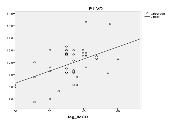
Figure 1: Correlation between peritumoral lymph vessel density with intratumoral mast cell density
Data of intratumoral lymph vessel density and intratumoral mast cell density was correlated in Pearson correlation test and statistical significance was found. (p< 0.05)
Table 8: Pearson correlation test between intratumoral lymph vessel density with intratumoral mast cell density
|
Characteristics |
Correlation coefficient(r) |
P value |
|
Intratumoral lymph vessel density and intratumoral mast cell density |
0.41 |
p< 0.05 |
Regression model shows a linear relationship between intratumoral lymph vessel density and intratumoral mast cell density.
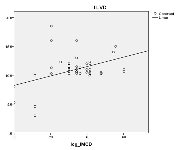
Figure 2: Correlation and regression between intratumoral lymph vessel density with intratumoral mast cell density.
Discussion
This cross-sectional study evaluated the correlation between intratumoral mast cell density (I-MCD), peritumoral mast cell density (P-MCD), intratumoral lymphatic vessel density (I-LVD), and peritumoral lymphatic vessel density (P-LVD), alongside the association of I-MCD and P-MCD with histological grades and molecular subtypes. The mean age of the 51 patients was 47.75 ± 12.79 years (range: 25-78 years), with the majority aged <50 years. This mean age is lower than that reported by another study (52 years) (5). However, the National Institute of Cancer Research and Hospital (NICRH) report of 5,255 patients similarly observed over 56% of cases occurring in women of reproductive age (15-44 years) (23). Age variations across studies reflect geographical, ethnic, lifestyle, and regional life expectancy differences. Tumor occurrence was higher in the left breast, mirroring broader literature on left-sided predominance and laterality ratio, including findings by another study (11). Infiltrating ductal carcinoma (IDC) was the predominant histological type (90.2%), followed by invasive lobular carcinoma (5.9%) and mixed types (3.9%), which aligns with a previous study (12). Grade 2 tumors predominated in this cohort (76.5%), consistent with reports by a few studies (13-15). In contrast, another study observed Grade 3 as the most frequent subtype (16). Most patients presented in N0 and N1 stages, agreeing with observations by some studies (17-19). Luminal tumors were the most common molecular subtype (55%), followed by triple-negative (31.4%) and HER2-enriched (13.7%), mirroring proportions reported by another observation (6). I-MCD correlated significantly with higher tumor grades, similar to reports by another study (20). Conversely, another study observed higher mast cell counts in Grade 1 tumors, highlighting intratumoral heterogeneity (14). P-MCD showed no correlation with tumor grade. I-MCD correlated significantly with the luminal subtype, consistent with findings by other studies (6,16). P-MCD showed no molecular subtype correlation in this cohort, although few studies reported positive associations between P-MCD and luminal tumors (24-25). A significant positive correlation was identified between I-MCD and both I-LVD and P-LVD, aligning with another observation (22). Mechanistically, activated mast cells release lymphangiogenic factors (VEGF-C, VEGF-D, NRP-1, NRP-2) that stimulate VEGFR-3. Discrepancies noted in other studies (12) suggest lymphangiogenic signals may also originate from stromal fibroblasts, macrophages, and tumor cells. Peritumoral LVD progressively increased with advanced N-stage (from 8.63 ± 2.63 in N0 to 11.60 ± 2.25 in N3; p < 0.05), whereas I-LVD showed no such association. This supports findings by another study is that peritumoral lymphatics play the primary role in tumor cell intravasation and lymphatic spread (26). Consequently, lymphangiogenesis pathways represent key targets for anti-metastatic therapy, including monoclonal antibodies and receptor tyrosine kinase inhibitors targeting VEGFR, PDGFR, KIT, and FLT3 signaling axes.
Conclusion
Intratumoral mast cell density is associated with increased grades of the invasive breast carcinoma and correlated positively with increased lymph vessel proliferation (both intratumoral and peritumoral) which further causes increased lymph node metastasis, thus associated with poor prognosis. Intratumoral mast cell density, however, is also associated with luminal subtypes of invasive breast carcinoma which has favorable treatment outcome.
Acknowledgements
The authors thank all technical staff of the Histopathology Lab in the Department of Pathology at BSMMU in Dhaka for their assistance with specimen processing and laboratory testing.
Conflicts of Interest
The authors declare no conflicts of interest.
References
- Sung H, Ferlay J, Siegel RL, et al. Global Cancer Statistics 2020: GLOBOCAN estimates of incidence and mortality worldwide for 36 cancers in 185 countries. CA: A Cancer Journal for Clinicians71 (2021): 209-249.
- Bray F, Ferlay J, Soerjomataram I, et al. Global cancer statistics 2018: GLOBOCAN estimates of incidence and mortality worldwide for 36 cancers in 185 countries. CA: A Cancer Journal for Clinicians68 (2018): 394-424.
- (Duplicate of #1 — see Entry 1 above)
- Kumar N, Gulati HK, et al. Most recent strategies targeting estrogen receptor alpha for the treatment of breast cancer: Comprehensive review. Molecular Diversity25 (2021): 603-624.
- Pyla R, Potekar R, Patil V, et al. Quantitative mast cell analysis and hormone receptor study (ER, PR and HER2/neu) in invasive carcinoma of breast. Indian Journal of Pathology and Microbiology63 (2020): 200-204.
- Keser SH, Kandemir NO, Ece D, et al. Relationship of mast cell density with lymphangiogenesis and prognostic parameters in breast carcinoma. The Kaohsiung Journal of Medical Sciences33 (2017): 171-180.
- Sin S-H, Kim Y, Eason A, et al. KSHV latency locus cooperates with Myc to drive lymphoma in mice. PLoS Pathogens11 (2015): Article e1005135.
- Ribatti D & Ranieri G. Tryptase, a novel angiogenic factor stored in mast cell granules. Experimental Cell Research332 (2015): 157-162.
- Ran S, Volk L, Hall K, et al. Lymphangiogenesis and lymphatic metastasis in breast cancer. Pathophysiology17 (2010): 229-251.
- Johansson A, Rudolfsson S, Hammarsten P, et al. Mast cells are novel independent prognostic markers in prostate cancer and represent a target for therapy. The American Journal of Pathology177 (2010): 1031-1041.
- Goldblum JR, Lamps LW, McKenney J, et al. Rosai and Ackerman's surgical pathology (11th ed.). Elsevier (2017).
- Anderson WF, Chu KC, ChangS, & et al. Comparison of age-specific incidence rate patterns for different histopathologic types of breast carcinoma. Cancer Epidemiology, Biomarkers & Prevention13 (2004): 1128-1135.
- Divyarani MN, Harendra Kumar ML, Swaroop Raj BV, et al. Quantitative analysis of mast cells in invasive ductal carcinoma. International Journal of Current Research and Academic Review2 (2014): 91-96.
- (Duplicate of #12 — see Entry 12 above)
- Christiansen A & Detmar M. Lymphangiogenesis and cancer. Genes & Cancer2 (2011): 1146-1158.
- Glajcar A, Szpor J, Pacek A, et al. The relationship between breast cancer molecular subtypes and mast cell populations in tumor microenvironment. Virchows Archiv470 (2017): 505-515.
- Kandemir NO, Barut F, Bektas S, et al. Can lymphatic vascular density be used in determining metastatic spreading potential of tumor in invasive ductal carcinomas? Pathology & Oncology Research18 (2012): 253-262.
- Moorman AM, Vink R, Heijmans HJ, et al. The prognostic value of tumour-stroma ratio in triple-negative breast cancer. European Journal of Surgical Oncology38 (2012): 307-313.
- Majorini MT, Cancila V, Rigoni A, et al. Infiltrating mast cell-mediated stimulation of estrogen receptor activity in breast cancer cells promotes the luminal phenotype. Cancer Research80 (2020): 2311-2324.
- Fakhrjou A, Naghavi-Behzad M, Montazeri V, et al. The relationship between histologic grades of invasive carcinoma of breast ducts and mast cell infiltration. South Asian Journal of Cancer5 (2016): 5-7.
- Jana S, Ghosh S, De A, Pal S, et al. Quantitative analysis and comparison of mast cells in breast carcinomas and axillary lymph nodes. Clinical Cancer Investigation Journal6 (2017): 214-218.
- Raica M, Cimpean AM, Popovici RA, et al. Mast cells stimulate lymphangiogenesis in the gingiva of patients with periodontal disease. In Vivo29 (2015): 29-34.
- National Institute of Cancer Research & Hospital (NICRH). (n.d.). Department of Medical Oncology. Retrieved September 20, (2026).
- Dalton DK, & Noelle RJ. The roles of mast cells in anticancer immunity. Cancer Immunology, Immunotherapy61 (2012): 1511-1520.
- Dabiri S, Huntsman D, Makretsov N, et al. The presence of stromal mast cells identifies a subset of invasive breast cancers with a favorable prognosis. Modern Pathology17 (2004): 690-695.
- Raica M, Cimpean AM, Ceauşu R, et al. Interplay between mast cells and lymphatic vessels in different molecular types of breast cancer. Anticancer Research33 (2013): 957-963.