Argyropoulou E1*, Anagnostou K2, Karampinas P3, Kontovazainitis P2
1Department of Orthopaedics and Traumatology, University General Hospital of Patras, 26504 Patras, Greece
210th Orthopaedic Department, Hygeia Hospital, 15123 Athens, Greece
3Department of Orthopaedic Surgery, KAT General Hospital, National & Kapodistrian University of Athens, 14561 Athens, Greece
*Corresponding Author: Argyropoulou Evangelia, Department of Orthopaedics and Traumatology, University General Hospital of Patras, 26504 Patras, Greece.
Received: 03 July 2026; Accepted: 15 July 2026; Published: 24 July 2026
Background: Proximal hamstring avulsion injuries, although relatively uncommon, represent a severe form of hamstring pathology with significant functional implications, particularly when diagnosis and treatment are delayed. While conservative management may be appropriate for partial tears with minimal retraction in low-demand patients, complete avulsions involving all three tendons typically warrant surgical intervention to restore function and alleviate symptoms. This case highlights the importance of timely diagnosis and appropriate management, demonstrating that even in delayed presentations, surgical repair combined with a structured, individualized rehabilitation program can result in favorable functional outcomes. Early recognition and intervention remain critical to preventing long-term morbidity and optimizing recovery.
Case Summary: A 59-year-old male presented 3.5 months after a right proximal hamstring injury initially misdiagnosed as a hamstring sprain. Conservative treatment failed to improve symptoms. MRI demonstrated proximal avulsion of the biceps femoris and semitendinosus tendons with 2.5–3 cm retraction, associated with a high-grade semimembranosus musculotendinous injury. Surgical repair was performed using three suture anchors. Postoperatively, the patient followed a protected, progressive rehabilitation protocol. At early follow-up (2.5 months), the patient reported resolution of pain and improved function. At 1.5-year follow-up, he demonstrated full knee range of motion, absence of pain and return to normal daily and recreational activities without limitations.
Conclusion: Proximal hamstring avulsion involving the biceps femoris and semitendinosus tendons, associated with high-grade semimembranosus musculotendinous injury is rare but severe injury with significant functional consequences, particularly in active individuals. Although early surgical repair yields the best outcomes, delayed diagnosis often leads to late presentations. This case demonstrates that delayed surgical repair may provide early symptomatic and functional improvement in selected patients with persistent disability after failed conservative treatment. Longer follow-up is required to assess final functional recovery and return to activity. Timely recognition and individualized management are critical for optimizing outcomes.
Hamstrings tendon; Hamstrings avulsion; Tendon injury; Rehabilitation
Hamstrings tendon articles; Hamstrings avulsion articles; Tendon injury articles; Rehabilitation articles
1. Introduction
Hamstring injuries are very common and can measure up to 30% of lower extremity injuries [1]. Except for young athletes, those injuries are prevalent in people around the age of 45-59 years [2]. Hamstring Avulsion Injuries (PHAIs) represents the most severe and uncommon derivative of hamstring injuries with a prevalence of 9% [3]. Complete proximal hamstring avulsion is usually defined as detachment of all three tendons from the ischial tuberosity (biceps femoris, semitendinosus and semimembranosus). However, partial or two-tendon avulsions with significant retraction may also cause substantial functional limitation and may require surgical treatment [4,5]. Usually, the hamstrings are being injured during hip hyperflexion and knee extension [6]. Deciding on the best treatment is a complex process and involves the degree of retraction, partial or total tear, lesion chronicity, and associated symptoms. The literature supports the nonoperative treatment for PHAIs with partial tears with < 2cm of retraction or patients with low functional demand [7,8]. Delaying surgery leads to residual loss of function, persistent pain, weakness with delay in return to sports and late sciatic nerve palsy [9,10]. The purpose of this case report is to evaluate the functional outcome of a patient with delayed proximal hamstring avulsion involving two tendons with associated semimembranosus musculotendinous injury, who underwent surgical management and structured, protected and individualized physical therapy.
2. Case Presentation
A 59-year-old male presented to our outpatient clinic 3.5 months following an injury to the hamstring tendons of the right thigh, sustained while attempting to stabilize his heavily loaded motorcycle. Over the intervening period, he had been evaluated by multiple orthopedic specialists and was misdiagnosed with a hamstring sprain. Consequently, he underwent conservative management, including rest, cryotherapy, and physical therapy, with no clinical improvement.
Due to persistent symptoms, he underwent magnetic resonance imaging (MRI), which revealed a proximal avulsion of the biceps femoris and semitendinosus tendons with 2.5–3 cm retraction, along with a high-grade sprain of the semimembranosus at the musculotendinous junction (Figure 1).
Clinically, the patient reported persistent posterior thigh pain, especially after walking, and demonstrated a palpable defect near the ischial tuberosity. He exhibited marked weakness in prone knee flexion compared to the contralateral side. Although weight-bearing was possible, gait was altered in an effort to avoid simultaneous hip flexion and knee extension due to functional hamstring deficiency. Stair climbing was significantly impaired. On examination, the popliteal angle was increased relative to the unaffected side. The patient had no significant past medical history, and his family history was unremarkable.
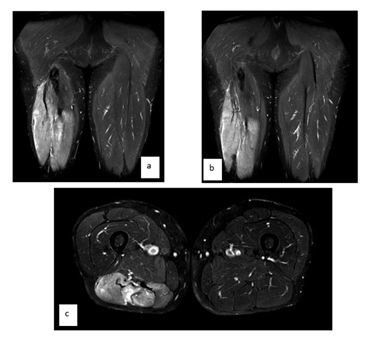
Figure 1: (a-b) Coronal T2-weighted fat-suppressed MRI demonstrating proximal avulsion of the biceps femoris and semitendinosus tendons with associated edema and hematoma in the posterior thigh, as well as asymmetry compared to the contralateral side. (c) Axial T2-weighted fat-suppressed MRI with edema in the posterior right thigh.
3. Surgical Technique
Surgical repair was indicated. Under general anesthesia, the patient was positioned prone. A transverse incision was made over the gluteal crease. After careful hemostasis using electrocautery, the gluteus maximus was retracted superiorly to protect the inferior gluteal neurovascular structures. The sciatic and posterior femoral cutaneous nerves were identified, tagged, and protected throughout the procedure.
Due to the chronicity of the injury, meticulous dissection was required to mobilize the tendons from surrounding scar tissue. Special attention was paid to preserving the sciatic nerve branches innervating the hamstring muscles. Tendon augmentation was not deemed necessary. Reattachment of the avulsed tendons was performed using three suture anchors at the ischial origin. One knotless suture anchor (Argo, CONMED corporation) and two all-suture anchors (TruShot™ with Y-Knot® All-In-One Soft Tissue Fixation System for Small Joint, CONMED Corporation) were used (Figure 2). The all-suture anchors were selected for their small footprint and shallow insertion depth, facilitating fixation in the ischial tuberosity while minimizing bone removal. This configuration allowed stable fixation with appropriate tendon tension while accommodating the chronic nature of the injury. Intraoperative dynamic assessment of the repair was conducted by gradually extending the knee under direct visualization and palpation to identify a safe range of motion for postoperative rehabilitation.
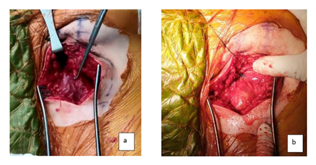
Figure 2: (a) Biceps femoris detachment from ischial tuberosity. (b) biceps femoris and semitendinosus reattachment with suture anchors.
4. Rehabilitation Protocol
Rehabilitation followed a structured, progressive protocol. During the first three weeks postoperatively, the patient remained in a hinged knee brace locked in full extension, with protected weight-bearing.
Gradual knee flexion was introduced thereafter under supervised physical therapy. At two months postoperatively, the patient achieved approximately 60o of knee flexion, progressing to 90o by three months.
Early rehabilitation focused on protected mobilization and active-assisted range of motion exercises, followed by gradual activation of the hamstring muscles. Unassisted movements against gravity were introduced in the intermediate phase.
Progressive resistance training was initiated at three months postoperatively. The patient returned to recreational sports activities at six months.
At 1.5-year follow-up, the patient demonstrated symmetric strength compared to the contralateral limb and no functional limitations in daily activities.
5. Discussion
Hamstring injuries are very common and can measure up to 30% of lower extremity injuries and 10-30% of all injuries in athletes [1,11]. Except for young athletes, those injuries are prevalent in people around the age of 45-59 years with older women being injured in activities of daily living, while men mostly during sports [2]. The injury can vary from a sprain of the muscle or musculotendinous junction to complete avulsions from the ischial tuberosity. Hamstring Avulsion Injuries (PHAIs) represents the most severe and uncommon derivative of hamstring injuries with a prevalence of 9% [3]. The definition of complete rupture is the avulsion of all three tendons, with first muscle to be injured being biceps femoris, followed by the semitendinosus and semimembranosus [4,5]. Hamstring injuries are at substantial risk of becoming chronic with reinjury rates ranging from 12 to 34% [12]. Usually, the hamstrings are being injured during hip hyperflexion and knee extension [6] and mostly during eccentric contractions and sports like dancing, judo, bull riding, soccer, rugby, waterskiin [13-16].
Multiple risk factors for PHAIs exists like previous hamstring injury, inadequate warm up, muscle fatigue, dehydration, decreased lower extremity flexibility, poor core stability, prior fluoroquinolone antibiotic use, and strength imbalances, with the previous injury being the most important one, as increases the risk up to 6 times [17].
Following hamstrings avulsion injury, the overall complication rate reaches 15%, with 10% local nerve [7,18] mostly from the sciatic nerve due to its close proximity to the hamstring origin at the ischial tuberosity [19]. There also is 1.7% of partial re-rupture in the cases with delayed intervention [20,21].
Proximal hamstring avulsions (PHAs) represent a challenging injury, particularly in active and athletic populations and involves the degree of retraction, type of tear (partial or total), lesion chronicity, and associated symptoms. The timing of intervention plays a critical role in determining both clinical outcomes and long-term function. Management can be broadly divided into early surgical intervention, delayed or late, and nonoperative treatment. Early surgical intervention is defined as treatment within 6 weeks of injury, while "delayed" and "late" interventions are those performed between 6 weeks to 6 months and beyond 6 months, respectively. Early surgery is consistently associated with superior clinical outcomes, reduced complication rates, and faster return to sport. In a large series by Subbu et al. [20], 96.4% of patients returned to sport, with early surgery enabling a return to play in an average of 16 weeks, which is 9 and 13 weeks faster than delayed and late interventions, respectively. Delayed diagnosis and intervention increase morbidity and lead to greater proximal hamstring complex retraction, embedding of the sciatic nerve, and obliteration of tissue planes. These changes make the procedure more technically challenging and increase the risk of sciatic nerve injury. Early surgery, by contrast, facilitates clear tissue identification, tension-free reattachment, and minimizes the need for postoperative bracing, allowing early mobilization and rehabilitation.
Nonoperative treatment like ice, physical therapy, and occasionally platelet-rich plasma injections, is generally reserved for lower-demand patients or partial tears with <2cm retraction [22]. Patients typically follow a rehabilitation timeline of minimum 4 months [6,21]. However, nonsurgical management is associated with suboptimal outcomes: up to 30% fail to return to their preinjury level of sport, and nearly 50% regret not undergoing surgery [8]. Persistent pain, weakness, poor endurance, and functional limitations are common, although some demonstrate isolated hamstring strength deficits without significant impairment on functional tests [13,23]. Nevertheless, long-term satisfaction and return to sport rates remain significantly lower than in operatively treated cohorts. Notably, 40% of non-surgically treated patients eventually require surgical intervention due to ongoing symptoms [24]. Lefevre et al. [21], in a long-term follow-up of 32 patients, confirmed that surgical management results in significantly improved functional outcomes, satisfaction, and return to sport rates (68.4% vs. 46.9%) as measured by the Perth Hamstring Assessment Score (PHAS).
Operative repair is indicated for complete three-tendon avulsions, osseous avulsions with >2 cm displacement, and partial tears that fail nonoperative management after at least 3 months [4,25]. While significantly retracted tears (>5 cm) have traditionally required open repair, endoscopic techniques have been successfully applied to cases with up to 15 cm of retraction [26]. Nonetheless, an open approach remains preferred for chronic or complex tears [25]. Bertiche et al. [22] recommend an open surgical approach with incision type guided by chronicity and retraction: a transverse buttock crease incision for acute repairs (<6 weeks), and a longitudinal incision for chronic cases. Both approaches can be used for tears with <5 cm retraction, though the longitudinal incision is the preferred default.
Numerous studies support surgical treatment for active individuals with complete proximal hamstring injuries. Patients report significantly better satisfaction, pain relief, strength, and endurance compared to those managed nonoperatively. Importantly, despite better subjective outcomes, single-leg hop distance and isokinetic torque measures may not differ significantly between groups. Nonetheless, the overall functional recovery, particularly return to sport and quality of life, strongly favors surgical management when performed early [9,23,27].
6. Conclusion
Delayed proximal hamstring avulsion injuries may be challenging because of scar formation, tendon retraction and proximity to the sciatic nerve. In this case, delayed surgical repair of a two-tendon proximal hamstring avulsion with associated semimembranosus musculotendinous injury resulted in sustained pain relief and functional recovery at 1.5-year follow-up. Nonetheless, surgical treatment, even when delayed, can still offer meaningful functional recovery and symptom relief, particularly when performed with appropriate technique and patient selection. Awareness of injury patterns, timely diagnosis, and individualized treatment planning remain essential to optimize outcomes in patients with proximal hamstring avulsion injuries.
7. Acknowledgements
Author contributions: Argyropoulou E designed research; Argyropoulou E and Kontovazainitis P performed research; Karampinas P contributed analytic tools; Anagnostou K analyzed data; Argyropoulou E wrote the paper and; Kontovazainitis P acquisition funding. All authors have read and agreed to the published version of the manuscript.
Informed consent statement: Informed consent was obtained from the patient for being included in the study. The patient has signed a written consent form.
Conflict-of-interest statement: The authors declare that they have no conflict of interest.
References
- Maniar N, Carmichael DS, Hickey JT, et al. Incidence and prevalence of hamstring injuries in field-based team sports: a systematic review and meta-analysis of 5952 injuries from over 7 million exposure hours. Br J Sports Med 57 (2023): 109-116.
- Irger M, Willinger L, Lacheta L, et al. Proximal hamstring tendon avulsion injuries occur predominately in middle-aged patients with distinct gender differences: epidemiologic analysis of 263 surgically treated cases. Knee Surg Sports Traumatol Arthrosc 28 (2020): 1221-1229.
- Koulouris G, Connell D. Evaluation of the hamstring muscle complex following acute injury. Skeletal Radiol 32 (2003): 582-589.
- Cohen SB, Rangavajjula A, Vyas D, et al. Functional results and outcomes after repair of proximal hamstring avulsions. Am J Sports Med 40 (2012): 2092-2098.
- Wood DG, Packham I, Trikha SP, et al. Avulsion of the proximal hamstring origin. J Bone Joint Surg Am 90 (2008): 2365-2374.
- Hofmann KJ, Paggi A, Connors D, et al. Complete avulsion of the proximal hamstring insertion: Functional outcomes after nonsurgical treatment. J Bone Joint Surg 96 (2014): 1022-1025.
- Bodendorfer BM, Curley AJ, Kotler JA, et al. Outcomes After Operative and Nonoperative Treatment of Proximal Hamstring Avulsions: A Systematic Review and Meta-analysis. Am J Sports Med 46 (2018): 2798-2808.
- Piposar JR, Vinod AV, Olsen JR, et al. High-Grade Partial and Retracted (<2 cm) Proximal Hamstring Ruptures: Nonsurgical Treatment Revisited. Orthop J Sport Med 5 (2017): 2325967117692507.
- Birmingham P, Muller M, Wickiewicz T, et al. Functional outcome after repair of proximal hamstring avulsions. J Bone Joint Surg Am 93 (2011): 1819-1826.
- Takami H, Takahashi S, Ando M. Late sciatic nerve palsy following avulsion of the biceps femoris muscle from the ischial tuberosity. Arch Orthop Trauma Surg 120 (2000): 352-354.
- Chang JS, Kayani B, Plastow R, et al. Management of hamstring injuries: current concepts review. Bone Joint J 102-B (2020): 1281-1288.
- Malliaropoulos N, Isinkaye T, Tsitas K, et al. Reinjury after acute posterior thigh muscle injuries in elite track and field athletes. Am J Sports Med 39 (2011): 304-310.
- Chakravarthy J, Ramisetty N, Pimpalnerkar A, et al. Surgical repair of complete proximal hamstring tendon ruptures in water skiers and bull riders: a report of four cases and review of the literature. Br J Sports Med 39 (2005): 569-572.
- Coughlin RP, Kay J, Shanmugaraj A, et al. Return to Sport After Surgical Management of Proximal Hamstring Avulsions: A Systematic Review and Meta-analysis. Clin J Sport Med 30 (2020): 598-611.
- Brooks JHM, Fuller CW, Kemp SPT, et al. Incidence, risk, and prevention of hamstring muscle injuries in professional rugby union. Am J Sports Med 34 (2006): 1297-1306.
- Sonnery-Cottet B, Archbold P, Thaunat M, et al. Proximal hamstring avulsion in a professional soccer player. Orthop Traumatol Surg Res 98 (2012): 928-931.
- Engebretsen AH, Myklebust G, Holme I, et al. Intrinsic risk factors for hamstring injuries among male soccer players: a prospective cohort study. Am J Sports Med 38 (2010): 1147-1153.
- Lawson JJ, Abraham EA, Imbergamo CM, et al. Systematic Review of Complications Associated With Proximal Hamstring Tendon Repair. Orthop J Sport Med 11 (2023): 23259671231199092.
- Miller SL, Webb GR. The proximal origin of the hamstrings and surrounding anatomy encountered during repair. Surgical technique. J Bone Joint Surg Am 90 Suppl 2 (2008): 108-116.
- Subbu R, Benjamin-Laing H, Haddad F. Timing of Surgery for Complete Proximal Hamstring Avulsion Injuries: Successful Clinical Outcomes at 6 Weeks, 6 Months, and After 6 Months of Injury. Am J Sports Med 43 (2015): 385-391.
- Lefèvre N, Moussa MK, El Otmani L, et al. Surgical Treatment of Proximal Hamstring Avulsion Injuries Compared With Nonsurgical Treatment: A Matched Comparative Study With a Mean Follow-up of >4 Years From the Proximal Hamstring Avulsion Surgery Cohort Study. Am J Sports Med 52 (2024): 2718-2727.
- Bertiche P, Mohtadi N, Chan D, et al. Proximal hamstring tendon avulsion: State of the art. J ISAKOS 6 (2021): 237-246.
- Harris JD, Griesser MJ, Best TM, et al. Treatment of proximal hamstring ruptures - a systematic review. Int J Sports Med 32 (2011): 490-495.
- Castillo-de-la-Peña J, Wong I. Endoscopic Repair of Proximal Hamstring Insertion With Sciatic Nerve Neurolysis. Arthrosc Tech 11 (2022): e789-e795.
- Yetter TR, Halvorson RT, Wong SE, et al. Management of Proximal Hamstring Injuries: Non-operative and Operative Treatment. Curr Rev Musculoskelet Med 17 (2024): 373-385.
- Maier J, Clark E, Laskovski J. Repair of Retracted Hamstring Tears with Hamstring Pulley Technique and Inferomedial Portal. Arthrosc Tech 12 (2023): e2273-e2280.
- Konan S, Haddad F. Successful return to high level sports following early surgical repair of complete tears of the proximal hamstring tendons. Int Orthop 34 (2010): 119-123.