Polma R1,2*, Bonitz L1,2, Bicsak A1,2, Soemmer C1,2, Schulze Wenning A1,2
1General Hospital Dortmund, Oral and Maxillofacial Surgery, Muensterstrasse 240, 44145 Dortmund, Germany
2University Witten Herdecke, Alfred-Herrhausen-Strasse 50, 58454 Witten, Germany
*Corresponding Author: Richard Polma, Department of Oral and Maxillofacial Surgery, University Witten-Herdecke
Hospital Dortmund, Münsterstr. 240, 44145 Dortmund, Germany.
Received: 02 September 2026; Accepted: 10 September 2026; Published: 15 September 2026
We present a case of 14-year-old female with the aneurysmal bone cyst of the palate, compare the imaging possibilities and discuss the therapy options.
Aneurysmal bone cyst, Literature review
Aneurysmal bone cyst articles; Literature review articles
Introduction
Aneurysmal bone cyst (ABC) is an expansive, destructive lesion of the bone which presents as nontender swelling. Histologically could be seen a mixture of fibroblasts, osteoid, histiocytes, osteoclast multinucleated giant cell and accompanied by high intraosseous pressure. It occurs most frequently in the second decade of life in the metaphyseal bone in children [1] , in the jaws is it rare [2]. Kaffe et al. summarize the 64 cases of ABC of the jaws with median age of 17, it occurred more often in the posterior mandible than maxilla (2,4:1), 53% were multilocular [3]. Guzmán et al. reported a destructive ABC by a 14 y.o. female of the maxillary sinus [4]. Richardson et al. analyzed 72 cases of the ABC in head and neck region: average age of the diagnosis was 19,1 years, the most often location was mandible and sinus [2]. Bataineh summarizes 30 cases of maxillary ABC without gender prevalence and estimate that the radiographic appearance is suggestive but not diagnostic [5].
The MRI remains the best imaging option despite not imaging a sublte bone erosion [6]. In our case, we compare the imaging possibilities – CT, MRI 1,5T and dental MRI 0,55T (Siemens MAGNETOM Free.Max Dental Edition, Siemens Healthineers, Forchheim, Germany) improved with artificial intelligence.
Case Report
A healthy 14-year-old female was referred to our clinic with swelling of the palate started 1 month ago. It was firstly treated as palatal abscess with incision und drainage by the dentist. After one week of non-healing wound was the patient sent from the ENT-doctor to our clinic. The patient takes no medication, has no allergy and no symptoms (pain, fever, anorexia). She underwent a labial upper frenectomy one month before our presentation.
The physical examination reveals a nontender, firm swelling of the hard and soft palate and small incision in the dorsal midline, slight hyperemic. The dental status was inconspicuous and there were no palpable cervical lymph nodes.
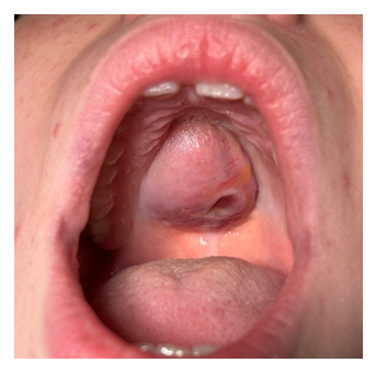
Figure 1: Clinical view.
It was performed a CT, MRI (1,5 Tesla) with gadolinium contrast and dental MRI scan (0,55 Tesla). It revealed a solid tumor mass in upper jaw, sharp demarcated, slight inhomogeneous. As it could be observed on figures 2-5, the CT scan can´t show the soft tissue situation sufficiently and it is suitable just for the bone resorption. The defect was 23 x 22 x 26mm. The MRI scan (1,5T) with gadolinium shows detailed the soft tissue defect 24 x 21 x 26mm. The new dental MRI (0,55T) shows the tumor with a very good quality without the necessary contrast enhancement and size of 22 x 20 x 25mm.
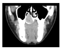
Figure 2: CT scan
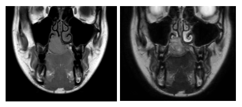
Figure 3,4: MRI 1,5T T1 and T2.
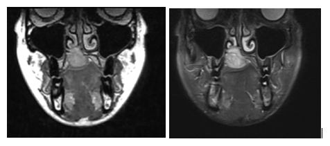
Figures 5,6: Dental MRI 0,55T T1 and T2.
After open biopsy, the tumor was removed in general anaesthesia. The cyst was removed and the defect was filled with iliac bone and covered with dressing plate on palatal mucosa. Postoperative histology confirms the aneurysmal bone cyst. After 10 days, the sutures were removed and the patient reported no problems.
Discussion
On the CT scan, we can identify the bone erosion but the structure of the tumor is unrecognizable. In contrast, the MRI sequences revealed also the inner structure, esp. T2 sequences. The dental MRI without enhancement shows very similar results to conventional 1.5T with enhancement.
Therapy options have been discusses. The gold standard remains the surgical curettage with adjuvant burring and eventual bone grafting. Another option is a sclerotherapy with polidocanol. Large inoperable lesion could treat with denosumab but it should lead to severe hypercalcemia [7]. It was also described a percutaneous doxycyklin injection in the extracranial skeleton [8] and arterial embolization of hard reachable location in combination with scletotherapy [9]. The recurrence of head and neck ABC is underreported because of rare occurrence, Richardson estimates 5,6% [2], Yu in contrast to 25% [10].
Another diagnosis includes a pleomorphic adenoma of the maxilla in transition zone between soft and hart palate [11] and couple of pediatric cases have been reported [3,5,6] with slightly higher occurrence by girls than boys [12]. It should be excluded also dental abscess, chondroblastoma, simple bone cyst, fibrous dysplasia, nonossifying fibroma, giant cell granuloma, osteosarcoma [7] or lymphoma.
Every nonhealing lesion should awake a suspicion of malignancy. Swelling of the palate could be consider as odontogenic (dental abscess, nasopalatinal cyst), but also (semi)malignacy as pleomorphic adenom, mucoepidermoid carcinoma, adenocarcinoma or lymphoma. The biopsy should be done in any case.
Resume
As advised above, the dental MRI 0.55T demonstrated sufficient tissue contrast to characterize the internal cystic architecture comparable quality with conventional 1,5 Tesla MRI. It suggests that low-field specialized systems can be a viable and accessible alternative for aneurysmal bone cyst imaging in the maxillofacial region.
Consent
Consent to publish the case report was obtained.
Conflicts of Interest
There is no conflict of interest.
Acknowledgements
This research received funding from the internal grant program (project IFF 2026-45) of the Faculty of Health at Witten/Herdecke University, Germany.
References
- Semenova LA, Malakhov OA, Zherdev KV. Aneurysmal bone cyst in children. Arkh Patol 72 (2010): 55-58.
- Richardson J, Litman E, Stanbouly D, et al. Aneurysmal Bone Cyst of the Head & Neck: A Review of Reported Cases in the Literature. J Stomatol Oral Maxillofac Surg 123 (2022): 59-63.
- Kaffe I, Naor H, Calderon S, et al. Radiological and Clinical Features of Aneurysmal Bone Cyst of the Jaws. Dentomaxillofac Radiol 28 (1999): 167-172.
- Guzmán G, Baeza O A, Araya O J, et al. Aneurysmal bone cyst of the maxilla. Report of one case. Rev Med Chil 133 (2005): 1355-1360.
- Bataineh AB. Aneurysmal Bone Cysts of the Maxilla: A Clinicopathologic Review. J Oral Maxillofac Surg 55 (1997): 1212-1216.
- Alsufyani NA, Altowaijri AA, Alshehri BM, et al. Systematic Review of Clinical and Radiographic Signs of Pediatric Pleomorphic Adenoma of Minor Salivary Glands. J Contemp Dent Pract 22 (2021): 1063-1068.
- van Geloven TPG, van de Sande MAJ, van der Heijden L. The Treatment of Aneurysmal Bone Cysts. Curr Opin Pediatr 35 (2023): 131.
- Shiels WE, Mayerson JL. Percutaneous Doxycycline Treatment of Aneurysmal Bone Cysts with Low Recurrence Rate: A Preliminary Report. Clin Orthop 471 (2013): 2675-2683.
- Bavan L, Wijendra A, Kothari A. Efficacy of Treatment Interventions for Primary Aneurysmal Bone Cysts: A Systematic Review. Bone Jt Open 2 (2021): 125-133.
- Yu L, Zhou J, Shi J. Clinicopathology and Recurrence Analysis of 44 Jaw Aneurysmal Bone Cyst Cases: A Literature Review. Front Surg 8 (2021).
- Waldron CA, el-Mofty SK, Gnepp DR. Tumors of the Intraoral Minor Salivary Glands: A Demographic and Histologic Study of 426 Cases. Oral Surg Oral Med Oral Pathol 66 (1988): 323-333.
- Daniels JSM, Ali I, Bakri IMA, et al. Pleomorphic Adenoma of the Palate in Children and Adolescents: A Report of 2 Cases and Review of the Literature. J Oral Maxillofac Surg 65 (2007): 541-549.