Talia Fradkin, Charles Carpenter, Angela Chen, Alexander Fethiere, Diego Avila Lessa Garcia*
University of Florida, Gainesville, Florida
Corresponding Author: Diego Avila Lessa Garcia, University of Florida, Gainesville, Florida
Received: 21 September 2026; Accepted: 28 September 2026; Published: 16 October 2026
Osteosarcoma is the most common primary malignant bone tumor in children and adolescents and most frequently arises in metaphysis of the long bones, particularly around the knee. Imaging has a central role throughout the clinical course, from initial diagnosis and staging to assessment of treatment response, surgical planning, and postoperative surveillance. We present the case of an 11-year-old girl with distal femoral osteosarcoma treated with neoadjuvant chemotherapy, wide surgical resection, and Van Nes rotationplasty. Initial radiographs demonstrated an aggressive mixed lytic and sclerotic metaphyseal lesion with cortical destruction and aggressive periosteal reaction. Magnetic resonance imaging demonstrated extensive intramedullary involvement with cortical breakthrough and a large extraosseous soft-tissue component, without neurovascular encasement or definite intra-articular extension. Whole-body bone scintigraphy and chest imaging demonstrated no evidence of additional disease. Following neoadjuvant chemotherapy, MRI demonstrated decreased soft-tissue enhancement and increased areas of nonenhancing tissue. Histopathologic examination of the resection specimen demonstrated greater than 90% tumor necrosis. Van Nes rotationplasty was selected as a durable limb-salvage reconstruction, with the primary goal of maximizing long-term mobility in this skeletally immature patient. Postoperative radiographs demonstrated satisfactory reconstruction and progressive osseous consolidation. At more than 5 years of follow-up, the patient remains free of local recurrence and distant metastatic disease. This case illustrates the complementary contribution of radiography, bone scintigraphy, MRI, and postoperative imaging in pediatric distal femoral osteosarcoma and demonstrates the role of imaging in following the patient from initial diagnosis through long-term surgical reconstruction.
Osteosarcoma; Distal femur; Pediatric oncology; Magnetic resonance imaging; Multimodality imaging; Rotationplasty; Limb salvage; Chemotherapy response
Osteosarcoma articles; Distal femur articles; Pediatric oncology articles; Magnetic resonance imaging articles; Multimodality imaging articles; Rotationplasty articles; Limb salvage articles; Chemotherapy response articles
Introduction
Osteosarcoma is the most common primary malignant bone tumor in children and adolescents, with a peak incidence during the adolescent growth spurt. The tumor most frequently arises in the metaphysis of long bones, with the distal femur representing one of the most common sites of involvement [1,2]. Modern treatment combines multiagent chemotherapy with complete surgical resection and requires close coordination among oncology, surgery, pathology, and radiology [3,4]. Imaging is central to this multidisciplinary process. Radiographs provide an initial assessment of bone destruction, osteoid matrix, cortical involvement, and periosteal response, whereas MRI is essential for local staging and surgical planning. MRI can define the longitudinal intramedullary extent of tumor, cortical breakthrough, extraosseous extension, joint involvement, and the relationship of the tumor to major neurovascular structures [5-8]. Systemic imaging is also required to evaluate for pulmonary metastases and additional skeletal disease, while careful assessment for skipping lesions is important because their presence may alter staging and treatment [9,10]. Following neoadjuvant chemotherapy, imaging may provide indirect evidence of treatment response through changes in tumor size, signal characteristics, enhancement, or diffusion. Nevertheless, histopathologic tumor necrosis remains the reference standard for treatment response assessment [11,12]. The subsequent surgical reconstruction is particularly challenging in skeletally immature patients. Van Nes rotationplasty provides a durable limb-salvage option in selected patients and allows the ankle to function as a mechanical knee following rotation of the distal limb segment [13-15]. We describe an 11-year-old girl with distal femoral osteosarcoma treated with neoadjuvant chemotherapy, wide resection, and Van Nes rotationplasty. The imaging findings are presented sequentially, emphasizing the contribution of each modality to diagnosis, staging, treatment assessment, surgical planning, and postoperative evaluation. The patient has remained free of local recurrence and distant disease for more than 5 years after treatment.
Case Presentation
An 11-year-old previously healthy girl presented with a six-week history of progressive pain and swelling of the distal left thigh. There was no reported history of trauma, fever, or weight loss. Physical examination demonstrated localized tenderness and mild swelling over the distal femur without a neurovascular deficit.
Initial anteroposterior and lateral radiographs of the left knee demonstrated an aggressive mixed lytic and sclerotic lesion centered in the distal femoral metaphysis. The lesion had ill-defined margins and associated cortical destruction, with an aggressive periosteal reaction and areas of mineralization within the adjacent soft tissues. The overall appearance was highly suspicious for osteosarcoma.
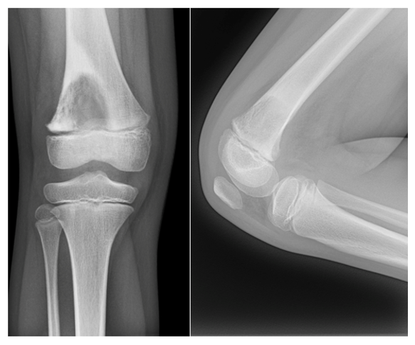
Figure 1: Initial radiographs. Anteroposterior and lateral radiographs demonstrate an aggressive mixed lytic and sclerotic lesion centered in the distal femoral metaphysis, with cortical destruction, aggressive periosteal reaction, and mineralization within the adjacent soft tissues.
Whole-body bone scintigraphy demonstrated intense radiotracer uptake corresponding to the distal femoral lesion without additional suspicious osseous foci. Staging chest imaging demonstrated no pulmonary metastatic disease.
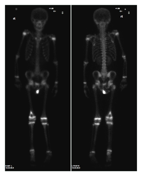
Figure 2: Whole-body bone scintigraphy. Anterior and posterior images demonstrate intense radiotracer accumulation at the distal femoral primary lesion without additional suspicious osseous foci.
MRI was subsequently performed for local staging. The lesion replaced the marrow of the distal femoral metaphysis and extended into the adjacent diaphysis. It demonstrated low-to-intermediate signal intensity on T1-weighted images and increased signal intensity on fluid-sensitive sequences. Cortical breakthrough was associated with a substantial extraosseous soft-tissue component. Despite the extensive local disease, the neurovascular bundle remained separated from the tumor without evidence of encasement. There was no definite intra-articular extension or additional skip lesion identified on the available MRI.
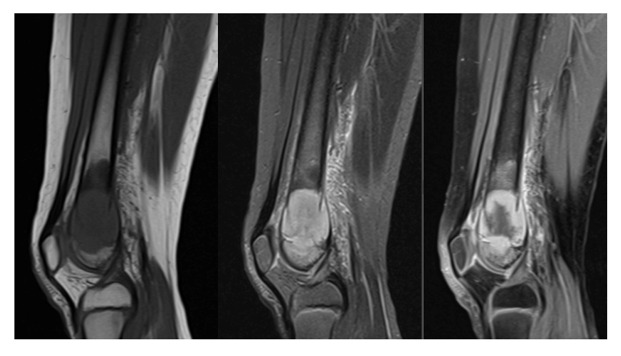
Figure 3: Sagittal MRI. Sagittal MR images demonstrate extensive marrow replacement involving the distal femoral metaphysis with extension into the diaphysis, cortical breakthrough, and a large extraosseous soft-tissue component.
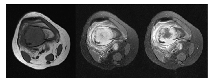
Figure 4: Axial MRI. Axial MR images demonstrate the extensive extraosseous component and heterogeneous tumor signal. The neurovascular structures remain separated from the tumor without evidence of encasement.
Image-guided core needle biopsy confirmed high-grade conventional osteoblastic osteosarcoma. The patient subsequently underwent neoadjuvant multiagent chemotherapy according to the MAP protocol. Following chemotherapy, follow-up MRI demonstrated reduction in the extraosseous soft-tissue component, decreased enhancement, and increased areas of nonenhancing tissue, findings considered compatible with treatment effect. Histopathologic examination after resection subsequently demonstrated greater than 90% tumor necrosis. Following neoadjuvant chemotherapy, the patient underwent wide resection of the distal femoral tumor. Given her young age and skeletal immaturity, Van Nes rotationplasty was selected as the reconstructive procedure. In this case, the primary objective was to provide durable functional reconstruction with the potential to maximize long-term mobility and independence. The distal limb segment was rotated approximately 180 degrees and reattached so that the ankle could function as a mechanical knee.
Postoperative Imaging and Long-Term Follow-up
Immediate postoperative radiographs demonstrated the expected reconstructed anatomy following wide tumor resection and Van Nes rotationplasty, with internal fixation and satisfactory alignment. Subsequent radiographic follow-up demonstrated progressive osseous bridging and consolidation of the reconstructed segment, with maintained alignment and eventual osseous union.
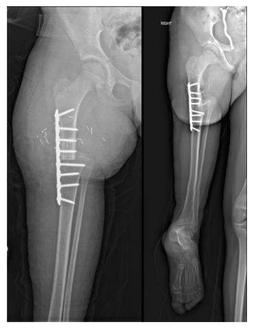
Figure 5: Postoperative radiographs following Van Nes rotationplasty. Radiographs demonstrate the reconstructed limb after wide resection of the distal femoral tumor and rotationplasty, with internal fixation and the expected altered anatomy. Serial follow-up demonstrated progressive osseous consolidation and stable reconstruction.
At more than 5 years after treatment, the patient remains free of local tumor recurrence and distant metastatic disease. The reconstruction has remained stable, with osseous integration and no reported oncologic evidence of disease during long-term follow-up.
Discussion
The diagnosis and management of osteosarcoma require integration of clinical, radiographic, nuclear medicine, MRI, pathologic, and surgical information. Although the diagnosis is established histologically, imaging determines the extent of disease and provides much of the anatomic information required for treatment planning. This case illustrates this process particularly well because the imaging findings can be followed from the initial aggressive lesion through local staging, treatment assessment, and postoperative reconstruction. The initial radiographs demonstrated several characteristic features of an aggressive osteogenic tumor, including mixed lytic and sclerotic components, cortical destruction, aggressive periosteal reaction, and soft-tissue mineralization. These findings appropriately prompted MRI for detailed local staging. Classic radiographic descriptions remain important because the pattern of bone destruction and osteoid matrix often establishes the initial diagnostic direction and helps distinguish osteosarcoma from other aggressive pediatric bone lesions [5]. MRI is particularly important because the apparent extent of osteosarcoma on radiographs does not necessarily reflect its full intramedullary or extraosseous extent. MRI provides superior assessment of tumor length, cortical penetration, soft-tissue extension, joint involvement, and the relationship to neurovascular structures [6-8]. In the present case, MRI demonstrated extensive local disease but preserved a plane between the tumor and the neurovascular bundle. This finding was directly relevant to surgical planning because successful limb salvage required complete tumor resection while maintaining an adequate margin around critical structures.
The absence of additional disease on staging evaluation was also relevant to the treatment strategy. Pulmonary metastases represent the most common site of distant spread at presentation, while skip lesions may occur within the same bone or an adjacent bone [9,10]. In this patient, whole-body bone scintigraphy showed no additional suspicious skeletal focus, and chest imaging showed no pulmonary metastases. MRI likewise did not demonstrate a skip lesion. Together, these findings supported a localized presentation amenable to multimodality treatment. The assessment of chemotherapy response represents another important role for imaging. In this case, the post-treatment MRI demonstrated decreased soft-tissue extension and enhancement with increasing areas of nonenhancing tissue. Such findings are compatible with treatment effect and are consistent with prior studies showing that MRI-derived changes can correlate with histologic response [11,12]. Nevertheless, imaging should not be interpreted as a replacement for histopathologic assessment. Histologic necrosis remains an important measure of response in osteosarcoma, and greater than 90% necrosis is generally considered a favorable histologic response [11,13]. In this patient, the resection specimen demonstrated greater than 90% tumor necrosis, providing pathologic confirmation of a substantial response to neoadjuvant chemotherapy. The choice of reconstruction represents an important feature of the case. Van Nes rotationplasty has a long history in the treatment of malignant tumors around the knee and remains a reconstructive option for selected skeletally immature patients [14-16]. The procedure converts the ankle into a functional knee by rotating the distal limb segment and attaching it to the remaining proximal femur or tibia after tumor resection. Although the resulting anatomy is unconventional, the reconstruction can provide a stable and durable limb with active control of the prosthetic knee.
In this patient, the principal consideration in selecting rotationplasty was the goal of maximizing long-term mobility. At 11 years of age, she had substantial remaining skeletal growth. A durable biologic reconstruction offered the potential for long-term function without dependence on repeated lengthening or revision of a growing endoprosthetic reconstruction. Long-term series of skeletally immature patients have demonstrated durable reconstructions and favorable functional outcomes, while gait studies have shown that patients can develop effective control of the reconstructed limb [17,18]. More recent studies have continued to document long-term functional and quality-of-life outcomes after rotationplasty [19,20]. The postoperative radiographs in this case are also important because the altered anatomy can initially be unfamiliar to radiologists. Recognition of the expected configuration is necessary to distinguish normal postoperative findings from complications. The immediate postoperative examination establishes the reconstructed anatomy and hardware position, while subsequent studies document osseous bridging, consolidation, and maintenance of alignment. In this patient, the long-term radiographic evolution was favorable, with progressive osseous union and stable reconstruction. The more than 5-year disease-free follow-up adds an important dimension to the case. The value of rotationplasty in pediatric sarcoma is not limited to limb preservation at the time of surgery; its rationale is to provide durable function over many years of expected activity and growth. The patient's absence of local recurrence or distant disease during prolonged follow-up, together with stable osseous reconstruction, illustrates the potential for an integrated oncologic and functional outcome.
From a radiologic perspective, the case emphasizes that imaging contributes throughout the entire treatment pathway rather than at a single diagnostic time point. Radiographs suggest the diagnosis, MRI establishes local extent and surgical relationships, systemic imaging evaluates metastatic disease, post-treatment MRI provides evidence of treatment effect, and serial radiographs document the evolution of the reconstruction. The complementary information provided by these modalities is particularly valuable when complex limb-salvage surgery is considered.
Conclusion
This case demonstrates the complementary role of multimodality imaging in the management of distal femoral osteosarcoma in a skeletally immature patient. Radiographs established the aggressive nature of the primary lesion, MRI defined its local extent and relationship to critical structures, bone scintigraphy and chest imaging supported the assessment of localized disease, and post-treatment MRI demonstrated changes compatible with chemotherapy response. Histopathologic examination confirmed greater than 90% tumor necrosis. The case also illustrates the radiologic evolution of Van Nes rotationplasty, from the immediate postoperative configuration through progressive osseous consolidation and long-term stable reconstruction. In this young patient, rotationplasty was selected primarily to provide a durable functional reconstruction and maximize long-term mobility. More than 5 years after treatment, the patient remains free of local recurrence and distant metastatic disease, with maintained osseous union. Sequential imaging therefore provides a visual and clinically relevant demonstration of the patient's course from diagnosis through long-term oncologic and reconstructive follow-up.
References
- Mirabello L, Troisi RJ, Savage SA. Osteosarcoma incidence and survival rates from 1973 to 2004: data from the Surveillance, Epidemiology, and End Results Program. Cancer 115 (2009): 1531-1543.
- Ritter J, Bielack SS. Osteosarcoma. Ann Oncol 21 (2010): vii320-vii325.
- Link MP, Goorin AM, Miser AW, et al. The effect of adjuvant chemotherapy on relapse-free survival in patients with osteosarcoma of the extremity. N Engl J Med 314 (1986): 1600-1606.
- Meyers PA, Schwartz CL, Krailo M, et al. Osteosarcoma: a randomized, prospective trial of the addition of ifosfamide and/or muramyl tripeptide to cisplatin, doxorubicin, and high-dose methotrexate. J Clin Oncol 23 (2005): 2004-2011.
- Murphey MD, Robbin MR, McRae GA, et al. The many faces of osteosarcoma. Radiographics 17 (1997): 1205-1231.
- Nguyen JC, Baghdadi S, Pogoriler J, et al. Pediatric osteosarcoma: correlation of imaging findings with histopathologic features, treatment, and outcome. Radiographics 42 (2022): 1196-1213.
- Panicek DM, Gatsonis C, Rosenthal DI, et al. CT and MR imaging in the local staging of primary malignant musculoskeletal neoplasms: report of the Radiology Diagnostic Oncology Group. Radiology 202 (1997): 237-246.
- Schima W, Amann G, Stiglbauer R, et al. Preoperative staging of osteosarcoma: efficacy of MR imaging in detecting joint involvement. AJR Am J Roentgenol 163 (1994): 1171-1175.
- Kaste SC, Pratt CB, Cain AM, et al. Metastases detected at the time of diagnosis of primary pediatric extremity osteosarcoma: imaging features. Cancer 86 (1999): 1602-1608.
- Sajadi KR, Heck RK Jr, Neel MD, et al. The incidence and prognosis of osteosarcoma skip metastases. Clin Orthop Relat Res (2004): 92-96.
- Bajpai J, Gamnagatti S, Kumar R, et al. Role of MRI in osteosarcoma for evaluation and prediction of chemotherapy response: correlation with histological necrosis. Pediatr Radiol 41 (2011): 441-450.
- Uhl M, Saueressig U, Koehler G, et al. Evaluation of tumour necrosis during chemotherapy with diffusion-weighted MR imaging: preliminary results in osteosarcomas. Pediatr Radiol 36 (2006): 1306-1311.
- Bielack SS, Kempf-Bielack B, Delling G, et al. Prognostic factors in high-grade osteosarcoma of the extremities or trunk: an analysis of 1,702 patients treated on neoadjuvant Cooperative Osteosarcoma Study Group protocols. J Clin Oncol 20 (2002): 776-790.
- Kotz R, Salzer M. Rotation-plasty for childhood osteosarcoma of the distal part of the femur. J Bone Joint Surg Am 64 (1982): 959-969.
- Cammisa FP Jr, Glasser DB, Otis JC, et al. The Van Nes tibial rotationplasty. A functionally viable reconstructive procedure in children who have a tumor of the distal end of the femur. J Bone Joint Surg Am 72 (1990): 1541-1547.
- Gottsauner-Wolf F, Kotz R, Knahr K, et al. Rotationplasty for limb salvage in the treatment of malignant tumors at the knee. A follow-up study of seventy patients. J Bone Joint Surg Am 73 (1991): 1365-1375.
- Hanlon M, Krajbich JI. Rotationplasty in skeletally immature patients. Long-term followup results. Clin Orthop Relat Res (1999): 75-82.
- Fuchs B, Kotajarvi BR, Kaufman KR, et al. Functional outcome of patients with rotationplasty about the knee. Clin Orthop Relat Res (2003): 52-58.
- Grimsrud C, Killen C, Murphy M, et al. Long-term outcomes of rotationplasty patients in the treatment of lower extremity sarcomas with cost analysis. J Clin Orthop Trauma 11 (2020): S149-S152.
- Filis P, Varvarousis D, Ntritsos G, et al. Rotationplasty outcomes assessed by gait analysis following resection of lower extremity bone neoplasms: a systematic review and meta-analysis. Bone Jt Open 4 (2023): 817-824.