Jan Bernardy PhD, DVM*, Rea Jarosova PhD
Veterinary Research Institute, Hudcova 296/70, 621 00 Brno-Medlánky, Czechia
*Corresponding Author: Jan Bernardy, Veterinary Research Institute, Hudcova 296/70, 621 00 Brno-Medlánky, Czechia.
Received: 22 July 2026; Accepted: 31 July 2026; Published: 07 August 2026
Background: Sequential hybrid protocols combining ablative and nonablative laser delivery have been proposed to achieve epidermal renewal and dermal remodeling within a single session, but are subject to operatordependent variability in timing and spatial registration between applications. ONEpulse technology addresses this limitation by integrating both wavelengths within a single co-registered pulse.
Methods: A single sow underwent two treatments on the ventral abdominal skin. One treatment used a sequential application of Er:Glass laser (1540 nm, 30 J/cm²) followed by Er:YAG laser (2940 nm, 18 J/cm²). The second treatment used ONEpulse technology to deliver both wavelengths within a single co-registered pulse at equivalent fluences. A baseline 6-mm punch biopsy was obtained, followed by two post-treatment biopsies at 2-month follow-up (one per treatment site). Samples were stained with hematoxylin and eosin, and epidermal area and collagen content were quantified using ImageJ software.
Results: Epidermal area increased from 54,654 μm² at baseline to 65,673 μm² after sequential treatment and to 89,090 μm² after ONEpulse treatment. The ONEpulse treatment was associated with a 35.7% greater epidermal area compared with the sequential treatment. Collagen content increased from 53.4% at baseline to 56.8% after sequential treatment, and to 75.2% after ONEpulse treatment. The ONEpulse treatment was associated with a 32.5% greater collagen content compared with the sequential treatment.
Conclusion: This pilot porcine study provides preliminary histological observations that ONEpulse delivery was associated with greater increases in epidermal area and collagen content compared with sequential Er:YAG and Er:Glass delivery at two months.
Laser resurfacing; Ablative systems; Non-ablative systems; Histology; ONEpulse technology
Laser resurfacing articles; Ablative systems articles; Non-ablative systems articles; Histology articles; ONEpulse technology articles
1. Introduction
Laser resurfacing remains a cornerstone of aesthetic and regenerative dermatology, offering a versatile approach to the management of photoaging, rhytides, dyschromias, and cicatricial deformities [1,3]. Current therapeutic platforms are broadly classified into ablative and non-ablative lasers, each governed by distinct laser-tissue interactions and producing complementary, yet anatomically discrete, biological effects [3].
Ablative lasers, including carbon dioxide (CO₂) and 2940-nm erbium:yttrium aluminium garnet (Er:YAG) systems, exploit the high absorption of laser energy by tissue water to induce rapid vaporization of the epidermis and superficial dermis [4,5]. This controlled injury initiates a wound-healing cascade involving fibroblast activation, extracellular matrix reorganisation, and reepithelialization, replacing a disorganized, photodamaged epidermis with a thickened, structurally uniform cellular architecture [6-11]. Clinically, these changes translate into improved surface texture, barrier restoration, and a more uniform complexion [3,11-13]. In contrast, non-ablative lasers, such as the 1540-nm erbium:glass (Er:Glass), generate microscopic coagulation zones within the dermis while preserving epidermal integrity, stimulating fibroblast-driven neocollagenesis and elastogenesis [14-16]. The resulting dermal remodeling is clinically associated with improved tissue firmness, enhanced elasticity, and reduction in rhytides [17,18].
Given the complementary nature of these mechanisms, hybrid protocols combining sequential ablative and non-ablative laser delivery have been proposed to achieve epidermal renewal and dermal remodeling within a single session [19,20]. Sequential delivery is, however, subject to operator-dependent variability in the timing and spatial registration between successive passes, which can influence the final clinical outcome [21,22].
ONEpulse technology was developed to address this limitation by integrating 2940-nm Er:YAG and 1540-nm Er:Glass wavelengths within a single coaxial optical pathway, delivering both laser energies to the same microscopic treatment zone in one pulse. In this configuration, the Er:Glass wavelength is delivered milliseconds before the Er:YAG wavelength within the same pulse. This differs from conventional sequential delivery, where the procedural workflow implies that the two wavelengths are separated by seconds to minutes. This intra-pulse energy delivery is hypothesized to create a photothermal synergy between the two wavelengths, potentially yielding a greater tissue remodeling response than sequential delivery.
The present study aimed to explore the histological effects of sequential Er:YAG and Er:Glass delivery compared with ONEpulse™ delivery in a porcine skin model, using epidermal area and dermal collagen content as outcome measures.
2. Materials and Methods
This exploratory pilot study was conducted at the Veterinary Research Institute (VRI, Brno, Czech Republic), working under Good Laboratory Practice (GLP) certification. A single animal (Sus scrofa f. domestica, sow (gilt), 75 kg, n = 1) served as the experimental model. The animal was housed at VRI for the duration of the study, with handling and welfare oversight provided by certified veterinary staff throughout.
General anesthesia was introduced by combination of dissociative anesthetics and alpha-2 agonists a TKX mixture, consisting of tiletamine (Zoletil, Virbac, FR), 4 mg/kg, ketamine (Narkamon, Bioveta a.s., CZ), 2 mg/kg and xylazine (Rometar, Bioveta a.s., CZ), 2 mg/kg, and continued by total intravenous anesthesia (TIVA) by propofol (Propofol 1% MCT/LCT Fresenius, 10 mg/kg/h) administered through an auricular vein cannula. Following anesthesia induction, the ventral abdominal skin was cleansed, shaved, and demarcated into three spatially distinct areas.
Two treatments were delivered using the EXOLASE ONE device (BTL Industries) to the ventral abdominal skin. The first treatment was the sequential treatment, which consisted of non-ablative Er:Glass (1540 nm, 30 J/cm²) followed immediately by ablative Er:YAG (2940 nm, 18 J/cm²) applied to the same area. The second treatment was the ONEpulse treatment, which delivered both wavelengths within a single spatially co-aligned pulse: Er:Glass (1540 nm, 30 J/cm²) followed immediately by Er:YAG (2940 nm, 18 J/cm²), with an intra-pulse temporal offset on the order of milliseconds. Each treatment lasted three minutes. Both treatment areas were spatially separated to prevent overlap.
Punch biopsies (6 mm) were collected at two time points. A single baseline biopsy was obtained from untreated ventral abdominal skin prior to any laser exposure. At two months post-treatment, one biopsy was collected from each treatment area, yielding one specimen per treatment. Biopsies were performed under aseptic conditions; the wounds were treated by surgical stapling. All specimens were fixed in 10% neutral-buffered formalin, embedded in paraffin, sectioned at 5 µm, and stained with hematoxylin and eosin (H&E) for assessment of tissue architecture.
Stained slides were digitized using a calibrated automated slide-scanning light microscope in bright-field mode, with spatial calibration confirmed using a stage micrometer. One representative section per specimen was selected for quantitative analysis using semiautomatic ImageJ software (version 1.54g, NIH, USA). Epidermal area was quantified by tracing the epidermal region in each section, with results expressed in µm². Collagen content was quantified using the Color Threshold tool in the Hue, Saturation, Brightness (HSB) color space. Images were calibrated to µm². HSB parameters were adjusted to selectively isolate eosinophilic (pink) regions representing the collagenous matrix, and the area of positive staining was expressed as a percentage of the total region of interest.
Given the single-animal, exploratory design of this study, no inferential statistical analysis was performed. All results are reported as absolute and relative differences between treatments.
3. Results
At baseline, the epidermal area measured 54,654 µm². Following sequential treatment, the epidermal area increased to 65,673 µm², representing an absolute increase of 11,019 µm² from baseline. Following ONEpulse treatment, the epidermal area increased to 89,090 µm², representing an absolute increase of 34,436 µm² from baseline. The ONEpulse treatment was associated with a 35.7% greater epidermal area compared with the sequential treatment.
Baseline collagen content accounted for 53.4% of the region of interest. Following sequential treatment, collagen content increased to 56.8%. Following ONEpulse treatment, collagen content increased to 75.2%. The ONEpulse treatment was associated with a 32.5% greater collagen content compared with the sequential treatment. See Figure 1. for the representative histologic visualisation of differences between the sequential and ONEpulse treatment.
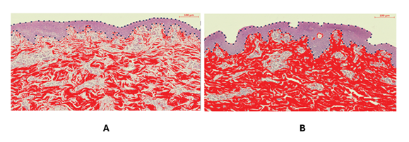
Figure 1: Representative histologic sections showing differences in epidermal area and collagen content between the sequential treatment (left) and the ONEpulse treatment (right). Staining with H&E; 40× magnification, scale bar 100 µm.
4. Discussion
This pilot study explored the histological effects of sequential Er:YAG and Er:Glass delivery compared with ONEpulse delivery in a porcine skin model, using epidermal area and dermal collagen content as outcome measures. Within this single-animal model, an increase in epidermal area was observed following both treatment settings, with the ONEpulse treatment associated with a 35.7% greater epidermal area compared with the sequential treatment. Similarly, an increase in collagen content was observed after both treatments, with the ONEpulse treatment associated with a 32.5% greater collagen content compared with the sequential treatment.
The increase in epidermal area observed following both treatments aligns with the well-documented biological response to ablative Er:YAG delivery, characterized by re-epithelialization and progressive epidermal thickening [6-10]. Similarly, the increase in collagen content observed following both treatments is consistent with the established biological response to non-ablative Er:Glass delivery, characterized by fibroblast-driven extracellular matrix remodeling [14-16]. Beyond these expected biological effects, however, the magnitude of the histological response differed markedly between the two treatments. This disparity supports the hypothesis of photothermal synergy between the two wavelengths, an interaction proposed to arise from their rapid consecutive intra-pulse delivery via ONEpulse technology. Two interconnected mechanisms are hypothesized as the basis of this synergy.
The first proposed mechanism relates to the epidermal response and arises directly from the delivery of the Er:Glass non-ablative wavelength immediately before the Er:YAG ablative wavelength. As the Er:Glass non-ablative wavelength penetrates through the epidermis into the dermis, it transiently deposits thermal energy within the epidermal tissue. This raises the local tissue temperature without causing ablation, pre-heating the tissue prior to Er:YAG delivery. This pre-heating brings the epidermal tissue closer to the vaporization threshold, and as a result, a greater proportion of the Er:YAG energy can be directed straight toward ablation rather than being spent on heating of unprepared tissue. This may produce a more consistent and uniform ablation across the treatment column, potentially accounting for the more pronounced epidermal area increase observed after the ONEpulse treatment. This proposed mechanism is based on prior physical and experimental studies suggesting that laser-induced temperature elevation just below the ablative threshold may alter thermal properties of the tissue, and thereby redirect the ablative energy [23-26].
The second proposed mechanism relates to the dermal collagen response. Because both the Er:Glass and Er:YAG lasers deposit thermal energy within the same tissue column, their concurrent contributions may lead to an accumulation of thermal energy within the dermis. This accumulation may elevate local tissue temperature beyond what the Er:Glass would generate independently, facilitating deeper heat diffusion into the dermis. This may increase the likelihood that the thermal stimulus reaches the fibroblast population, thereby promoting collagen remodeling, consistent with the greater increase in collagen content observed after ONEpulse treatment. This proposed mechanism is grounded in prior studies describing the heat accumulation that creates a thermal gradient within the tissue column [19,23,27].
Underlying both proposed mechanisms is the temporal and spatial unification of the two wavelengths within the same tissue column throughout treatment. In conventional sequential delivery, by contrast, the two wavelengths are applied in separate passes, introducing operator-dependent variability in both the timing and spatial registration between passes [21,22]. This variability may prevent the ablative and non-ablative energies from consistently acting on the same tissue column, thereby reducing the likelihood of the photothermal interactions described above. Furthermore, longer intervals between passes may allow the initial thermal elevation to dissipate before the second wavelength is applied, limiting their photothermal synergy. The broader principle that the timing of energy delivery can influence biological tissue response is not without precedent in the literature.
Indirect support has been reported in other energy-based systems. Harari et al. [28] found that simultaneous microwave energy delivery in liver tissue produced larger and more confluent ablation zones with higher peripheral temperatures compared with sequential delivery. While not directly comparable to laser resurfacing, this finding suggests that the timing of energy delivery may influence the biological tissue response [28]. The present findings can be further contextualized within the laser resurfacing literature. The epidermal area increase measured after the sequential treatment is broadly consistent with published histological data from ablative resurfacing studies at comparable follow-up intervals [29,30]. Similarly, the collagen increase observed after the sequential treatment falls within the range reported in non-ablative resurfacing studies at similar timepoints [31-33]. To our knowledge, however, no histological studies have directly compared ONEpulse technology with sequential ablative and non-ablative laser delivery, making the present study the first to provide preliminary comparative histological observations between these two approaches.
As hybrid laser resurfacing protocols combining ablative and non-ablative delivery continue to gain clinical interest for their ability to address multiple tissue compartments within a single session, there is growing demand for approaches that offer greater reproducibility and efficiency of outcomes [34,35]. From a clinical perspective, delivery systems that can achieve greater epidermal renewal and dermal collagen deposition within a single session, while reducing operator-dependent variability, may offer advantages in terms of procedural consistency, with the potential to translate into more reproducible improvements in skin texture, laxity, and scarring. Conceptually, if confirmed in adequately powered clinical studies, ONEpulse technology may provide a more reproducible approach to combined ablative and non-ablative laser resurfacing than conventional sequential delivery.
This study has notable strengths. First, porcine skin is widely regarded as a reliable preclinical surrogate for human dermatology due to its structural and physiological similarity to human skin, and has been used extensively in laser resurfacing research [36]. Second, the intra-animal design, in which both treatments were applied to the same subject under identical systemic and environmental conditions, controls for inter-subject biological variability and provides a direct within-subject comparison.
As with any exploratory pilot study, several limitations should be considered when interpreting these findings. The single-animal design, while appropriate for hypothesis-generating observations of this nature, precludes inferential statistical analysis and limits the generalizability of the results. Additionally, direct quantitative comparison of the findings in this study is limited by the methodological heterogeneity across studies. Furthermore, the observed differences between treatments may partly reflect local anatomical variation and should therefore be interpreted with appropriate caution. Similarly, a single 6-mm punch biopsy per treatment area may not fully capture the spatial distribution of the biological response across the entire treated zone. Another limitation is that the dermal collagen content was estimated from H&E-stained sections, rather than using a collagen-specific stain. In the future, studies should incorporate larger cohorts to enable inferential analysis and histological safety endpoints. Ultimately, studies in human subjects with validated clinical outcome measures will be necessary to determine whether the histological observations here are reproducible and translate into clinically meaningful differences between ONEpulse and sequential dual-wavelength delivery.
5. Conclusions
This pilot porcine study provides preliminary histological observations that ONEpulse delivery was associated with greater increases in epidermal area and collagen content compared with sequential delivery after two months post-treatment. These findings provide a histological basis for hypothesis generation and support further investigation.
Author Contributions:
J.B. acts as a clinical investigator for BTL. R.J. is an associate of Veterinary Research Institute.
Funding:
No funds, grants, or other support was received.
Data Availability Statement:
The data sets generated and/or analyzed during the study are not publicly available due to confidentiality reasons, but are available upon reasonable request from the corresponding author.
Acknowledgments:
The author has no acknowledgements to declare.
Conflicts of Interest:
All authors act as clinical investigators for BTL Industries. However, no funding or financial support was received for the research, authorship, or publication of this article.
Statement on welfare of animals:
All procedures were conducted in accordance with accepted standards for animal welfare and in compliance with applicable national regulations.
References
- Meaike JD, Agrawal N, Chang D, et al. Noninvasive Facial Rejuvenation. Part 3: Physician-Directed—Lasers, Chemical Peels, and Other Noninvasive Modalities. Semin Plast Surg 30 (2016): 143-150.
- Anderson RR, Donelan MB, Hivnor C, et al. Laser treatment of traumatic scars with an emphasis on ablative fractional laser resurfacing: consensus report. JAMA Dermatol 150 (2014): 187-193.
- Mirza HN, Mirza FN, Khatri KA. Outcomes and adverse effects of ablative vs nonablative lasers for skin resurfacing: A systematic review of 1093 patients. Dermatol Ther 34 (2021): e14432.
- Verma N, Yumeen S, Raggio BS. Ablative Laser Resurfacing. In: StatPearls. StatPearls Publishing (2025).
- Riggs K, Keller M, Humphreys TR. Ablative laser resurfacing: high-energy pulsed carbon dioxide and erbium:yttrium-aluminum-garnet. Clin Dermatol 25 (2007): 462-473.
- Guo H, Zhang X, Li H, et al. Dynamic panoramic presentation of skin function after fractional CO₂ laser treatment. iScience 26 (2023): 107559.
- Debeuf MEPH, Rauwenhoff MHP, van Geel M, et al. Biomolecular Changes Upon Ablative Laser Therapy of the Skin: A Scoping Review. Int J Dermatol.
- Orringer JS, Kang S, Johnson TM, et al. Connective tissue remodeling induced by carbon dioxide laser resurfacing of photodamaged human skin. Arch Dermatol 140 (2004): 1326-1332.
- Alberts B, Johnson A, Lewis J, et al. Epidermis and Its Renewal by Stem Cells. In: Molecular Biology of the Cell. 4th Edition. Garland Science (2002).
- Suter MM, Schulze K, Bergman W, et al. The keratinocyte in epidermal renewal and defence. Vet Dermatol 20 (2009): 515-532.
- Bao M, Gempeler M, Campiche R. Melanosome Transport and Processing in Skin Pigmentation: Mechanisms and Targets for Pigmentation Modulation. Int J Mol Sci 26 (2025): 8630.
- Gilchrest BA. A review of skin ageing and its medical therapy. Br J Dermatol 135 (1996): 867-875.
- Del Rosso JQ, Kircik L. Skin 101: Understanding the Fundamentals of Skin Barrier Physiology—Why is This Important for Clinicians? J Clin Aesthetic Dermatol 18 (2025): 7-15.
- Helbig D, Moebius A, Simon JC, et al. Nonablative skin rejuvenation devices and the role of heat shock protein 70: results of a human skin explant model. J Biomed Opt 15 (2010): 038002.
- Alam M, Hsu TS, Dover JS, et al. Nonablative laser and light treatments: histology and tissue effects—a review. Lasers Surg Med 33 (2003): 30-39.
- Orringer JS, Voorhees JJ, Hamilton T, et al. Dermal matrix remodeling after nonablative laser therapy. J Am Acad Dermatol 53 (2005): 775-782.
- Fournier N, Mordon S. Nonablative remodeling with a 1,540 nm erbium:glass laser. Dermatol Surg 31 (2005): 1227-1235.
- Dahan S, Lagarde JM, Turlier V, et al. Treatment of neck lines and forehead rhytids with a nonablative 1540-nm Er:glass laser: a controlled clinical study combined with the measurement of the thickness and the mechanical properties of the skin. Dermatol Surg 30 (2004): 872-879.
- Clementi A, Cannarozzo G, Guarino L, et al. Sequential Fractional CO₂ and 1540/1570 nm Lasers: A Narrative Review of Preclinical and Clinical Evidence. J Clin Med 14 (2025): 3867.
- Kim S, Cho KH. Clinical trial of dual treatment with an ablative fractional laser and a nonablative laser for the treatment of acne scars in Asian patients. Dermatol Surg 35 (2009): 1089-1098.
- Clementi A, Cannarozzo G, Guarino L, et al. Combined Laser Strategies for Scar Treatment: A Comprehensive Review of Synergistic Protocols. Bioengineering 12 (2025).
- Maheshwari A, Manstein N, Warner-Levy J, et al. Optimized Distance Holders Improve Precision in Fractional Laser Treatment. Lasers Surg Med 57 (2025): 618-624.
- Lukac M, Zorman A, Lukac N, et al. Characteristics of Non-Ablative Resurfacing of Soft Tissues by Repetitive Er:YAG Laser Pulse Irradiation. Lasers Surg Med 53 (2021): 1266-1278.
- Lukač M, Košir J, Žel T, et al. Influence of tissue desiccation on critical temperature for thermal damage during Er:YAG laser skin treatments. Lasers Surg Med 56 (2024): 107-118.
- El-Khalil H, Alzanina M, Lweesy K, et al. Investigation of laser pulsing parameters' importance in Er:YAG laser skin ablation: a theoretical study conducted via newly developed thermo-mechanical ablation model. Int J Hyperthermia 36 (2019): 612-623.
- Wang-Evers M, Blazon-Brown AJ, HaWissel L, et al. Assessment of a 3050/3200 nm fiber laser system for ablative fractional laser treatments in dermatology. Lasers Surg Med 54 (2022): 851-860.
- Ma J, Yang X, Sun Y, et al. Theoretical analysis on thermal treatment of skin with repetitive pulses. Sci Rep 11 (2021): 9958.
- Harari CM, Magagna M, Bedoya M, et al. Microwave Ablation: Comparison of Simultaneous and Sequential Activation of Multiple Antennas in Liver Model Systems. Radiology 278 (2016): 95-103.
- Jimenez LM, Oliver MA, Keyloun JW, et al. Laser-treatment of Hypertrophic Scar Induces Change to Epidermal Histoarchitecture Correlating to Improved Epidermal Barrier Function. J Burn Care Res 43 (2022): S147.
- Jimenez LM, Oliver MA, Keyloun JW, et al. Laser Treatment of Hypertrophic Scar in a Porcine Model Induces Change to Epidermal Histoarchitecture That Correlates to Improved Epidermal Barrier Function. J Burn Care Res 44 (2023): 758-768.
- Mordon S, Capon A, Creusy C, et al. In vivo experimental evaluation of skin remodeling by using an Er:Glass laser with contact cooling. Lasers Surg Med 27 (2000): 1-9.
- Fournier N, Dahan S, Barneon G, et al. Nonablative Remodeling: Clinical, Histologic, Ultrasound Imaging, and Profilometric Evaluation of a 1540 nm Er:Glass Laser. Dermatol Surg 27 (2001): 799.
- Patriota RCR. Estudo do laser Erbium Glass fracionado não ablativo no tratamento do fotoenvelhecimento cutâneo: avaliação clínica, histopatológica, microscopia eletrônica e imuno-histoquímica. Tese. Universidade de São Paulo (2013).
- Haykal D, Cartier H, Goldberg D, et al. Advancements in laser technologies for skin rejuvenation: A comprehensive review of efficacy and safety. J Cosmet Dermatol 23 (2024): 3078-3089.
- Pour Mohammad A, Gholizadeh Mesgarha M, Seirafianpour F, et al. A systematic review and meta-analysis of efficacy, safety, and satisfaction rates of laser combination treatments vs laser monotherapy in skin rejuvenation resurfacing. Lasers Med Sci 38 (2023): 228.
- Kong R, Bhargava R. Characterization of porcine skin as a model for human skin studies using infrared spectroscopic imaging. The Analyst 136 (2011): 2359-2366.