Mohamad Rakka*, Sorin Cimpean, Pieter Demetter, Ion Surdeanu, Abdelilah Mehdi
Department of General and Digestive Surgery, Iris Hospitals South, Free University of Brussels, Belgium
*Corresponding author: Mohamad Rakka, Department of General and Digestive Surgery, Iris Hospitals South, Free University of Brussels, Belgium
Received: 14 July 2021; Accepted: 20 July 2021; Published: 21 July 2021
Introduction: Ectopic liver tissue is a rare entity. It can be found in various organs of the body but the most common reported location is the gallbladder. The incidence is varying between 0.27% and 0.47%. Most of the cases are asymptomatic and diagnosed intra-operatively or during autopsy. We report a case of Gallbladder-Associated ectopic liver tissue that was encountered during a laparoscopic cholecystectomy.
Case presentation: A 40-year-old female patient was admitted to the hospital for laparoscopic cholecystectomy for symptomatic gallstones, intra-operatively we found an ectopic liver tissue attached to the gallbladder wall. Cholecystectomy was performed and the patient was discharged home the day after surgery in good condition. The histopathological examination of the specimen was confirmed to be an ectopic liver tissue without signs of malignant transformation.
Discussion: Ectopic liver tissue is a rare congenital anomaly. It is defined as the presence of liver tissue outside the native liver. It occurs due to a failure of embryological liver development and it can be found in various organs of the body but the most common reported location is the gallbladder. Ectopic liver tissue is found to be functionally handicapped and more vulnerable to carcinogens and it is strongly associated with the development of hepatocellular carcinoma.
Conclusion: Although it is a rare condition, ectopic liver tissue may pose a challenge to surgeons. When encountered, a surgical excision with histopathological examination is recommended because of its serious complications and the potential risk of malignant transformation.
Ectopic liver, Hepatocellular carcinoma, Gallbladder
1. Introduction
Ectopic liver tissue is a rare entity [1]. It occurs due to abnormal embryological development that is represented by the presence of liver tissue outside the native liver. It can be found in various organs of the body but the most common reported location is the gallbladder [2]. Its incidence is varying between 0.27% and 0.47% [3]. To date, the total number of reported cases is 91 cases with the first case reported in 1925 [4,5]. Most of the cases are asymptomatic and are diagnosed at laparoscopy, laparotomy or during an autopsy [6]. Few reported cases are diagnosed preoperatively [7,8]. Herein we report a rare case of gallbladder-associated ectopic liver tissue that was found incidentally during laparoscopic cholecystectomy for symptomatic gallstones and we discuss the presentation, diagnosis and the options of management of this rare entity.
Case 1
A 40-year-old female patient, presented to our outpatient clinic with a 2-month history of recurrent, postprandial right upper quadrant abdominal pain with occasional episodes of nausea. Her past medical history was only remarkable for hypertension. She is nonsmoker, occasionally alcoholic, with no known food and drug allergies. Physical examination revealed a negative Murphy sign. The laboratory evaluations of patient were in normal range. Abdominal ultrasound showed multiple gallstones without evidence of acute cholecystitis. So, the patient was scheduled for laparoscopic cholecystectomy. Intra-operatively, an about 2 cm mass-like liver tissue was encountered. It was attached to the gallbladder wal (Figure 1).
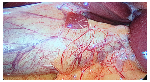
Figure 1: Laparoscopic view of gallbladder-associated ectopic liver tissue
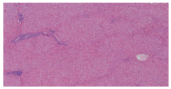
Figure 2: Microscopic view (Hematoxylin and Eosin staining, 20x) confirmed the ectopic liver tissue without pathological features
Cholecystectomy was performed using the standard procedure to include the mass and the specimen was removed and sent for histopathological examination. The postoperative course was uneventful and she was discharged home on the first postoperative day in good condition.The histopatological examination revealed this mass to be ectopic liver parenchyma with normal hepatocytes (Figure 2).
2. Discussion
Ectopic liver tissue is a rare congenital anomaly [1]. It is defined as the presence of liver tissue outside the native liver and is divided into four main types: Ectopic liver tissue that is not connected to the main liver, and is usually attached to the gallbladder wall (as it was in our case), microscopic liver tissue found in the gallbladder wall, a large accessory liver lobe connected to the main liver by a pedicle, small accessory liver lobe which is not connected to the main liver [9]. The incidence of ectopic liver tissue is varying between 0.27% and 0.47% [3]. It can be found in different body organs but the most common location is associated with the gallbladder [2]. Several theories have been proposed to explain the development of the ectopic liver tissue at different body sites; it may occurs as a result of regression or atrophy of the bridge to the main liver, or migration of ectopic liver during the embryological development [11]. Concerning the blood supply of gallbladder associated- ectopic liver tissue, three vascular patterns have been described in the literature: Artery arising from the cystic artery, vascular pedicle arising from the liver parenchyma, or in a form of a mesentery lying from the hepatic site to the ectopic liver tissue [12]. Therefore, if such tissue is encountered during laparoscopic cholecystectomy, the vascular pedicle should be dissected and clipped before the gallbladder dissection to prevent serious bleeding. Ectopic liver tissue-like the native liver tissue- usually has normal histological architecture and it is susceptible to the same pathologies such as fatty changes, cholestasis, hemosiderosis, cirrhosis and malignant transformation [13]. Several studies have been shown that the ectopic liver tissue is strongly associated with hepatocellular carcinoma [2-14]. It is theorized that the high incidence of HCC in the ectopic liver tissue is due to the defect in the functional structure which makes the ectopic liver tissue handicapped and more vulnerable to carcinogens [15]. The incidence of HCC is varying between 46% of ectopic liver tissue encountered outside the mother liver and 2.4% of gallbladder- associated ectopic liver tissue15. In fact, this difference in incidence is due to that the gallbladder- associated ectopic liver tissue occur later during the embryogenesis and therefore, it is well-differentiated than the other ectopic locations [16]. Gallbladder-associated ectopic liver tissue is rarely symptomatic and is usually encountered during laparoscopy (as it was in our case), laparotomy or autopsy [6]. Thus, preoperative diagnosis of ectopic liver tissue is rare and only a few cases have been reported [7,8]. However, when symptoms occur, the main complaint is recurrent right upper quadrant pain due to complications such as torsion, intra-peritoneal bleeding or obstruction [8]. Due its rarity, the small size and the lack of awareness, ectopic liver tissue is usually not recognized by the radiologist (as in the present case); however, it can appear as a soft tissues tumor on the imaging studies [17]. When it is suspected preoperatively, percutaneous biopsy should be avoided due to the risk of bleeding and malignancy [2]. Studies have recommended that when ectopic liver tissue is encountered, it should be excised and sent for histopathological examination because of its serious complications and the potential risk of malignant transformation [8]. Our specimen did not show any evidence of malignant degeneration when excised.
3. Conclusion
To conclude, although it is a rare entity, ectopic liver tissue can be found in different body organs but the most common location is associated with the gallbladder and it may pose a challenge to surgeons when encountered. Surgical excision is strongly recommended with proper histopathological examination to exclude malignant transformation.
References
- Kubota, Keiichi, et al. Ectopic hepatocellular carcinoma arising from pancreas: a case report and review of the literature. World Journal of Gastroenterology 31 (2007): 42-70.
- Martinez, Carlos Augusto Real, et al. Gallbladder-associated ectopic liver: A rare finding during a laparoscopic cholecystectomy. International journal of surgery case reports 4 (2013): 312-315.
- Arslan, Yusuf, et al. Rare entity: ectopic liver tissue in the wall of the gallbladder-a case report. World Journal of Clinical Cases 12 (2014): 924.
- Akbulut, Sami, et al. Ectopic liver tissue (choristoma) on the gallbladder: A comprehensive literature review. World Journal of Gastrointestinal Surgery 12 (2020): 534.
- Cullen, Thomas S. Accessory lobes of the liver: an accessory hepatic lobe springing from the surface of the gallbladder. Archives of Surgery11 (1925): 718-764.
- Avdaj, Afrim, et al. Case report of ectopic hepatic tissue, a rare finding during a laparoscopic cholecystectomy. International journal of surgery case reports68 (2020): 100-103.
- Lundy J, Johnson E, Edwards K, et al. Laparoscopic management of gallbladder associated ectopic liver 9 (2005): 485-487.
- Termos, Salah, et al. Gallbladder-associated symptomatic hepatic choristoma: Should you resect?. International journal of surgery case reports 41 (2017): 243-246.
- Collan Y, Hakkiluoto A, Hästbacka J. Ectopic liver. Annales Chirurgiae et Gynaecologiae 67 (1978): 18-25.
- Sakarya Y, Erhan H, Aydede E, et al. Ectopic liver(choristoma) associated with the gallbladder encountered during laparoscopic cholecystectomy: a case report, Surg Endosc 16 (2002): 11-16.
- Triantafyllidis L, Papapavlou N, et al. Ectopic liver tissue attached to the gallbladder wall: a case report, Cases J 2 (2009): 67-86.
- Bal S, Yilmaz B, Yavas, et al. A rare condition: ectopic liver tissue with itsunique blood supply encountered during laparoscopic cholecystectomy. Int J Surg Case Rep 9 (2015): 47-50.
- Dettmer, Matthias, Gieri C, et al. Alpha 1-antitrypsin retention in an ectopic live. Diagnostic pathology 6 (2011): 1-4.
- Liu KL, Ho MC, Chen PJ. Ectopic liver with hepatocellular carcinoma in the peritoneum. Am J Roentgenol 188 (2007): 206-217.
- Arakawa, Masahiro, et al. Propensity of ectopic liver to hepatocarcinogenesis: case reports and a review of the literature. Hepatology 29 (1999): 57-61.
- Griniatsos, John, Ali R, et al. Two cases of ectopic liver attached to the gallbladder wall. HPB 4 (2002): 191-194.
- Hamdani, Sandra D, Richard L. Ectopic liver simulating a mass in the gallbladder wall: imaging findings. American journal of roentgenology 162 (1994): 647-648.