Pankaj Yadav1, Alissa Hofmann1, Jay S. Mishra1, Sathish Kumar1,2,*
1Department of Comparative Biosciences, School of Veterinary Medicine, University of Wisconsin, Madison, WI 53706, USA.
2Department of Obstetrics and Gynecology, School of Medicine and Public Health, University of Wisconsin, Madison, WI 53792, USA.
*Corresponding author: Sathish Kumar, DVM, PhD Professor, Department of Comparative Biosciences and Obstetrics and Gynecology, University of Wisconsin-Madison, 2015 Linden Drive, Madison, WI 53706, USA.
Received: 06 October 2026; Accepted: 12 October 2026; Published: 15 October 2026
Aim:
This study investigated mechanisms underlying perfluorooctane sulfonate (PFOS)-associated fetal growth restriction. Specifically, we asked whether PFOS directly impairs transcellular transfer of substrates representing three major nutrient classes—glucose, long-chain fatty acid, and system A-mediated neutral amino acid transport—across the placental epithelial barrier, and examined the transporters involved.
Materials and Methods:
Syncytialized human BeWo b30 monolayers cultured on collagen-coated Transwell inserts were exposed to PFOS (10 and 50 μM) for 24 h. Transcellular transport of [³H]-2-deoxy-D-glucose, [1-¹?C]-linoleic acid, and the system A-specific substrate L-[¹?C]MeAIB was quantified. Transporter expression was assessed by digital PCR and Western blot. Timed-pregnant Sprague-Dawley rats received PFOS (10 or 50 mg/L, corresponding to 1.2 and 6.1 mg/kg/day) in drinking water from gestational day (GD) 4 to 20 (n = 5 dams/group). Maternal intake and body weight, fetal and placental weights, and labyrinth zone SNAT and CD31 expression were quantified.
Results:
In BeWo monolayers, PFOS did not alter transcellular transport of 2-deoxy-D-glucose or linoleic acid but reduced MeAIB transport by 24% (10 μM) and 28% (50 μM). SNAT2 mRNA and protein were dose-dependently downregulated; SNAT1 mRNA was reduced only at 50 μM, with no change in SNAT1 protein, and SNAT4 was expressed at very low levels in this model. In pregnant rats, PFOS reduced fetal weight at both exposures (9% and 15% at 10 and 50 mg/L) and reduced placental and labyrinth zone weights, without altering maternal water intake, feed intake, gestational weight gain, or litter size. Quantitative immunohistochemistry showed dose-dependent reductions in labyrinth zone SNAT2 (60% and 79% at 10 and 50 mg/L) and SNAT1 immunostaining, with no change in SNAT4, together with reduced CD31- positive area (19% and 33%).
Conclusions:
Gestational PFOS exposure impairs placental system A-mediated neutral amino acid transport, as measured with the system A-specific substrate MeAIB, without detectable effects on 2-deoxy-D-glucose or linoleic acid transfer under identical conditions, and is accompanied by downregulation of the stress-responsive syncytiotrophoblast transporter SNAT2. In vivo, this epithelial transport defect coincides with reduced labyrinth zone vascularization. Together, these epithelial and vascular changes describe a plausible route by which a persistent environmental contaminant constrains maternal-fetal nutrient supply.
PFAS; Placental transport; System A; Fetal growth restriction; SNAT2; Labyrinth vascularization
PFAS articles; Placental transport articles; System A articles; Fetal growth restriction articles; SNAT2 articles; Labyrinth vascularization articles
Introduction
Perfluorooctane sulfonate (PFOS), a persistent environmental contaminant and member of the broader per- and polyfluoroalkyl substances (PFAS) family, is consistently detected in maternal and cord blood and has been epidemiologically linked to fetal growth restriction [1-3]. Understanding whether PFOS causes fetal growth restriction, and through which toxicological mechanisms, is of critical public health importance because exposure is essentially universal among pregnant women [4, 5]. PFAS are widely present in environmental media and enter the human body through dietary intake, skin contact, inhalation, and contaminated drinking water; they accumulate in tissues because of a strong proteinophilic affinity for albumin [4, 5]. Groundwater and soils near aqueous film-forming foam (AFFF) use sites, such as civilian and military airports, act as persistent environmental reservoirs of contamination [6]. Because pregnant women increase daily water intake to support gestation, they represent an exceptionally susceptible population exposed to these chemicals through drinking water.
Among the many PFAS congeners, we focused on PFOS for three reasons. First, it remains one of the most abundant and most frequently detected PFAS in human serum, including maternal and cord blood, and is a dominant contributor to total body burden in population surveys [1, 4]. Second, its toxicokinetics are among the most unfavorable of the legacy PFAS: PFOS is essentially non-metabolized, binds avidly to albumin and fatty acid-binding proteins, and has a serum elimination half-life of approximately 3.8-5.4 years in humans [7], so that historical exposures continue to be reflected in current maternal body burdens. Third, PFOS is the archetypal long-chain perfluoroalkyl sulfonate: it is listed under Annex B of the Stockholm Convention [8] and is regulated in U.S. drinking water at an enforceable maximum contaminant level of 4 ng/L [9]. Despite these phase-outs, legacy PFOS remains detectable in human blood and ecosystems worldwide. PFOS therefore represents the compound for which human exposure data, regulatory relevance, and mechanistic uncertainty currently intersect most acutely.
Normal fetal growth and development depend on the capacity of the placenta to transport essential maternal nutrients to the developing fetus. This transfer is a highly specialized process that delivers glucose, amino acids, and fatty acids to meet the exponential metabolic demands of late gestation [10-12]. Each nutrient class relies on a distinct transport mechanism: glucose crosses by facilitated diffusion through GLUT isoforms [13, 14], long-chain fatty acids by protein-facilitated diffusion involving FATPs and FABPs [15, 16], and neutral amino acids by secondary active, sodium-coupled transporters, of which the system A family (SNAT1, SNAT2, SNAT4; SLC38A1/2/4) generates the transmembrane gradients that drive net amino acid accumulation [17]. Because these mechanisms differ in their energetic requirements and regulation, they need not be equally vulnerable to a given toxicant—a possibility that has not been tested directly for PFOS.
While gestational PFOS exposure has been linked to reduced fetal weight and altered placental development in both animal models and human cohorts [18-21], the pathways through which PFOS impairs maternal-fetal nutrient delivery remain unresolved. In the rodent placenta, the labyrinth zone is the primary site of maternal-fetal nutrient exchange, whereas the junctional zone serves mainly endocrine functions [22]. We previously reported that gestational PFOS exposure in rats reduces labyrinth zone weight and area without affecting the junctional zone [23], suggesting that PFOS may selectively compromise the structural and functional capacity for nutrient transfer. This observation led us to ask whether PFOS directly targets the nutrient transport machinery within this exchange region.
Emerging evidence suggests PFAS may disrupt placental amino acid handling. Metabolomic studies, including the Boston Birth Cohort, have linked prenatal PFAS exposure to altered cord blood amino acid profiles [24]. Independently, reduced placental system A activity and SNAT2 abundance are consistent features of human intrauterine growth restriction (IUGR) [25-28], and placenta-specific Slc38a2/SNAT2 knockdown is sufficient to cause fetal growth restriction in mice [29]. Animal studies report that gestational PFOS exposure causes fetal growth restriction and placental abnormalities, including altered vascularization and transporter expression [21, 23, 30, 31]. It remains unknown whether the PFOS-induced reduction in fetal weight reflects a direct inhibitory effect of PFOS on epithelial nutrient transport or is secondary to other systemic or tissue-level gestational changes. In the intact maternal-fetal unit, gestational PFOS exposure decreases uterine artery blood flow, increases uteroplacental vascular resistance, and produces maternal-fetal endocrine and steroidogenic changes [23, 31-33], any of which could reduce nutrient delivery without a primary transporter defect. Prior in vivo work has reported broad deficits across multiple nutrient classes at high doses [30], but such exposures are often accompanied by maternal systemic toxicity or generalized degeneration of the exchange barrier, which can mask nutrient-specific vulnerabilities.
We tested the hypothesis that PFOS directly impairs transcellular nutrient transfer at the syncytiotrophoblast barrier, and used three substrate-specific assays, run in parallel under identical conditions, to determine empirically whether the deficit was generalized or restricted to a particular transport mechanism. For this, we employed a multi-tiered approach: direct transport measurements in a validated human trophoblast model (BeWo b30 cells) paired with in vivo evaluation in pregnant rats. Differentiated, syncytialized BeWo b30 monolayers on Transwell inserts permit quantitative assessment of transcellular nutrient transport under controlled conditions, isolating direct effects on the epithelial barrier without confounding maternal vascular, hemodynamic, or endocrine influences [34, 35]. We then examined pregnant rats to determine how these epithelial effects integrate with maternal systemic parameters, labyrinth zone transporter expression, and CD31 microvascular density. Specifically, we: (1) measured transcellular transport of glucose ([³H]-2-deoxy-D-glucose), a long-chain fatty acid ([1-¹⁴C]-linoleic acid), and the system A-specific substrate MeAIB (L-[¹⁴C]methylaminoisobutyric acid) in differentiated BeWo monolayers after PFOS exposure; (2) quantified mRNA and protein expression of the system A transporter subtypes by digital PCR and Western blot; and (3) assessed placental transporter expression, vascularization (CD31), and fetal growth in PFOS-exposed pregnant rats.
Materials and Methods
Cell Culture and Treatments
Human choriocarcinoma-derived BeWo clone b30 cells (ABC-TC535S; AcceGen, Fairfield, NJ) were cultured in Ham’s F-12K medium (ATCC, Washington, DC) supplemented with 10% fetal bovine serum (FBS; Invitrogen, Newark, DE) and 1% penicillin-streptomycin at 37 °C in a 5% CO2 humidified atmosphere, and were passaged with 0.25% trypsin-EDTA at ~80% confluency. To generate differentiated, syncytialized monolayers, cells were seeded at 100,000 cells/cm² onto polyester Transwell inserts (0.4 µm pore size, 1.12 cm² surface area) pre-coated with 0.01% w/v human placental collagen type IV. Syncytialization was induced on day 3 post-seeding with 20 µM forskolin (Sigma-Aldrich) for 72 h. Differentiation was validated on day 6 by quantifying mRNA expression of human chorionic gonadotropin beta (β-hCG) and human placental lactogen (hPL) and by measuring soluble hCG secretion using an electrochemiluminescence immunoassay [36]. Monolayer confluence and tight-junction integrity were assessed by transepithelial electrical resistance (TEER) immediately before and after all transport assays using an epithelial volt/ohm meter, and monolayer integrity was verified by trypan blue exclusion (0.4% dye added apically), confirming no transfer to the basal compartment over 30 min.
Following verification of differentiation on day 6, syncytialized monolayers were exposed to PFOS (10 or 50 µM; Sigma-Aldrich) or vehicle (0.1% DMSO) in Ham’s F-12K medium containing 1% FBS for 24 h, after which transport assays, RNA isolation, and protein extraction were performed. Concentrations were selected to be sub-lethal and non-cytotoxic: MTS assays showed that BeWo viability and membrane integrity are unaffected by PFOS at concentrations up to 200 µM (data not shown), consistent with prior reports in human trophoblasts [37-39]. Working below the cytotoxic threshold allows interrogation of active transcellular transport without the confounding influence of necrosis or membrane disintegration. These concentrations (10 and 50 µM; 5 and 25 µg/mL) exceed serum PFOS levels in the general population (typically low ng/mL, i.e., <0.05 µM) and in highly exposed communities (up to several hundred ng/mL, ~0.2-1 µM), and approach the upper range reported in historical occupational cohorts (low µg/mL, ~2-6 µM) [7]. They fall within the range widely used for mechanistic in vitro work with PFOS in placental cells [37-39]. Local tissue concentrations of PFOS in the placenta also exceed corresponding serum concentrations because of tissue partitioning and protein binding [40, 41].
Transcellular Nutrient Transport Assays
Following 24 h treatment with vehicle or PFOS, transport assays were initiated by replacing apical medium with F-12K containing 1% FBS and one of the following radiolabeled tracers (all from Moravek Inc., Brea, CA): [³H]-2-deoxy-D-glucose (1 µCi/mL) for glucose transport; L-[¹⁴C]methylaminoisobutyric acid (MeAIB; 1 µCi/mL) for system A amino acid transport; or [1-¹⁴C]-linoleic acid (1 µCi/mL) for long-chain fatty acid transport. MeAIB is a non-metabolizable substrate transported specifically by system A and is therefore a selective readout of system A activity rather than of total amino acid transfer [17]. The basal reservoir contained tracer-free medium. Aliquots (100 µL) were collected from the basal compartment at 0.5, 1, 2, 4, 8, 12, and 24 h, immediately replaced with fresh medium, and mixed with scintillation cocktail (PerkinElmer, Waltham, MA). Radioactivity was quantified using a Tri-Carb 2100TR Liquid Scintillation Counter (PerkinElmer) with automatic quench correction. After the final time point, cells were washed and lysed, and intracellular radioactivity was measured. Transport was calculated as cumulative basal radioactivity normalized to cellular protein [36]. Each tracer was assayed in four independent experiments (n = 4 inserts per group per experiment).
Animal Studies
All animal procedures were approved by the Animal Care and Use Committee at the University of Wisconsin-Madison (IACUC #V005847) and conducted in accordance with the NIH Guide for the Care and Use of Laboratory Animals. Timed-pregnant Sprague-Dawley rats were obtained from Envigo (Indianapolis, IN) (plug date = GD 1). After acclimatization, dams were randomly assigned on GD 4 to control or treatment groups (n = 5 dams per group). PFOS was administered via drinking water at 10 or 50 mg/L from GD 4 through GD 20; control animals received vehicle alone.
Dose selection was based on published gestational PFOS exposure paradigms in rats rather than on equivalence to any external human exposure concentration, because interspecies differences in elimination make drinking water concentrations non-comparable between rats and humans. PFOS serum elimination half-life in rats is on the order of weeks to a few months (reported range ~7-82 days, varying with sex, dose, and study design) [42], compared with 3.8-5.4 years in humans [7]; substantially higher external exposure is therefore required in rodents to approach comparable steady-state tissue burdens. The 10 and 50 mg/L drinking water concentrations correspond to approximately 1.2 and 6.1 mg/kg body weight/day based on measured water intake and contemporaneous body weight, within the range used in published rat developmental toxicity studies of PFOS [23, 31, 33, 43]. Exposures should therefore be regarded as experimental hazard-identification concentrations rather than as equivalents of any specific human exposure scenario.
All animals were housed individually under controlled temperature (22 ± 2 °C) and a 12 h light-dark cycle, with standard rodent chow (Teklad global diet #TD.94045, Envigo) and water provided ad libitum. Maternal body weight, water intake, and feed consumption were recorded daily. On GD 21, dams were euthanized under deep isoflurane anesthesia and a laparotomy was performed. The gravid uterus was excised, and individual fetal weights, placental weights, and the wet weights of the placental labyrinth and junctional zones were determined.
Immunohistochemistry
Paraffin-embedded rat placentas were sectioned at 6 µm through the mid-sagittal plane to expose the labyrinth and junctional zones. Sections were deparaffinized, rehydrated, and treated with 1.5% H2O2 in PBS to quench endogenous peroxidase activity. Antigen retrieval was performed in 10 mM sodium citrate buffer (pH 6.0) at 98 °C for 15 min. Sections were blocked with 5% bovine serum albumin and incubated overnight at 4 °C with primary antibodies against SNAT1, SNAT2, and SNAT4 (mouse monoclonal; Santa Cruz Biotechnology, catalog# sc-137032, sc-166366, sc-515125; 1:200) and the endothelial marker CD31 (mouse monoclonal; Abcam, ab24590; 1:200). After TBST washes, secondary detection used biotinylated anti-mouse IgG (1:500) for 30 min, followed by streptavidin-horseradish peroxidase (ABC Elite Kit, Vector Laboratories) for 30 min and visualization with diaminobenzidine (DAB) chromogen. Nuclei were counterstained with hematoxylin. Sections were dehydrated, cleared, and mounted with Permount™ (Electron Microscopy Sciences, Hatfield, PA). Negative controls in which the primary antibody was omitted were processed in parallel for every run.
For quantitative analysis, five animals per group with two sections each were evaluated from the labyrinth zone, the primary site of maternal-fetal nutrient exchange [44, 45]. Images were captured with a Keyence BZ-X700 microscope using standardized illumination, exposure, and white-balance settings held constant across all slides within a staining run. The percent DAB-positive area above a fixed threshold was quantified in ImageJ (NIH) by an investigator blinded to the treatment group. Values from the two sections were averaged within each animal before analysis, with the dam as the experimental unit.
Digital PCR Analysis
Total RNA was extracted from BeWo cells using the RNeasy Mini Kit (QIAGEN, Valencia, CA). RNA concentration and purity were assessed by NanoDrop (ThermoFisher Scientific); samples with A260/A280 ratios between 1.8 and 2.0 were used. RNA integrity was confirmed by agarose gel electrophoresis. RNA (1 µg) was reverse transcribed using the iScript cDNA Synthesis Kit (Bio-Rad Laboratories, Hercules, CA). Digital PCR was performed on a QIAcuity One system (Qiagen) using 20 ng cDNA per reaction in 96-well nanoplates (8,500 partitions/well). Reactions contained gene-specific primers (0.8 µM each; sequences in Table 1) and 4 µL 3× EvaGreen PCR Master Mix (Qiagen). Cycling conditions were 95 °C for 2 min; 40 cycles of 95 °C for 15 s, 60 °C for 15 s, 72 °C for 15 s; 40 °C for 5 min. Data were analyzed using QIAcuity Software Suite v2.0. Expression is reported relative to GAPDH, with absolute GAPDH copy number reported separately to confirm stability of the reference transcript.
Table 1: Primer sequences for human SNAT genes.
|
Gene |
Forward Primer |
Reverse Primer |
|
SNAT1 |
5’-AAGAAACAGGCTGCATGGTGTA-3’ |
5’-CCCTGTGGTGCCAAAGACTT-3’ |
|
SNAT2 |
5’-TTCAGTTGGTGGCGTCATAGT-3’ |
5’-AATCAACATAGAAGCTGCAGATGC-3’ |
|
SNAT4 |
5’-AGGTGGTGGTGTTGATTGGCTTG-3’ |
5’-TTTGGAATGGCAGACCCGTGTAAC-3’ |
|
GAPDH |
5’-CCTGCACCACCAACTGCTTAG-3’ |
5’-TCTTCTGGGTGGCAGTGATG-3’ |
Western Blot Analysis
BeWo cells were homogenized in ice-cold RIPA buffer (Cell Signaling Technology, Danvers, MA) with protease and phosphatase inhibitors (Sigma-Aldrich). Lysates were centrifuged at 14,000 × g for 10 min at 4 °C, and protein concentrations were determined by BCA assay (Thermo Scientific). Samples (30 µg) were prepared in NuPAGE LDS sample buffer with reducing agent (Invitrogen), heated at 70 °C for 10 min, and resolved on 4-12% Bis-Tris gels at 110 V for 2 h. Proteins were transferred to PVDF membranes (Millipore, Billerica, MA) by semi-dry transfer at 20 V for 1 h. Membranes were blocked with 5% non-fat milk in TBST for 1 h, then incubated overnight at 4 °C with primary antibodies against SNAT1, SNAT2, SNAT4 (Santa Cruz Biotechnology; 1:500) and β-actin (Cell Signaling, #4070; 1:1000). After TBST washes, membranes were incubated with HRP-conjugated secondary antibodies for 1 h. Bands were detected using Pierce ECL or, for weaker signals, Femto substrate (Thermo Scientific) and visualized on an Odyssey® XF Imaging System (LI-COR Biosciences). Densitometry was performed in ImageJ, with protein levels normalized to β-actin and expressed as fold-change relative to vehicle controls.
Statistical Analysis
Data are presented as mean ± SEM. Normality was assessed using the Shapiro-Wilk test; data met assumptions for parametric testing. For multiple group comparisons, one-way ANOVA was performed followed by Bonferroni post hoc tests. Time-course transport data were analyzed by repeated-measures two-way ANOVA (treatment × time) with Bonferroni post hoc tests. For in vitro studies, n denotes independent experiments (n = 4 per group). For animal studies, the dam/litter was the experimental unit (n = 5 per group); values from pups within each litter were averaged before analysis to account for litter effects. Analyses were conducted using GraphPad Prism (San Diego, CA), with significance set at P < 0.05.
Results
In Vitro Transcellular Nutrient Transport
Exposure of syncytialized BeWo monolayers to PFOS (10 and 50 µM; equivalent to 5 and 25 µg/mL) for 24 h did not significantly alter transcellular transport of [³H]-2-deoxy-D-glucose (Fig. 1A) or [1-¹⁴C]-linoleic acid (Fig. 1B).
In contrast, transcellular transport of the system A-specific substrate L-[¹⁴C]-MeAIB was significantly reduced by PFOS (Fig. 1C). The transport deficit was detectable by 4 h and persisted through 24 h, with 10 µM and 50 µM PFOS decreasing cumulative basolateral MeAIB delivery at 24 h by 24% (P < 0.05) and 28% (P < 0.05), respectively.
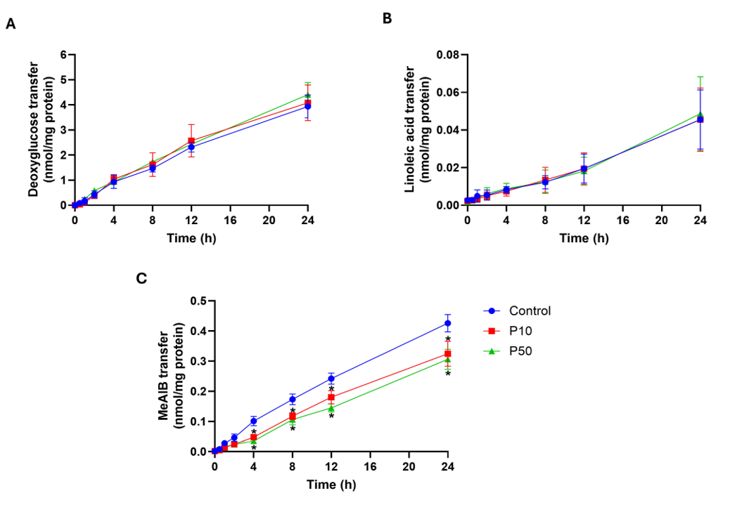
Figure 1: PFOS reduces system A-mediated MeAIB transport without altering 2-deoxy-D-glucose or linoleic acid flux in differentiated BeWo monolayers. BeWo b30 cells were syncytialized on Transwell inserts and exposed to vehicle (Control) or PFOS at 10 µM (P10) or 50 µM (P50) for 24 h. Apical-to-basal transfer of [³H]-2-deoxy-D-glucose (A), [1-¹⁴C]-linoleic acid (B), and the system A-specific substrate L-[¹⁴C]MeAIB (C) was measured over 24 h by liquid scintillation counting and normalized to cellular protein (nmol/mg protein). Glucose and linoleic acid transfer did not differ between groups at any time point. MeAIB transfer was reduced by both P10 and P50 relative to Control from 4 h onward. Data are mean ± SEM from four independent experiments. Time-course data were analyzed by repeated-measures two-way ANOVA (treatment × time) with Bonferroni-adjusted comparisons. *P < 0.05 versus Control at the indicated time point.
In Vitro Gene Expression of System A Transporters
To determine whether the reduction in system A transport reflected changes in transporter expression, digital PCR was used to quantify SNAT1, SNAT2, and SNAT4 mRNA following PFOS exposure (Fig. 2). SNAT4 was detected at very low copy numbers across all treatment groups, precluding reliable quantitative analysis of PFOS effects on this transcript. SNAT1 mRNA was significantly decreased only at 50 µM (P < 0.05), with no change at 10 µM. SNAT2 mRNA was significantly reduced at both 10 µM and 50 µM relative to vehicle control (P < 0.05). GAPDH copy number remained stable across all groups.
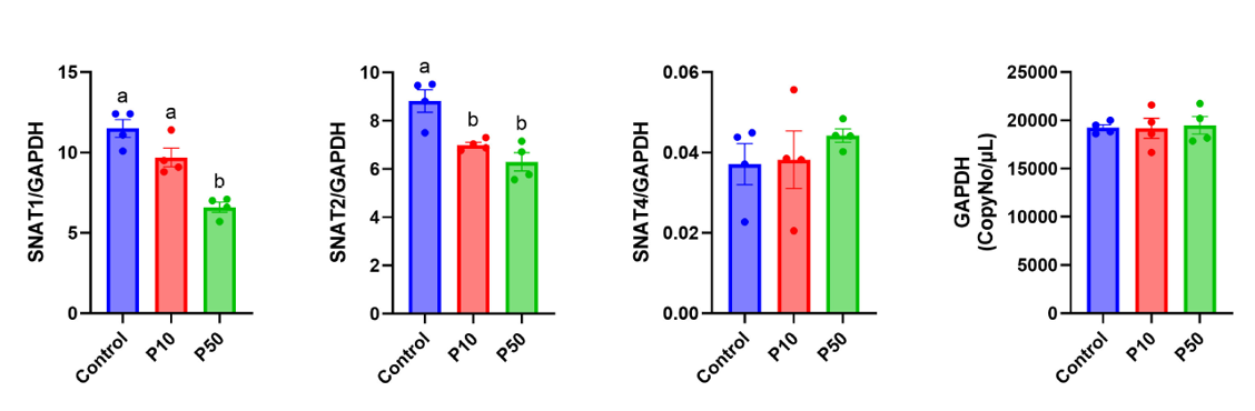
Figure 2: PFOS differentially alters system A transporter transcript abundance in BeWo monolayers. Digital PCR analysis of SNAT1, SNAT2, SNAT4, and GAPDH after 24 h exposure to vehicle (Control), 10 µM PFOS (P10), or 50 µM PFOS (P50). Expression of SNAT1, SNAT2, and SNAT4 is shown relative to GAPDH, with absolute GAPDH copy number per µL shown separately to document that the reference transcript was stable across groups. SNAT1 was reduced only at P50, whereas SNAT2 was reduced at both concentrations; SNAT4 was detected at very low copy numbers, precluding quantitative analysis. Bars represent mean ± SEM and points represent independent experiments (n = 4/group). Groups with different letters differ at P < 0.05 by one-way ANOVA with Bonferroni-adjusted comparisons; the absence of letters indicates no significant group difference.
In Vitro Protein Expression of System A Transporters
Western blotting showed that SNAT1 protein was unchanged at both PFOS concentrations despite the reduction in SNAT1 mRNA at 50 µM. In contrast, SNAT2 protein was significantly reduced in a dose-dependent manner at both 10 µM and 50 µM (P < 0.05), consistent with the mRNA data. SNAT4 protein was unchanged at either concentration (Fig. 3).
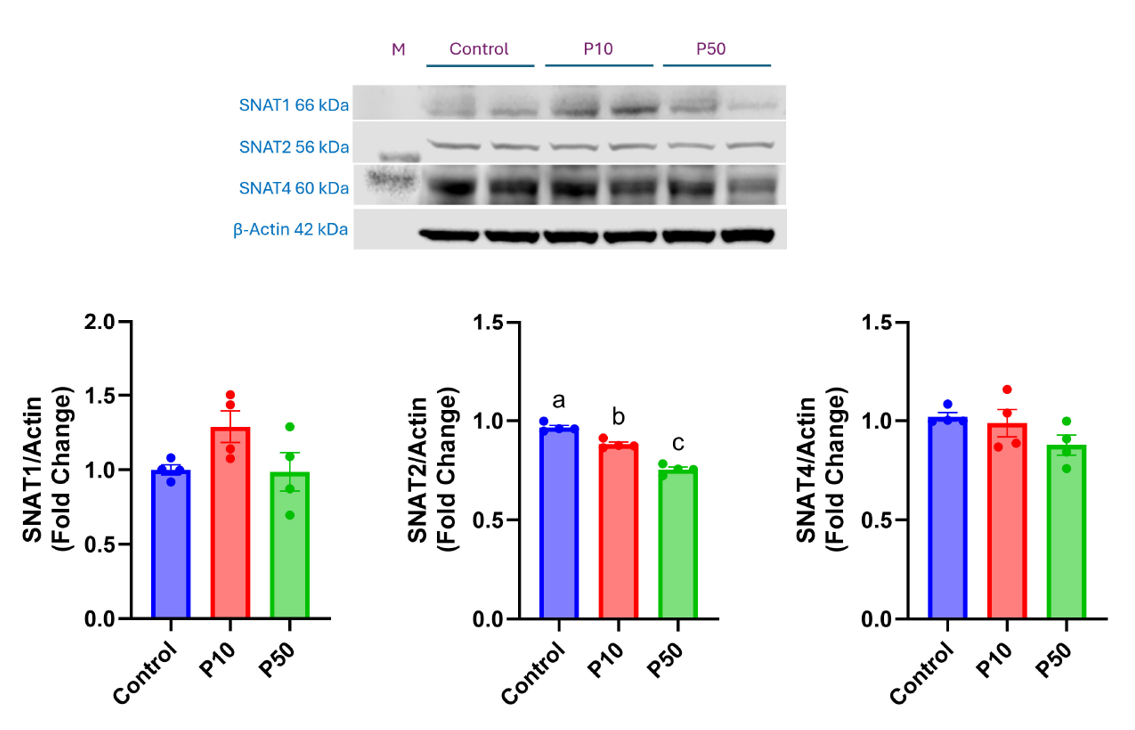
Figure 3: PFOS exposure is associated with lower SNAT2 protein abundance in BeWo monolayers. Representative immunoblots and densitometric analyses of SNAT1, SNAT2, and SNAT4 after 24 h exposure to vehicle (Control), 10 µM PFOS (P10), or 50 µM PFOS (P50). Protein abundance was normalized to β-actin and expressed as fold change relative to Control. SNAT2 was reduced dose-dependently; SNAT1 and SNAT4 did not differ between groups. Bars represent mean ± SEM and points represent independent experiments (n = 4/group). Groups with different letters differ at P < 0.05 by one-way ANOVA with Bonferroni-adjusted comparisons; the absence of letters indicates no significant group difference. M, molecular-weight marker.
Maternal and Fetal Outcomes
Exposure of pregnant Sprague-Dawley rats to PFOS in drinking water (10 and 50 mg/L) from GD 4 to 20 reduced fetal, placental, and labyrinth zone weights (Table 2). Mean water intake (124.1 ± 20, 119.9 ± 17, and 121.8 ± 18 mL/kg/day; P = 0.99) and feed intake (67.1 ± 7.5, 69.8 ± 8.2, and 65.8 ± 7.9 g/kg/day; P = 0.93) were comparable across groups, corresponding to mean PFOS intakes of 1.20 ± 0.19 and 6.09 ± 0.90 mg/kg/day in the 10 and 50 mg/L groups. Mean fetal weight was reduced at both exposures (P = 0.0002), by 9% at 10 mg/L (4.78 ± 0.09 vs. 5.25 ± 0.12 g; P = 0.012) and by 15% at 50 mg/L (4.46 ± 0.06 g; P = 0.0002). The number of live fetuses per litter was unaffected (P = 0.91). Mean placental weight was likewise reduced (P = 0.001), by 15% at 50 mg/L (0.50 ± 0.02 vs. 0.59 ± 0.006 g; P = 0.001). Separation of the placental zones showed that this change was confined to the exchange region: labyrinth zone weight was significantly reduced (P = 0.0007), by 12% at 50 mg/L (0.30 ± 0.006 vs. 0.34 ± 0.005 g; P = 0.0006), whereas junctional zone weight was unchanged (P = 0.46), reproducing our earlier findings in this model [23].
Maternal gestational weight gain did not differ significantly between groups (117.7 ± 10.2, 110.2 ± 8.5, and 100.1 ± 9.2 g; P = 0.43), although the 50 mg/L group showed a 15% numerical decrease. No dam in any group showed mortality, morbidity, or clinical signs of toxicity.
Table 2: Maternal, fetal, and placental outcomes following gestational PFOS exposure.
|
Outcome |
Control (n = 5 dams) |
PFOS 10 mg/L (n = 5 dams) |
PFOS 50 mg/L (n = 5 dams) |
Overall P value |
|
Maternal outcomes |
||||
|
Gestational body weight gain, GD 4-21 (g) |
117.7 ± 10.2 |
110.2 ± 8.5 |
100.1 ± 9.2 |
0.43 |
|
Mean water intake, GD 4-20 (mL/kg/day) |
124.12 ± 20 |
119.89 ± 17 |
121.75 ± 18 |
0.99 |
|
Mean feed intake, GD 4-20 (g/kg/day) |
67.12 ± 7.5 |
69.78 ± 8.2 |
65.75 ± 7.9 |
0.93 |
|
Estimated PFOS intake, GD 4-20 (mg/kg/day) |
0 |
1.20 ± 0.19 |
6.09 ± 0.90 |
— |
|
Fetal outcomes |
||||
|
Live fetuses per litter |
11.0 ± 1.8 |
11.8 ± 2.0 |
10.6 ± 1.9 |
0.91 |
|
Mean fetal weight per litter (g) |
5.25 ± 0.12a |
4.78 ± 0.09b |
4.46 ± 0.06b |
0.0002 |
|
Placental outcomes |
||||
|
Mean placental weight per litter (g) |
0.59 ± 0.006a |
0.54 ± 0.009a,b |
0.50 ± 0.02b |
0.001 |
|
Mean labyrinth-zone weight per litter (g) |
0.34 ± 0.005a |
0.32 ± 0.005a,b |
0.30 ± 0.006b |
0.0007 |
|
Mean junctional-zone weight per litter (g) |
0.18 ± 0.007 |
0.17 ± 0.008 |
0.18 ± 0.003 |
0.46 |
Data are mean ± SEM with the dam/litter as the experimental unit (n = 5 dams per group); fetal and placental measurements were averaged within each litter before analysis. P values are from one-way ANOVA. Within a row, values not sharing a superscript letter differ at P < 0.05 by Bonferroni-adjusted post hoc comparison; rows without letters showed no significant pairwise differences. Estimated PFOS intake was calculated from measured water intake and the nominal drinking water concentration and was not subjected to group comparison.
In Vivo Placental Vascularization and System A Transporter Expression
Immunohistochemical analysis of placental tissue from PFOS-exposed dams showed significant reductions in SNAT1 and SNAT2 immunostaining throughout the labyrinth zone at both concentrations (P < 0.05; Fig. 4). The effect was most pronounced for SNAT2, with reductions in percent DAB-positive area of approximately 60% and 79% at 10 and 50 mg/L, respectively. SNAT4 immunostaining was unchanged.
Parallel analysis of vascularization showed that CD31 immunostaining, a marker of endothelial cells, was also reduced in a dose-dependent manner (19% and 33% at 10 and 50 mg/L; P < 0.05; Fig. 4). Representative photomicrographs confirmed reduced CD31, SNAT1, and SNAT2 staining in PFOS-exposed placentas with unchanged SNAT4 (Fig. 4); negative controls processed in parallel confirmed antibody specificity.
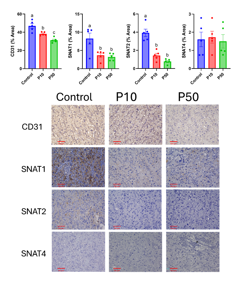
Figure 4: PFOS reduces labyrinth zone amino acid transporter expression and CD31-positive area in vivo. Pregnant rats received vehicle (Control) or PFOS at 10 mg/L (P10) or 50 mg/L (P50) in drinking water from GD 4 through GD 20. Upper panels: percent DAB-positive area for CD31 (endothelial marker), SNAT1, SNAT2, and SNAT4 in the placental labyrinth zone. CD31, SNAT1, and SNAT2 were reduced at both exposures; SNAT4 did not differ between groups. Bars represent mean ± SEM and points represent individual dams (n = 5 dams/group, two sections per animal, averaged within animal before analysis). Lower panels: representative 20× photomicrographs; brown indicates DAB-positive staining and blue indicates hematoxylin counterstain. All images were acquired using identical microscope settings. Groups with different letters differ at P < 0.05 by one-way ANOVA with Bonferroni-adjusted comparisons; the absence of letters indicates no significant group difference.
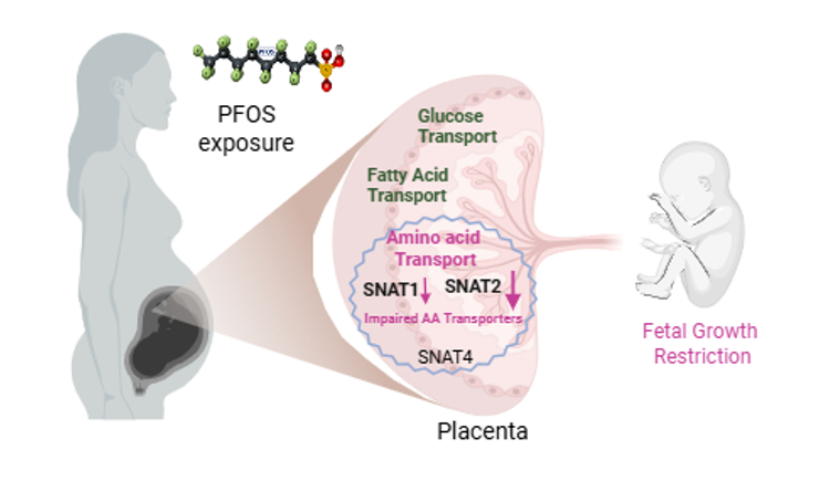
Figure 5: Proposed mechanism by which gestational PFOS exposure constrains maternal–fetal amino acid supply. Maternal PFOS exposure reaches the placenta, where it impairs system A–mediated neutral amino acid transport across the syncytiotrophoblast, measured here with the system A–specific substrate MeAIB. Transfer of 2-deoxy-D-glucose and of the long-chain fatty acid linoleic acid was unaffected under the same conditions. The transport deficit is accompanied by reduced SNAT2 abundance in differentiated BeWo monolayers and reduced SNAT1 and SNAT2 immunostaining in the rat placental labyrinth zone, with SNAT4 unchanged in both models. Constrained amino acid delivery to the fetal compartment is proposed to contribute to the fetal growth restriction observed in vivo. The schematic depicts the trophoblast transport arm of the phenotype; the concurrent reduction in labyrinth zone CD31-positive area is not shown. Causality between reduced SNAT2 abundance and the transport deficit was not tested directly.
Discussion
This study demonstrates that PFOS impairs system A-mediated amino acid transport and alters placental labyrinth vascular endpoints through concurrent trophoblast and vascular effects. Using differentiated human BeWo b30 monolayers, we found that PFOS reduced transcellular transport of the system A-specific substrate MeAIB without affecting the measured transfer of 2-deoxy-D-glucose or linoleic acid under identical experimental conditions. In pregnant rats, gestational PFOS exposure was associated with reduced labyrinth-zone SNAT2 and CD31 immunostaining, reduced placental and labyrinth zone weights, and reductions in fetal weight of 9% and 15% at 10 and 50 mg/L. These findings are consistent with human cord-blood metabolomic data showing altered amino acid profiles in PFAS-exposed pregnancies [24] and identify placental system A transport as a potentially sensitive target of gestational PFOS exposure. The proposed mechanism is summarized in Figure 5.
MeAIB is a nonmetabolizable substrate transported predominantly by system A; therefore, reduced MeAIB flux reflects impaired system A activity rather than a generalized reduction in placental amino acid transport. Human and rodent placentas express several additional amino acid transport systems, including system L (LAT1/LAT2), system y⁺L, and system B⁰,⁺ [17], which were not evaluated here and could be affected independently or secondarily through changes in the intracellular neutral amino acid pool generated in part by system A activity. Because system L functions as an obligatory exchanger dependent on intracellular substrate, impaired system A activity could indirectly limit uptake of essential branched-chain and aromatic amino acids, although this possibility requires direct testing. Similarly, the absence of an effect on [1-¹⁴C]-linoleic acid transfer indicates preservation of transport for this specific long-chain fatty acid and should not be generalized to all fatty acid species, since placental fatty acid transfer involves multiple transport and binding proteins with distinct substrate preferences, including for docosahexaenoic and arachidonic acids [15, 16]. Determining whether PFOS affects system L- and system y⁺L-mediated transport, or the transfer of other physiologically important fatty acids, is a priority for future work.
PFOS reduced MeAIB transport by 24-28% at 10 and 50 µM, whereas 2-deoxy-D-glucose and linoleic acid transfer were unchanged. This substrate-specific response, together with preserved transepithelial electrical resistance and complete dye exclusion, argues against loss of monolayer integrity or nonspecific membrane disruption as the primary explanation for the transport deficit. The differentiated BeWo Transwell system permits measurement of transcellular nutrient flux in the absence of maternal vascular, hemodynamic, and endocrine influences [34, 35], and the reduction in MeAIB transport therefore supports a direct effect of PFOS on the trophoblast transport phenotype, although it does not establish direct molecular interaction between PFOS and a specific transporter. The appearance of the deficit within 24 h is consistent with a relatively rapid effect on transporter abundance or activity rather than a secondary consequence of altered placental development, and its magnitude is comparable to transport deficits reported in models in which suppression of placental amino acid transport precedes or contributes to fetal growth restriction [29, 46, 47]. Whereas previous BeWo studies have focused primarily on PFAS transfer across the trophoblast barrier [48], these findings indicate that PFOS can also impair a functional nutrient-transport pathway within that barrier.
These results do not demonstrate that the fetus receives adequate glucose or lipid substrates in vivo, nor do they establish a generalized fetal amino acid deficiency. Rather, they identify a plausible mechanism by which PFOS could alter the quantity or composition of amino acids delivered to the fetal compartment. Cord-plasma metabolomic studies from the Boston Birth Cohort have associated prenatal PFAS exposure with altered amino acid profiles, including perturbations in arginine, proline, and histidine metabolism [24]. Because cord-plasma metabolite concentrations integrate placental transport, fetal metabolism, and maternal supply, these associations cannot be localized to the placenta, but the present data provide a biologically plausible placental mechanism that could contribute to them.
Digital PCR revealed clear differences among the three system A isoforms. SNAT2 mRNA was reduced at both concentrations, whereas SNAT1 mRNA was reduced only at 50 µM. SNAT4 transcripts were present at very low copy number, consistent with reports that SNAT4 contributes more to system A activity in the first-trimester placenta than at term [49, 50]; the low SNAT4 expression in BeWo cells limits detection of SNAT4-specific effects in this model. Protein analyses extended the transcript findings: SNAT2 protein abundance decreased across PFOS concentrations in BeWo cells, and a corresponding in vivo pattern was observed, with labyrinth-zone SNAT2-positive area decreasing by approximately 60% and 79% at 10 and 50 mg/L PFOS, respectively. SNAT4 protein abundance was not detectably altered in either model.
SNAT1 showed a discordant response: protein abundance was preserved in BeWo cells despite reduced mRNA at 50 µM, while the SNAT1-positive area was lower in placentas from PFOS-exposed rats. This difference could reflect post-transcriptional regulation in BeWo cells or the influence of the more complex in vivo environment, in which endocrine, vascular, inflammatory, and metabolic signals may affect transporter abundance; because post-transcriptional compensation was not directly evaluated, this interpretation remains a hypothesis. For SNAT2, the concordant reductions in mRNA and protein are consistent with altered transcription, mRNA stability, translation, or protein turnover. SNAT2 is responsive to amino acid availability, osmotic stress, and endocrine signals [51-53], properties that may increase its sensitivity to PFOS-induced cellular perturbation. Causality was not tested directly: because no SNAT2 gain- or loss-of-function experiments were performed, reduced SNAT2 abundance should be described as the molecular change most closely associated with the transport deficit rather than as its proven cause.
The rat data indicate that PFOS affects placental function through multiple pathways. Dose-related reductions in CD31-positive area are consistent with reduced endothelial area or altered vascular organization within the labyrinth zone, although CD31 immunostaining alone does not directly measure vessel density, vascular surface area, or perfusion, so the precise nature of this vascular phenotype remains undefined. Reduced endothelial area and lower transporter abundance could work together to limit nutrient exchange, and sufficiently severe vascular impairment could affect multiple nutrient classes in vivo. Human cohort studies reporting PFAS-associated alterations in placental arterial vascular surface area and architecture support the translational relevance of this possibility [40, 54]. PFAS have also been reported to alter endothelial proliferation and gene expression and to perturb angiogenic signaling in placental trophoblast [55, 56]. Because BeWo monolayers contain no vascular component, reduced MeAIB transport shows that PFOS can alter trophoblast transport independently of vascular changes; the integrated in vivo phenotype likely reflects both trophoblast-intrinsic effects and changes in placental vascular organization, whose relative contributions the present study cannot resolve.
Our findings differ in several respects from those of Wan et al. [30], who reported that gestational PFOS exposure in mice reduced maternal-fetal transfer of both glucose and amino acid analogs, decreased SNAT4 protein abundance, and did not alter placental zone areas. Wan et al. administered PFOS by daily oral gavage at 1-3 µg/g body weight from GD 4.5 to GD 17.5, whereas the present study used continuous drinking-water exposure corresponding to approximately 1.2 and 6.1 mg/kg/day; although the daily doses are of the same order, with the low dose overlapping theirs and the high dose approximately twice their highest, bolus and continuous exposure produce different concentration-time profiles, which could matter for a transporter such as SNAT2 that responds dynamically to cellular stress and nutrient availability. Species differences may also contribute. Although rats and mice both have hemochorial placentas, their placental architecture, trophoblast organization, and responses to xenobiotic exposure are not identical [57], and species-specific differences in the abundance, localization, or gestational regulation of SNAT isoforms could account for the predominant SNAT4 response reported in mice and the SNAT2-associated response observed in rats and human-derived BeWo cells. In addition, an intact animal includes vascular, hemodynamic, endocrine, and metabolic influences absent from an isolated trophoblast monolayer, and glucose transfer in vivo further depends on maternal glycemia and the maternal-fetal concentration gradient. A broad, multi-substrate deficit in vivo is therefore compatible with a more restricted trophoblast-intrinsic effect, and the two studies are best viewed as complementary: both implicate system A transport in PFOS-induced placental dysfunction, whereas the present in vitro approach isolates a trophoblast-associated component of a potentially broader in vivo phenotype.
The molecular basis for the reduction in SNAT2 remains unresolved. The amphiphilic properties of PFOS permit interactions with proteins and membrane microdomains that could modify transporter localization, membrane organization, or intracellular signaling [41], and PFOS and other PFAS can modulate peroxisome proliferator-activated receptor signaling, which influences metabolic and transporter gene expression [58-61]. Experiments evaluating SLC38A2 promoter activity, mRNA stability, protein trafficking, and potential PPAR-dependent transcriptional responses are needed to define the mechanism linking PFOS exposure to reduced SNAT2 abundance.
The exposure concentrations require careful consideration. The in vitro concentrations of 10 and 50 µM, corresponding to approximately 5 and 25 µg/mL, exceed serum PFOS concentrations reported in the general population and remain higher than concentrations observed in many highly exposed communities [7]. They were used in an acute, 24-h mechanistic design intended to identify direct effects on trophoblast transport in the absence of maternal endocrine, hemodynamic, and toxicokinetic variables. The in vivo drinking-water concentrations of 10 and 50 mg/L are likewise several orders of magnitude higher than the U.S. enforceable maximum contaminant level of 4 ng/L [9] and should be interpreted as experimental hazard-identification concentrations rather than simulations of environmental exposure. Direct comparison of drinking-water concentrations across species has limited toxicological value because internal dose depends on water consumption, absorption, distribution, placental accumulation, and elimination, and PFOS is eliminated substantially more rapidly in rats than in humans, in whom the serum half-life is measured in years [7, 42]. Maternal serum and placental PFOS concentrations would provide a more appropriate basis for cross-species comparison; because these were not measured here, the present study does not claim equivalence to occupationally or environmentally exposed human populations, and measuring them, along with longer and lower-concentration exposures, is a priority for subsequent work with this model.
PFOS manufacture and use have been largely phased out in North America and Europe, and PFOS is listed under Annex B of the Stockholm Convention [8]. Nevertheless, its environmental persistence, bioaccumulation, and long human elimination half-life sustain exposure during contemporary pregnancies [7], and the present findings best characterize a potential mechanism of toxicity associated with legacy PFOS exposure. An important extension will be to determine whether replacement PFAS—including short-chain sulfonates such as PFBS, fluoroether acids such as HFPO-DA, and fluorotelomer sulfonates such as 6:2 FTS—produce similar effects. High-throughput screening indicates substantial heterogeneity in the effects of structurally diverse PFAS on trophoblast function [37], and PFBS has been reported to perturb angiogenic and stress responses in human syncytiotrophoblast [39]. Whether these compounds affect SNAT2 or system A-mediated transport, and whether their shorter biological half-lives result in lower placental burdens at comparable external exposures, remain open questions.
Gestational PFOS exposure produced no evidence of systemic maternal toxicity. Feed and water intake were unaltered, gestational weight gain did not differ significantly between groups, and no animal showed mortality, morbidity, or clinical signs of toxicity. The modest numerical decrease in maternal weight gain at the highest exposure is consistent with reduced conceptus mass rather than with a maternal effect: maternal weight at GD 21 includes the litter, fetal growth at this stage is exponential and approaches approximately 1 g per day [62], and the combined fetal and placental mass per litter was approximately 12 g lower at 50 mg/L than in controls, accounting for most of the 17.6 g difference in gestational weight gain. The effects of PFOS on fetal growth in this model are therefore unlikely to be secondary to reduced nutritional intake by the dam.
PFAS cross the placental barrier, with reported fetal-to-maternal concentration ratios for PFOS commonly ranging from 30-50%, although transfer efficiency varies among populations, matrices, and gestational stages [63-65]. Impaired placental transport could therefore occur concurrently with direct fetal exposure, combining chemical insult with altered nutrient availability. Placental nutrient-transport endpoints, including transporter abundance and amino acid profiles in matched maternal and fetal samples, may therefore complement conventional measures of developmental toxicity, although these applications remain hypotheses requiring validation in independent experimental and human cohorts.
Several limitations should be considered. First, the association between reduced SNAT2 abundance and impaired system A transport does not establish causality. To establish necessity or sufficiency, SNAT2 knockdown would be needed to determine whether its loss reproduces the PFOS phenotype, and SNAT2 overexpression to determine whether it rescues MeAIB transport. The proposed role of SNAT2 is supported by the concordance among MeAIB transport, SNAT2 mRNA, and SNAT2 protein measurements, and by independent evidence that placenta-specific Slc38a2 knockdown causes fetal growth restriction in mice [29]; nevertheless, SNAT1 and other system A isoforms may also contribute to MeAIB transport, and the data should not be read as demonstrating that SNAT2 alone mediates the observed effect. Second, the BeWo model is a simplified representation of the multicellular placental barrier. Forskolin-induced fusion can be incomplete, producing a mixture of multinucleated syncytial structures and residual mononuclear cells [66] that may differ in PFOS sensitivity or transporter abundance, and a mixed monolayer may also differ from mature primary syncytiotrophoblast in its fatty acid transport machinery, potentially contributing to the absence of an observed effect on linoleic acid transfer. BeWo cells are additionally derived from a choriocarcinoma and do not reproduce all properties of primary trophoblast; primary cells, placental explants, three-dimensional models, and trophoblast-endothelial co-cultures would address these constraints.
Third, the study did not include human placental tissue or clinical specimens, so its relevance to human pregnancy rests on model systems and concordance with published cohort observations [24, 40, 54]; analysis of banked human placentas with matched maternal and cord PFAS measurements would provide a logical translational extension. Fourth, although we measured fetal and placental weights, we did not measure fetal amino acid concentrations or in vivo placental amino acid transfer; these measurements would strengthen the sequence linking PFOS exposure, transporter changes, altered nutrient delivery, and fetal growth. Finally, we examined a single gestational window and only two exposure concentrations; longitudinal designs incorporating early gestation and additional lower-dose groups are needed to determine whether susceptibility changes as the relative contributions of system A isoforms evolve, and to define the concentration-response relationship and a no-observed-effect level.
In conclusion, gestational PFOS exposure impaired placental system A-mediated neutral amino acid transport, as measured using the system A-specific substrate MeAIB, without detectable effects on 2-deoxy-D-glucose or linoleic acid transfer under the same in vitro conditions. Reduced MeAIB transport was accompanied by lower SNAT2 abundance in differentiated BeWo monolayers and reduced SNAT2-positive area in rat placental labyrinth tissue; in vivo, these transporter-associated changes coincided with reduced CD31-positive area, suggesting that trophoblast transport and placental vascular alterations may act concurrently to constrain maternal-fetal nutrient exchange. The concordant SNAT2 response across a human-derived trophoblast model and intact rat placenta identifies SNAT2 as the molecular correlate most closely associated with the transport deficit, although its causal role remains to be established. Whether PFOS affects other amino acid transport systems, additional fatty acid species, or the replacement PFAS now in use remains unknown. Within these limitations, the findings identify placental system A transport—and SNAT2 in particular—as a candidate target of developmental PFOS toxicity.
Funding
Financial support from the National Institutes of Health (NIH) through grant R01ES033345, awarded to Sathish Kumar, is greatly appreciated. The content is solely the authors’ responsibility and does not necessarily represent the official views of the NIH. The funding agency was not involved in the design, analysis, or interpretation of the data reported.
Declarations
Conflict of interest:
The authors declare no conflict of interest.
CRediT authorship contribution statement: Pankaj Yadav: Writing – original draft, Methodology, Investigation. Alissa Hofmann: Writing – review & editing. Jay S. Mishra: Writing – review & editing. Sathish Kumar: Writing – review & editing, Conceptualization, Supervision.
Data availability statement:
The authors confirm that the data supporting the findings of this study are available within the article.
References
- Xu, Z., et al., Association study of umbilical cord serum per- and polyfluoroalkyl substances (PFAS) exposure with birth outcomes: A meta-analysis. Ecotoxicol Environ Saf, 309 (2026): p.119725.
- Wu, M., et al., Prenatal exposure to legacy and emerging PFAS and sex-specific associations with fetal growth: Evidence from a Chinese birth cohort. Environ Res, 285 (2025): p.122488.
- Yang, Y., et al., Association of prenatal exposure to perfluoroalkyl and polyfluoroalkyl substances with fetal growth trajectories. Environ Res, 274 (2025): p.121331.
- Botelho, J.C., et al., Per- and polyfluoroalkyl substances (PFAS) exposure in the U.S. population: NHANES 1999-March 2020. Environ Res, 270 (2025): p.120916.
- National Academies report. Guidance on PFAS Exposure, Testing, and Clinical Follow-Up. 2022: Washington (DC).
- Liu, M., et al., Per- and Polyfluoroalkyl Substances in Contaminated Soil and Groundwater at Airports: A Canadian Case Study. Environ Sci Technol, 56 (2022): p.885-895.
- Olsen, G.W., et al., Half-life of serum elimination of perfluorooctanesulfonate, perfluorohexanesulfonate, and perfluorooctanoate in retired fluorochemical production workers. Environ Health Perspect, 115 (2007): p.1298-1305.
- United Nations Environment Programme, Stockholm Convention on Persistent Organic Pollutants: listing of perfluorooctane sulfonic acid, its salts and perfluorooctane sulfonyl fluoride in Annex B. (2009), Geneva.
- S. Environmental Protection Agency, PFAS National Primary Drinking Water Regulation. Fed Regist, 89 (2024): p.32532-32757.
- Winterhager, E. and A. Gellhaus, Transplacental Nutrient Transport Mechanisms of Intrauterine Growth Restriction in Rodent Models and Humans. Front Physiol, 8 (2017): p.951.
- Jones, H.N., T.L. Powell, and T. Jansson, Regulation of placental nutrient transport–a review. Placenta, 28 (2007): p.763-774.
- Brett, K.E., et al., Maternal-fetal nutrient transport in pregnancy pathologies: the role of the placenta. Int J Mol Sci, 15 (2014): p.16153-16185.
- Sibiak, R., et al., Fetomaternal Expression of Glucose Transporters (GLUTs)-Biochemical, Cellular and Clinical Aspects. Nutrients, 14 (2022).
- Mueckler, M., Facilitative glucose transporters. Eur J Biochem, 219 (1994): p.713-725.
- Duttaroy, A.K., Transport of fatty acids across the human placenta: a review. Prog Lipid Res, 48 (2009): p.52-61.
- Larque, E., et al., Expression pattern of fatty acid transport protein-1 (FATP-1), FATP-4 and heart-fatty acid binding protein (H-FABP) genes in human term placenta. Early Hum Dev, 82 (2006): p.697-701.
- Shimada, H., T.L. Powell, and T. Jansson, Regulation of placental amino acid transport in health and disease. Acta Physiol (Oxf), 240 (2024): p.e14157.
- Ingaramo, P.I. and M.L. Zenclussen, The Placenta as a Target Organ for Poly- and Perfluoroalkyl Substances (PFASs): Molecular Mechanisms of Toxicity. Int J Mol Sci, 27 (2026).
- Xia, Y., et al., Real-life per- and polyfluoroalkyl substances mixture impairs placental function: insights from a trophoblast spheroid model. Environ Res, 287 (2025): p.123037.
- Blake, B.E. and S.E. Fenton, Early life exposure to per- and polyfluoroalkyl substances (PFAS) and latent health outcomes: A review including the placenta as a target tissue and possible driver of peri- and postnatal effects. Toxicology, 443 (2020): p.152565.
- Cheng, Z., et al., Impact of PFOS on maternal and placental lipid metabolism and decreased placental vascularization in a murine model. J Hazard Mater, 501 (2026): p.140636.
- Woods, L., V. Perez-Garcia, and M. Hemberger, Regulation of Placental Development and Its Impact on Fetal Growth-New Insights From Mouse Models. Front Endocrinol (Lausanne), 9 (2018): p.570.
- Dangudubiyyam, S.V., J.S. Mishra, and S. Kumar, Perfluorooctane sulfonic acid modulates expression of placental steroidogenesis-associated genes and hormone levels in pregnant rats. Reprod Toxicol, 118 (2023): p.108390.
- Li, Z., et al., Cord plasma metabolomic signatures of prenatal per- and polyfluoroalkyl substance (PFAS) exposures in the Boston Birth Cohort. Environ Int, 194 (2024): p.109144.
- Mando, C., et al., SNAT2 expression and regulation in human growth-restricted placentas. Pediatr Res, 74 (2013): p.104-110.
- Jansson, T., et al., Down-regulation of placental Cdc42 and Rac1 links mTORC2 inhibition to decreased trophoblast amino acid transport in human intrauterine growth restriction. Clin Sci (Lond), 134 (2020): p.53-70.
- Pantham, P., et al., Down-Regulation of Placental Transport of Amino Acids Precedes the Development of Intrauterine Growth Restriction in Maternal Nutrient Restricted Baboons. Biol Reprod, 95 (2016): p.98.
- Chen, Y.Y., et al., Increased ubiquitination and reduced plasma membrane trafficking of placental amino acid transporter SNAT-2 in human IUGR. Clin Sci (Lond), 129 (2015): p.1131-1141.
- Vaughan, O.R., et al., Placenta-specific Slc38a2/SNAT2 knockdown causes fetal growth restriction in mice. Clin Sci (Lond), 135 (2021): p.2049-2066.
- Wan, H.T., et al., Effects of In Utero Exposure to Perfluorooctane Sulfonate on Placental Functions. Environ Sci Technol, 54 (2020): p.16050-16061.
- Dangudubiyyam, S.V., et al., Maternal perfluorooctane sulfonic acid exposure during rat pregnancy causes hypersensitivity to angiotensin II and attenuation of endothelium-dependent vasodilation in the uterine arteries. Biol Reprod, 107 (2022): p.1072-1083.
- Mishra, J.S., et al., Perfluoroalkyl substances (PFAS) exposure and preeclampsia risk: Impaired angiogenesis through suppression of VEGF signaling. Reprod Toxicol, 132 (2025): p.108827.
- Dangudubiyyam, S.V., et al., Restoring Angiotensin Type 2 Receptor Function Reverses PFOS-Induced Vascular Hyper-Reactivity and Hypertension in Pregnancy. Int J Mol Sci, 24 (2023).
- Ghaffarian, R. and S. Muro, Models and methods to evaluate transport of drug delivery systems across cellular barriers. J Vis Exp, (2013): p.e50638.
- Bode, C.J., et al., In vitro models for studying trophoblast transcellular transport. Methods Mol Med, 122 (2006): p.225-239.
- Gopalakrishnan, K., et al., Hyperandrogenism diminishes maternal-fetal fatty acid transport by increasing FABP4-mediated placental lipid accumulation. Biol Reprod, 107 (2022): p.514-528.
- Blake, B.E., B.P. Rickard, and S.E. Fenton, A High-Throughput Toxicity Screen of 42 Per- and Polyfluoroalkyl Substances (PFAS) and Functional Assessment of Migration and Gene Expression in Human Placental Trophoblast Cells. Front Toxicol, 4 (2022): p.881347.
- Hofmann, A., et al., PFOS Impairs Mitochondrial Biogenesis and Dynamics and Reduces Oxygen Consumption in Human Trophoblasts. J Environ Sci Public Health, 7 (2023): p.164-175.
- Pham, A., J. Zhang, and L. Feng, Exposure to perfluorobutane sulfonate and perfluorooctanesulfonic acid disrupts the production of angiogenesis factors and stress responses in human placental syncytiotrophoblast. Reprod Toxicol, 98 (2020): p.269-277.
- Giorgetti, A., et al., Per- and polyfluoroalkyl substances (PFAS) in placental compartments: Histopathological and toxicological data integration in an Italian cohort. Environ Res, 279 (2025): p.121846.
- Zhao, L., et al., Insight into the binding model of per- and polyfluoroalkyl substances to proteins and membranes. Environ Int, 175 (2023): p.107951.
- Chang, S.C., et al., Comparative pharmacokinetics of perfluorooctanesulfonate (PFOS) in rats, mice, and monkeys. Reprod Toxicol, 33 (2012): p.428-440.
- Dangudubiyyam, S.V., et al., Perfluorooctane sulfonic acid (PFOS) exposure during pregnancy increases blood pressure and impairs vascular relaxation mechanisms in the adult offspring. Reprod Toxicol, 98 (2020): p.165-173.
- Belkacemi, L., et al., Altered placental development in undernourished rats: role of maternal glucocorticoids. Reprod Biol Endocrinol, 9 (2011): p.105.
- Novak, D., et al., SNAT expression in rat placenta. Placenta, 27 (2006): p.510-516.
- Rosario, F.J., et al., Reduction of In Vivo Placental Amino Acid Transport Precedes the Development of Intrauterine Growth Restriction in the Non-Human Primate. Nutrients, 13 (2021).
- Nathanielsz, P.W., Decreased placental amino acid transport and intrauterine growth restriction: which is the chicken and which is the egg? J Physiol, 576 (2006): p.649.
- Chen, H., et al., Transplacental transport of per- and polyfluoroalkyl substances (PFAS): Mechanism exploration via BeWo cell monolayer model. J Hazard Mater, 466 (2024): p.133205.
- Desforges, M., et al., The SNAT4 isoform of the system A amino acid transporter is functional in human placental microvillous plasma membrane. J Physiol, 587 (2009): p.61-72.
- Desforges, M., et al., SNAT4 isoform of system A amino acid transporter is expressed in human placenta. Am J Physiol Cell Physiol, 290 (2006): p.C305-312.
- Jakobsen, S., M. Pedersen, and C.U. Nielsen, Structure-activity relationship of amino acid analogs to probe the binding pocket of sodium-coupled neutral amino acid transporter SNAT2. Amino Acids, 56 (2024): p.64.
- Hoffmann, T.M., et al., Effects of Sodium and Amino Acid Substrate Availability upon the Expression and Stability of the SNAT2 (SLC38A2) Amino Acid Transporter. Front Pharmacol, 9 (2018): p.63.
- Velazquez-Villegas, L.A., et al., Transcriptional regulation of the sodium-coupled neutral amino acid transporter (SNAT2) by 17beta-estradiol. Proc Natl Acad Sci U S A, 111 (2014): p.11443-11448.
- Chowdhury, S.F., et al., PFAS alters placental arterial vasculature in term human placentae: A prospective pregnancy cohort study. Placenta, 149 (2024): p.54-63.
- Ashby, A., et al., The Effects of Perfluorooctanesulfonic acid (PFOS) on Human Umbilical Vein Endothelial Cells (HUVECs) Proliferation and Gene Expression and its Implications on Fetal Development. MicroPubl Biol, (2024).
- Poteser, M., et al., Perfluoroctanoic acid (PFOA) enhances NOTCH-signaling in an angiogenesis model of placental trophoblast cells. Int J Hyg Environ Health, 229 (2020): p.113566.
- Furukawa, S., N. Tsuji, and A. Sugiyama, Morphology and physiology of rat placenta for toxicological evaluation. J Toxicol Pathol, 32 (2019): p.1-17.
- Kanitz, M.H. and C. Striley, Differential Activation of Peroxisome Proliferator-Activated Receptor Alpha by Per- and Polyfluoroalkyl Substances. Int J Toxicol, (2025): p.10915818251393212.
- Soderstrom, S., et al., Agonistic and potentiating effects of perfluoroalkyl substances (PFAS) on the Atlantic cod (Gadus morhua) peroxisome proliferator-activated receptors (Ppars). Environ Int, 163 (2022): p.107203.
- Chen, Z., et al., PPARgamma stimulates expression of L-type amino acid and taurine transporters in human placentas: the evidence of PPARgamma regulating fetal growth. Sci Rep, 5 (2015): p.12650.
- Sheikh, K., et al., Beyond lipids, pharmacological PPARalpha activation has important effects on amino acid metabolism as studied in the rat. Am J Physiol Endocrinol Metab, 292 (2007): p.E1157-1165.
- Witlin, AG., et al., Placental and fetal growth and development in late rat gestation is dependent on adrenomedullin. Biol Reprod, 67 (2002): p.1025-1031.
- Noren, E., et al., Transplacental transfer efficiency of perfluoroalkyl substances (PFAS) after long-term exposure to highly contaminated drinking water: a study in the Ronneby Mother-Child Cohort. J Expo Sci Environ Epidemiol, 35 (2025): p.445-453.
- Xu, Y., et al., Early-life exposure to per- and polyfluoroalkyl substances: Analysis of levels, health risk and binding abilities to transport proteins. Eco Environ Health, 3 (2024): p.308-316.
- Kang, H., et al., Placental Transfer and Composition of Perfluoroalkyl Substances (PFASs): A Korean Birth Panel of Parent-Infant Triads. Toxics, 9 (2021).
- Orendi, K., et al., The choriocarcinoma cell line BeWo: syncytial fusion and expression of syncytium-specific proteins. Reproduction, 140 (2010): p.759-766.