Gustavo Eduardo Sánchez Martínez*ORCID ID
General Surgery Department Hospital General, Dr. Carlos Canseco. Tampico, Tamaulipas, 89130, Mexico.
*Corresponding Author: Gustavo Eduardo Sánchez Martínez, General Surgery Department Hospital General de Cd. Mante “Dr Emilio Martínez Manatou”. 89880 Tamaulipas, Mexico.
Received: 31 August 2026; Accepted: 07 September 2026; Published: 14 October 2026
Introduction and Importance: Penetrating cutting trauma to the face involving the auricle and parotid gland is uncommon and poses significant reconstructive challenges due to the risk of vascular injury, salivary fistula, cartilage exposure, and aesthetic deformity. Prompt exploration and layered anatomical repair are essential for optimal functional and cosmetic outcomes.
Case Presentation: A 40-year-old male was brought to the emergency department after sustaining a saber (cutlass-type) wound extending from the posterior aspect of the auricle to the mid-cheek (zygomatic region). On arrival he was hemodynamically stable but intoxicated, conscious and responsive, with active bleeding. Emergency operative exploration was performed. Vascular control was achieved by ligation of bleeding vessels. The parotid gland was found transected through its mid-portion and was repaired. The auricle was partially destroyed; layered repair with nylon 4-0 was carried out. Final facial wound closure was performed and a Penrose
drain was placed. The patient remained under hospital observation for 72 hours with a favorable course and was discharged.
Clinical Discussion: Management of complex facial cutting injuries requires systematic assessment of critical structures (facial nerve, parotid duct and parenchyma, cartilage). Isolated parenchymal injury of the parotid is best treated by meticulous capsule repair, drainage, and pressure dressing to minimize sialocele risk. Auricular lacerations with cartilage involvement demand precise realignment and complete soft-tissue coverage to prevent chondritis and deformity. Early primary repair within the first hours yields the best results.
Facial trauma, Cutting injury, Saber wound, Parotid gland injury, Auricular reconstruction, Soft-tissue reconstruction
Facial trauma articles; Cutting injury articles; Saber wound articles; Parotid gland injury articles; Auricular reconstruction articles; Soft-tissue reconstruction articles
Introduction and importance
Facial soft-tissue trauma caused by sharp weapons (knives, sabers, machetes) can produce extensive lacerations that simultaneously involve multiple anatomical subunits, including the auricle, parotid gland, and facial soft tissues. These injuries are relatively rare compared with blunt trauma or gunshot wounds, yet they carry a high risk of functional and aesthetic sequelae if not managed promptly and meticulously [1,2].
The parotid gland and its duct lie in a vulnerable zone of the cheek; transection may lead to sialocele, fistula, or infection if the capsule is not adequately repaired [3,4]. Likewise, the auricle, with its thin skin and exposed cartilage, is susceptible to chondritis, necrosis, and permanent contour deformity when cartilage remains uncovered [5,6].
We present a case of hemifacial saber trauma with simultaneous mid-parotid transection and partial auricular destruction, successfully managed by emergency exploration, vascular control, and layered primary reconstruction. This report is intended to highlight practical technical points and reinforce the value of early definitive repair in complex cutting facial injuries.
Case presentation
A 40-year-old male patient was transported to the emergency department after suffering a cutting injury inflicted by a saber. The wound extended from the posterior aspect of the pinna across the preauricular and zygomatic region to the mid-cheek (hemifacial distribution).
On arrival the patient was hemodynamically stable, conscious, and responsive, although in an intoxicated state. Active bleeding from the wound was evident. No obvious facial-nerve deficit was recorded at the initial examination. After rapid primary survey and basic resuscitation, the decision was made to transfer the patient immediately to the operating room for exploration and definitive repair under general anesthesia.
Operative findings and procedure:
Under general anesthesia, thorough irrigation and exploration of the wound were performed. Active bleeding vessels were identified and ligated to achieve hemostasis (damage-control vascular phase). The parotid gland was found to be transected approximately through its mid-portion; the glandular parenchyma and capsule were carefully reapproximated. The auricle showed partial destruction involving approximately half of its substance; the remaining cartilage and soft tissue were realigned and repaired in layers using nylon 4-0 sutures, ensuring complete coverage of any exposed cartilage. Final layered closure of the facial soft tissues was completed and a Penrose drain was left in place to prevent fluid collection.
Postoperatively the patient was kept under close hospital observation for 72 hours. The clinical course was favorable, with no signs of infection, salivary leakage, or wound dehiscence. The drain was removed and the patient was discharged with instructions for wound care, antibiotic coverage, and outpatient follow-up.
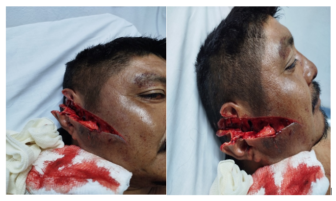
Figure 1: Preoperative clinical photograph of the hemifacial wound.
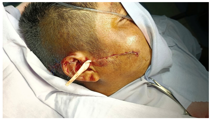
Figure 2: Intraoperative view showing parotid transection and auricular injury.
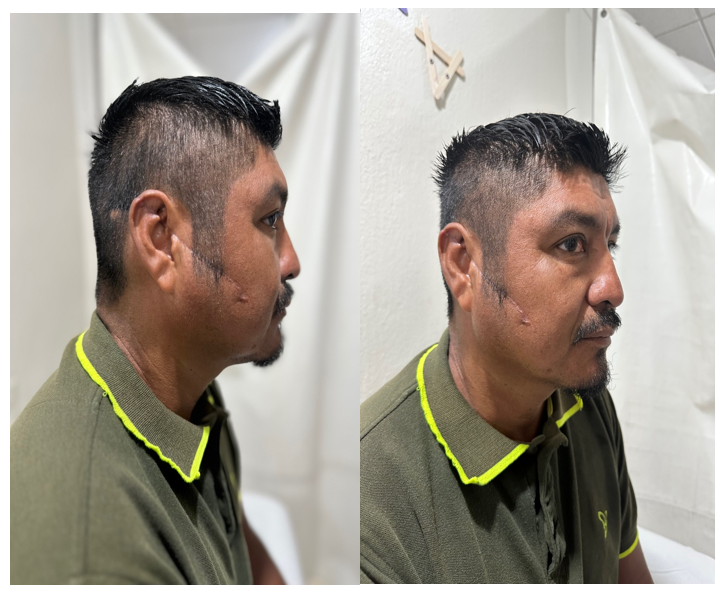
Figure 3: Immediate postoperative appearance after layered repair and Penrose drain placement.
Clinical discussion
Complex cutting injuries of the face require a systematic approach that prioritizes life-threatening hemorrhage, followed by identification and repair of critical structures (facial nerve branches, parotid duct and parenchyma, cartilage frameworks) [1,3]. In the present case, the wound trajectory crossed the classic “danger zone” for parotid injury (line from tragus to mid-upper lip), making glandular and possible ductal injury highly probable [3,4].
When only the parotid parenchyma is lacerated and the duct remains intact, primary repair of the capsule with fine absorbable sutures, placement of a drain, and a temporary pressure dressing constitute the standard of care and significantly reduce the risk of sialocele or fistula [3,4,7]. In our patient the gland was transected at mid-level; careful approximation of the capsule and the use of a Penrose drain followed this principle and were associated with an uneventful recovery.
Auricular reconstruction after partial traumatic destruction follows well-established principles: minimal debridement of viable tissue, precise realignment of cartilage (preferably with long-lasting absorbable or fine non-absorbable sutures through the perichondrium), and absolute coverage of any exposed cartilage by skin to prevent chondritis and late deformity [5,6,8,9]. Primary closure within the first 24 hours remains the preferred strategy whenever possible and yields high success rates even in complex lacerations [5,6].
The absence of clinically evident facial-nerve deficit at presentation allowed primary soft-tissue repair without the need for immediate nerve exploration or microsurgical neurorrhaphy. Nevertheless, a high index of suspicion and serial clinical examination remain mandatory in every deep cheek laceration [3].
This case illustrates that, even in the setting of alcohol intoxication and active bleeding, rapid transfer to the operating theatre, systematic exploration, and adherence to layered anatomical repair principles can produce a favorable short-term outcome after extensive hemifacial cutting trauma.
Methods
This case report has been reported in line with the SCARE 2023 Criteria [10].
Conclusion
Early emergency exploration with meticulous vascular control, primary repair of the transected parotid parenchyma, and layered reconstruction of the partially destroyed auricle resulted in a favorable clinical course after complex saber-induced hemifacial trauma. Prompt anatomical restoration remains the cornerstone of successful management of such injuries.
Declarations
Patient consent:
Written informed consent was obtained from the patient for publication of this case report and any accompanying images.
Ethical approval:
Not required for a single case report according to institutional guidelines.
Funding:
This research did not receive any specific grant from funding agencies in the public, commercial, or not-for-profit sectors.
Author contribution:
Dr. Gustavo Eduardo Sánchez Martínez. Redaction of case report. Clinical and surgical management.
Conflict of interest:
The authors declare no conflict of interest.
References
- McElwee TJ, Poche JN, Sowder JC, et al. Management of Acute Facial Nerve and Parotid Injuries. Facial Plast Surg 37 (201): 490-499.
- Le DL, Vu DHD, Vu NL, et al. Case report: Complex craniofacial trauma due to knife injuries – surgical management and outcomes. Vietnam Med J 550 (2025).
- Woźniczko K, Molga-Magusiak M, Rzepakowska A, Bartoszewicz R. Extensive cut wound of the face – case report. Pol Otorhinolaryngol Rev (2021).
- Pande S. Challenges encountered during the management of pediatric facial soft tissue injury from a mobile battery blast: A case report. Clin Case Rep 9 (2021): e04753.
- Goldberg A, Kerr R. Management of traumatic auricular avulsion injury – partial and complete. Curr Opin Otolaryngol Head Neck Surg 33 (2025): 262-265.
- Complex Ear Lacerations. StatPearls [Internet]. Treasure Island (FL): StatPearls Publishing (2026).
- Management of Traumatic Soft Tissue Injuries of the Face. Plast Reconstr Surg Glob Open / related reviews 2021-2022.
- Huded V, Gautam G. Management of Post-traumatic Ear Reconstruction in a Tertiary Care Center: A Retrospective Study. Cureus 18 (2026): e105893.
- Introducing a novel technique for auricular replantation following subtotal amputation combining Sexton and Mladick pocket methods: a case report. J Med Case Rep 19 (2025): 569.
- Sohrabi C, Mathew G, Maria N, et al. Collaborators. The SCARE 2023 guideline: updating consensus Surgical Case Report (SCARE) guidelines. Int J Surg 109 (2023): 1136-1140.