Shashi Kant*1, Rajesh Kumar1, Sanjay Kumar1, Rekha Kumari1, Anurag Kumar1
1All India Institute of Medical Sciences, Patna, India
*Corresponding Author: Shashi Kant, All India Institute of Medical Sciences, Patna, India.
Received: 14 July 2026; Accepted: 20 July 2026; Published: XX- July 2026
Abstract:Patients undergoing redo surgery for head and neck malignancies frequently present with anticipated difficult mask ventilation and tracheal intubation because of facial disfigurement, postoperative tissue loss, distorted upperairway anatomy, and impaired mask seal. Failed tracheal intubation in such patients is associated with significant morbidity and mortality, making meticulous preoperative airway assessment and planning essential. We report the case of a man in his 60s with carcinoma of the buccal mucosa who presented for deltopectoral flap reconstruction following previous surgery and tracheostomy. Although there was no tumour involving the trachea or laryngeal airway, postoperative anatomical distortion, enema, purulent secretions, and facial tissue loss resulted in an anticipated physiologically and anatomically difficult airway. Awake nasal fibreoptic intubation was planned as the primary airway management technique. However, visualisation was limited by secretions and an oedematous epiglottis. A C-MAC video laryngoscope was introduced to improve visualisation, facilitate suctioning, and guide advancement of the fibreoptic bronchoscope. The hybrid technique enabled successful awake nasal tracheal intubation while maintaining spontaneous ventilation. This case highlights the limitations of fibreoptic bronchoscopy when visualisation is compromised and demonstrates that combining a C-MAC video laryngoscope with an intubating video endoscope can be an effective strategy for securing the airway in selected patients with severely distorted upper-airway anatomy.
Awake Fiberoptic Intubation; C-Mac Video Laryngoscopy; Conscious Sedation; Difficult Mask Ventilation; Fiberoptic Bronchoscopy
Awake Fiberoptic Intubation articles; C-Mac Video Laryngoscopy articles; Conscious Sedation articles; Difficult Mask Ventilation articles; Fiberoptic Bronchoscopy articles
Introduction
Airway management remains one of the most critical skills in anesthetic practice and can be particularly challenging in patients with distorted upper-airway anatomy. Conditions such as head and neck malignancies, previous surgeries, tracheostomy, tissue loss, and restricted neck movement may significantly complicate both mask ventilation and tracheal intubation [1]. Therefore, anesthesiologists must be proficient in multiple airway management techniques, including video laryngoscopy, fiberoptic bronchoscopy, and front-of-neck access (FONA) procedures, to safely manage anticipated difficult airways [2]. Awake fiberoptic intubation is widely considered the gold standard for securing the airway in patients with predicted difficult intubation because it preserves spontaneous ventilation and airway reflexes during the procedure. However, blood, secretions, edema, and distorted anatomy may impair bronchoscopy visualization and limit its effectiveness [3]. In such situations, the combination of airway devices may improve visualization and facilitate successful intubation. Several reports have demonstrated the successful use of video laryngoscopy in conjunction with fiberoptic bronchoscopy in patients with challenging airway anatomy, including severe obesity, neck contractures, supraglottic masses, and other complex airway abnormalities [3]. We report the successful management of a severely distorted airway in a patient with buccal mucosa carcinoma using a combined awake video laryngoscope-fiberoptic bronchoscope-guided nasal intubation technique.
Case Presentation
We describe the case of a man in his 60s with carcinoma of the buccal mucosa and a known history of hypertension who was scheduled for deltopectoral flap reconstruction with split-thickness skin graft closure. The patient had previously undergone surgery for carcinoma of the buccal mucosa, during which an elective tracheostomy was performed to facilitate perioperative airway management. He required postoperative mechanical ventilation and was subsequently decannulated before hospital discharge. At the time of the present surgery, there was no tumor involving the trachea or laryngeal airway. The anticipated difficult airway resulted from postoperative facial tissue loss, distorted upper-airway anatomy, edema, purulent secretions, and postsurgical changes rather than from intratracheal tumor involvement. Four weeks later, during the preoperative evaluation, the patient was moderately built and nourished, with poor functional capacity (<4 metabolic equivalents [METs]) and a breath-holding time of 10 seconds while maintaining adequate oxygen saturation on room air. Airway examination revealed marked distortion of the right angle of the mouth with soft tissue debris and purulent discharge, a modified Mallampati grade IV airway, exposed cervical soft tissues with loose sutures, and minimal air leak through the healed tracheostomy scar. Frontal and lateral examination demonstrated severe distortion of the upper airway and cervical soft tissues, although the mouth opening remained adequate for insertion of a video laryngoscope (Figure 1).
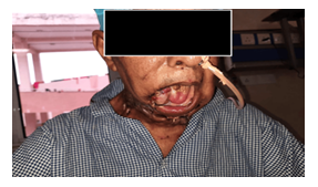
Figure 1: Frontal view showing retrognathia and distorted upper airway anatomy, which could be difficult to negotiate using a fiberoptic bronchoscope alone.
Based on the altered airway anatomy and clinical findings, difficult face-mask ventilation and tracheal intubation were anticipated. A stepwise airway management strategy was formulated. The patient and his family were counselled regarding the potential risks, including airway compromise, the possible need for emergency tracheostomy, postoperative mechanical ventilation, and intensive care unit (ICU) admission. Written informed consent was obtained. Awake nasal fiberoptic intubation was planned as the primary airway management technique because it was considered the safest and most feasible option available at our institution. Preoperatively, the patient received nebulization with 4 mL of 4% lignocaine in oxygen. Intramuscular glycopyrrolate was administered 30 minutes before the procedure as an ant sialagogue. Additional airway topicalization was achieved with 10% lignocaine spray applied to the faucial pillars. The tracheostomy scar was cleaned and covered with sterile gauze. The patient was preoxygenated with a high-flow nasal cannula delivering heated, humidified oxygen at 40 L/min through the left nostril. The nasal mucosa was prepared with xylometazoline drops, and serial dilatation of the right nostril was performed using a nasopharyngeal airway to facilitate passage of the endotracheal tube. A fiberoptic bronchoscope (Ambu a Scope 3) preloaded with a 7.0-mm flex metallic endotracheal tube was introduced through the right nostril. Although the epiglottis was identified, it was surrounded by debris and purulent secretions, which significantly limited advancement of the bronchoscope. In addition, the epiglottis appeared edematous and relatively immobile, making navigation into the glottic opening difficult. Therefore, a C-MAC video laryngoscope with a size 4 blade was introduced to improve visualization. Initial laryngoscopy revealed a Percentage of Glottic Opening (POGO) score of 0%, and the epiglottis could not be adequately elevated. After meticulous suctioning and clearance of the debris, visualization improved, and the epiglottis became sufficiently mobile to facilitate advancement of the bronchoscope.
The fiberoptic bronchoscope was then successfully advanced into the trachea up to the level of the carina. Under simultaneous visualization with the video laryngoscope and fiberoptic bronchoscope, the endotracheal tube was gently railroaded over the bronchoscope using a twisting motion, resulting in successful tracheal intubation (Figure 2).
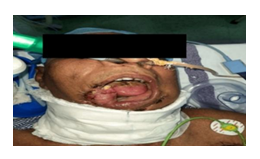
Figure 2: Awake nasal fiberoptic intubation performed using a combined video laryngoscope fiberoptic bronchoscope technique to facilitate successful tracheal intubation in a distorted airway.
Correct tube placement was confirmed by bilateral equal air entry and capnography, following which general anaesthesia was induced and the patient was placed on mechanical ventilation.
Discussion
Patients with head and neck malignancies frequently present with distorted airway anatomy, which makes airway management particularly challenging. In our patients, previous tracheostomy, extensive facial tissue loss, retrognathia, exposed neck structures, and purulent secretions created a high likelihood of difficult mask ventilation and tracheal intubation. Consequently, the maintenance of spontaneous ventilation during airway instrumentation is essential. Awake fiberoptic intubation remains the preferred approach for anticipated difficult airways because it allows continuous oxygenation while preserving the airway reflexes. Nevertheless, fiberoptic bronchoscopy has recognized limitations. Secretions, blood, edema, and anatomical distortion can obscure the view and hinder the advancement of the bronchoscope. In the present case, visualization of the glottic structures was compromised by purulent secretions and an edematous, relatively immobile epiglottis, which made it difficult to advance the bronchoscope. Video laryngoscopy has emerged as a valuable adjunct in difficult airway management, improving the visualization of the upper airway and facilitating the suctioning of secretions. Ji et al. described successful use of a C-MAC video laryngoscope in combination with a fiberoptic bronchoscope during airway management, demonstrating the complementary advantages of both devices [2]. Aziz et al. reported a 92% success rate for video laryngoscopy as a rescue technique following failed direct laryngoscopy, highlighting its usefulness as an effective rescue airway device [4]. The McGrath MAC video laryngoscope has also been successfully combined with other airway adjuncts in patients with challenging airway anatomy because its design resembles that of a conventional Macintosh laryngoscope, resulting in a relatively short learning curve and ease of use [5]. Similarly, previous reports have shown that combining video laryngoscopy and fiberoptic bronchoscopy can improve airway visualization and increase the likelihood of successful intubation in patients with severe obesity, neck contractures, and supraglottic masses [6,7]. In our patient, the introduction of the C-MAC video laryngoscope enabled the removal of secretions, improved visualization of the epiglottis, and facilitated the guidance of the fiberoptic bronchoscope toward the glottic opening. The combined video laryngoscope-fiberoptic bronchoscope technique ultimately allowed successful awake nasal intubation while maintaining spontaneous ventilation and avoiding emergency front-of-neck airway access. This case highlights the importance of having multiple airway strategies available and demonstrates that combining video laryngoscopy with fiberoptic bronchoscopy can be an effective and safe approach in patients with severely distorted airway anatomy
Conclusion
The combined use of fiberoptic bronchoscopy and video laryngoscopy can be used as an effective approach to facilitate tracheal intubation in the presence of distorted airway anatomy and obscured visualization.
References
- Sharma D, Kim LJ, Ghodke B. Successful airway management with combined use of Glidescope videolaryngoscope and fiberoptic bronchoscope in a patient with Cowden syndrome. Anesthesiology 113 (2010): 253-255.
- Ji S, Song J, Kim SK, et al. Fiberoptic bronchoscope and C-MAC video laryngoscope assisted nasal-oral tube exchange: two case reports. J Dent Anesth Pain Med 17 (2017): 219-223.
- Chung MY, Park B, Seo J, et al. Successful airway management with combined use of McGrath® MAC video laryngoscope and fiberoptic bronchoscope in a severe obese patient with huge goiter: a case report.Korean J Anesthesiol 71 (2018): 232-236.
- Aziz MF, Brambrink AM, Healy DW, et al. Success of intubation rescue techniques after failed direct laryngoscopy in adults: a retrospective comparative analysis from the Multicenter Perioperative Outcomes Group. Anesthesiology 125 (2016): 656-666.
- Karalapillai D, Darvall J, Mandeville J, et al. A review of video laryngoscopes relevant to the intensive care unit. Indian J Crit Care Med 18 (2014): 442-452.
- Park CD, Lee HK, Yim JY, et al. Anesthetic management for a patient with severe mentosternal contracture: difficult airway and scarce venous access: a case report. Korean J Anesthesiol 64 (2013): 61-64.
- Choi GS, Park SI, Lee EH, et al. Awake Glidescope® intubation in a patient with a huge and fixed supraglottic mass: a case report. Korean J Anesthesiol 59 (2010): 26-29.