Dr. Lokesh Singh*, Dr. Gita Devi, Dr. Uma Debi, Dr. Vikas Bhatia
Department of Radio diagnosis, PGIMER Chandigarh, India
*Corresponding Author: Dr. Lokesh Singh, Department of Radio diagnosis, PGIMER Chandigarh, India
Received: 09 April 2020; Accepted: 24 April 2020; Published: 28 April 2020
CECT; Clinical examination; Imaging; Reidel’s lobe
Clinical Image
A 34 year old man underwent CECT abdomen for complains of vague right lower quadrant region pain abdomen for last 24 days. Clinical examination revealed no palpable lump/mass, no abnormal bowel sounds, pain had no relation with meals. CECT examination revealed presence of a tongue like, inferior projection of right lobe of liver beyond the level of inferior cartilage. Projection measured 7.8 mm (AP) × 30 mm (TR) × 42 mm (CC) dimensions. No mass lesion noted within the projection. Mild ileo cecal thickening and pulled up IC junction seen with subcentimetric mesenteric lymph nodes. Other findings were cholelithasis and elongated non inflamed retrocecal appendix abutting Reidel’s lobe of liver. Rest of imaging findings were unremarkable.
Reidel’s lobe is an anatomic variation described by Reidel in 1888 [1]. The incidence of Riedel’s lobe varies in the general population (3.3%-31%) but typical case is rare [2-4]. It is important to be aware of the complications due to Reidel’s lobe notably malignancy and torsion. Our patient did not present with any palpable mass and significant symptoms, subsequently was referred to Gastroenterology for further evaluation.
The presence of Riedel’s lobe is presented with minor symptoms of acute or recurrent abdominal discomfort, nausea, constipation, or bloating caused by extrinsic compression or episode of torsion [5-7]. Cross sectional imaging namely CT, MRI with Ultrasonography plays a major role in establishing diagnosis and follow up of these patients and forms the mainstay in evaluation.
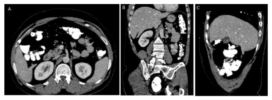
Figure 1A-1C: Transaxial CECT image showed tongue like projection (1A) projecting downwards below the inferior costal cartilage as shown in coronal (1B) and sagittal (1C) closely abutting retrocecal non inflamed appendix in (1A) and (1C).
Conflicts of interest
Nil
Funding
Nil
References
- Riedel I. Ueber den zungenfrmigen Fortsatz des rechten Leberlappens und seine pathognostische Bedeutung für die Erkrankung der Gallenblase nebst Bemerkungen über Gallensteinoperationen. Berliner klinische Wochenschrift 25 (1888): 577-602.
- Sham R, Sain A, Silver L. Hypertrophic Riedel's lobe of the liver. Clin Nucl Med3 (1978): 79-81.
- Baum S, Locko RC, d’Avignon MB. Functional anatomy and radionuclide imaging: Riedel's lobe of the liver. Anat Clin4 (1982): 121-123.
- Gillard JH, Patel MC, Abrahams PH, et al. Riedel's lobe of the liver: fact or fiction? Clin Anat11 (1998): 47-49.
- Glennison M, Salloum C, Lim C, et al. Accessory liver lobes: anatomical description and clinical implications. Jour Visc Surg 151 (2014): 451-455.
- Christos Savopoulos. Riedel’s Lobe of the Liver- A Case Report. Medicine 94 (2015): 1-3.
- Juferdy Kurniawan. Riedel’s Lobe: Clinical Importance of a Rare Variant in Liver Morphology. Acta Med Indones - Indones J Intern Med 49 (2017): 57-62.