Sophia Steinlein1,2, Annette M Staiger1,2, Katrin S. Kurz2,3, Viola Poeschel4,5, Gerhard Held6, Roland E Kontermann7, German Ott2, Heike Horn1,2*, Claudia Kalla1,2
1Dr. Margarete Fischer-Bosch Institute of Clinical Pharmacology, Stuttgart and University of Tuebingen, Tuebingen, Germany
2Department of Clinical Pathology, Robert-Bosch-Krankenhaus, Stuttgart, Germany
3Department of Clinical Pathology, Klinikum Wels-Grieskirchen, Wels, Austria
4Department of Internal Medicine 1, Saarland University Medical School, Homburg/Saar, Germany
5Department of Clinical Research, University Hospital Basel and University of Basel, Basel, Switzerland
6Internal Medicine 1, Westpfalz-Klinikum Kaiserslautern, Kaiserslautern, Germany
7Institute of Cell Biology and Immunology and Stuttgart Research Center for Systems Biology, University of Stuttgart, Stuttgart, Germany
*Corresponding author: Heike Horn. Dr. Margarete Fischer-Bosch Institute of Clinical Pharmacology, Stuttgart and University of Tuebingen, Tuebingen, Germany. Department of Clinical Pathology, Robert-Bosch-Krankenhaus, Stuttgart, Germany.
Received: 31 July 2026; Accepted: 07 August 2026; Published: 12s August 2026
Therapy resistance in diffuse large B-cell lymphoma (DLBCL) is mediated, in part, by stromal protection of malignant B cells. Carnitine palmitoyltransferase 1A (CPT1A) is a key regulator of fatty acid transport into mitochondria, including fatty acids supplied by stromal cells, thereby supporting lymphoma cell metabolism. We investigated the CPT1A inhibitor ST1326 for its ability to eliminate lymphoma cells and enhance chemotherapy efficacy in the presence of stromal cells. CPT1A expression in DLBCL patient samples was assessed by immunohistochemistry. The therapeutic efficacy of ST1326 was evaluated in 13 DLBCL cell lines by MTT assay. ATP levels, lipid accumulation, and intracellular doxorubicin were measured using CellTiter-Glo, ORO staining and flow cytometry, respectively. Stromal co-culture models of DLBCL cell lines with HS-5 stromal cells or primary DLBCL-CAFs, combined with the Chou–Talalay analysis, were used to assess stromal-mediated protection and drug synergy. CPT1A was expressed in 51% (95/186) of primary DLBCL cases. ST1326 reduced viability in all 13 DLBCL cell lines without affecting non-malignant cells and retained activity in stromal co-cultures. Suppression of fatty acid transport into mitochondria by ST1326 resulted in ATP depletion and cytoplasmic lipid accumulation. The combination of ST1326 with vincristine or doxorubicin enhanced their cytotoxic efficacy and increased the intracellular accumulation of doxorubicin, an effect that persisted in the presence of stromal cells. Targeting the fatty acid metabolism using ST1326 effectively eliminates lymphoma cells in the presence of stromal cells and increases the efficacy of standard chemotherapeutic agents, thus providing a new candidate approach in the treatment of DLBCL.
Carnitine palmitoyltransferase 1A; DLBCL; Stromal microenvironment; ST1326; R-CHOP; Fatty acid oxidation.
Carnitine palmitoyltransferase 1A articles; DLBCL articles; Stromal microenvironment articles; ST1326 articles; R-CHOP articles; Fatty acid oxidation articles.
Introduction
Diffuse large B-cell lymphoma (DLBCL) is the most common type of malignant lymphoma in the Western hemisphere. Gene expression profiling has identified two major DLBCL subtypes based on their cell of origin (COO): germinal center B-cell-like (GCB) and activated B-cell-like (ABC), as well as a third "unclassified" group [1]. Several studies have suggested that patients with ABC-DLBCL exhibit significantly poorer survival than those with GCB-DLBCL [2]. Currently, first-line immunochemotherapy (R-CHOP) fails to achieve durable responses in one third of the patients. Therapy resistance is mediated partly by ineffective elimination of B-cells protected by metabolic tumor-stroma interactions.3 Bystander cells supply free fatty acids to neoplastic B-cells, thereby providing an alternative energy source through fatty acid β-oxidation (FAO) and contributing to chemotherapy resistance against anti-cancer drugs, such as doxorubicin and vincristine [3,4]. Moreover, an altered fatty acid metabolism was uncovered as a major oncogenic factor in DLBCL [5,6].
Carnitine palmitoyltransferase 1A (CPT1A) is a protein that catalyzes the rate-limiting step of FAO, conversion of acyl-CoA to acylcarnitine, to support cancer cell metabolism and energy production (Figure 1A).
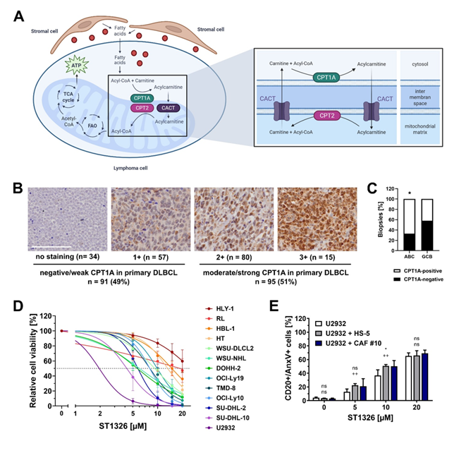
Figure 1: CPT1A protein is expressed in DLBCL and inhibition of CPT1A impairs DLBCL cell viability. (A) CPT1A catalyzes the entry of fatty acids, partly provided by stromal cells, into mitochondria by loading fatty acyl-groups onto carnitine. The carnitine shuttle system includes the membrane proteins carnitine palmitoyltransferase 1A and 2 (CPT1A/CPT2) and the transmembrane carnitine/acylcarnitine carrier protein (CACT). The illustration was created with BioRender.com. (B) CPT1A protein expression was assessed in 186 primary DLBCL specimens on tissue microarray format by evaluating the intensity of intracellular immunostaining: 0/no staining; 1+/faint; 2+/moderate; 3+/strong staining in at least 20% tumor cells. Scale bar, 100 µm. (C) Moderate/strong CPT1A protein expression was more frequent in activated B-cell-like (ABC)-DLBCL than in germinal center B-cell-like (GCB) subtype 67% [28/42] vs. 42% [18/43]; P = 0.03, two-sided Fisher’s exact test). (D) CPT1A inhibitor induced a concentration-dependent decrease of DLBCL cell viability. Cells were incubated with ST1326 (5-20 µM) for 72 h in culture medium containing 10% FBS. Viability was assessed using a MTT assay. Relative viability was calculated by normalizing absorbance values to those from cells grown in media without drug after background subtraction. Data points, mean ±SD of independent triplicate experiments. (E) In the presence of stromal cells, ST1326 remained effective in killing DLBCL cells. DLBCL cell line U2932 was cultured alone or in co-culture with HS-5 (grey) or cancer-associated fibroblasts isolated from primary DLBCL specimens (CAF, dark blue) and incubated with ST1326 for 72 h. The percentage of apoptotic DLBCL cells (CD20+/AnxV+ cells) was assessed by flow cytometry (mean ±SD, n = 3). Statistical significance was calculated using the Student t test (unpaired, 2-tailed; U2932+HS-5, ns≥0.05, ++p<0.01; U2932+CAF#11, ns≥0.05, *p<0.05).
Targeting CPT1A has shown remarkable anti-cancer activity in hematological neoplasms: the CPT1A inhibitor ST1326 decreased cell growth of lymphoma and leukemia cells in preclinical studies (Burkitt’s lymphoma, chronic lymphatic leukemia, acute myeloid leukemia (AML), acute lymphoblastic leukemia) [7-10]. In DLBCL, CPT1A is a component of the prognostic lymphoma-associated macrophage interaction gene expression signature (LAMIS), being associated with the protective impact of the microenvironment on therapy response [11]. In keeping with that, CPT1A overexpression has recently been identified as a prognostic mitochondria-related gene in DLBCL [12].
To date, no study exits exploring the clinical potential of a targeted CPT1A therapy in DLBCL. Thus, we investigated the CPT1A inhibitor ST1326 (Teglicar) for its ability to eliminate lymphoma cells in the presence of stromal bystander cells and to sensitize resistant cells to the chemotherapeutic agents doxorubicin and vincristine.
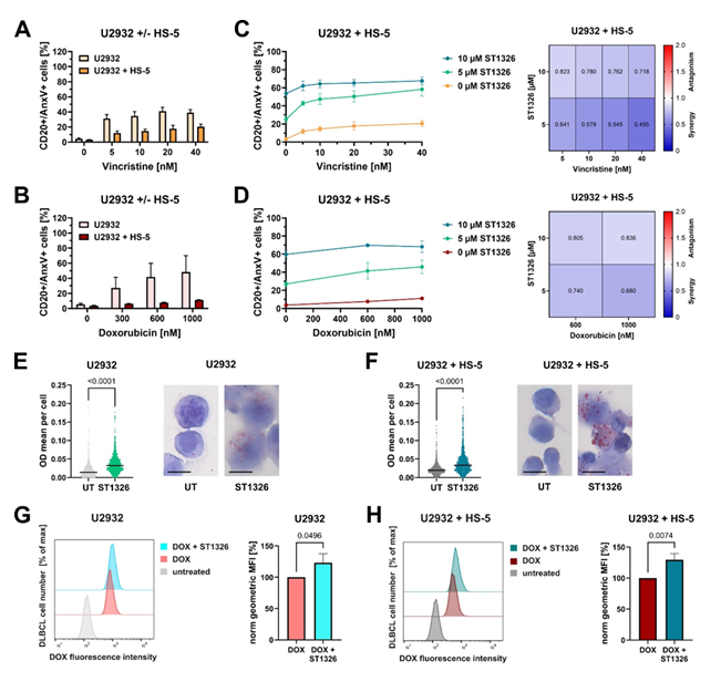
Figure 2: ST1326 enhances doxorubicin and vincristine efficacy in DLBCL cells in the presence of stromal cells via inhibition of FAO und transporter-mediated efflux of doxorubicin (A, B) Stromal protection decreased the chemotherapeutic sensitivity of DLBCL cells to vincristine and doxorubicin. DLBCL cell line U2932, cultured alone or in combination with stromal cells HS-5 for 36 h, was treated with indicated concentrations of vincristine (A) or doxorubicin (B) for a further 36 h. Apoptotic DLBCL cells (CD20+/AnxV+ cells) were determined by flow cytometry (mean ±SD, n = 3). (C, D) ST1326 increased the cytotoxic effect of vincristine and doxorubicin in DLBCL cells co-cultured with stromal cells. U2932 cells were cultivated alone or in combination with stromal cells HS-5 and incubated with indicated concentrations of ST1326 for 72 h. 36 h prior end of therapy, ascending concentrations of vincristine (C) or doxorubicin (D) were added. The percentage of apoptotic DLBCL cells (CD20+/AnxV+ cells) was determined by flow cytometry (mean ±SD, n = 3). Combination analysis was performed using CompuSyn algorithm generating a combination index (CI) of drug combinations (n = 3). (E, F) Cytoplasmic lipid accumulation in DLBCL cells induced by CPT1A inhibition. Hematoxylin and Oil red O (ORO) staining of U2932 cells after 72 h treatment with 10 µM ST1326 in mono- (E) or co-culture with stromal cells HS-5 (F). Oil Red O staining highlights the cytoplasmatic lipid accumulations as red droplets. Scale bar, 10 µm. Red staining was evaluated via the OD mean per cell with QuPath. N = 850 – 1200 cells, out of one experiment. (G, H) ABC transporter-mediated efflux of doxorubicin was reduced by ST1326 treatment. DLBCL cells of U2932 in mono- (G) or co-culture with stromal cells HS-5 (H) were treated with 10 µM ST1326 for 36 h, before addition of 3 µM doxorubicin for further 3 h. Fluorescence intensity of doxorubicin in DLBCL cells was detected by flow cytometry. Histogram profiles of the doxorubicin fluorescence intensity were obtained by gating in FSC/SSC (left panel). Geometric mean of fluorescence intensity (MFI) was determined using FlowJo Software and normalized to the MFI of DLBCL cells treated with doxorubicin exclusively (right panel) (mean ±SD, n = 3). (E-H) Statistical significance was calculated using the Student t test (unpaired, 2-tailed).
Materials and Methods
Formalin-fixed, paraffin-embedded (FFPE) tumor samples from patients diagnosed with DLBCL and enrolled in prospective clinical trials of the DSHNHL/GLA (Deutsche Studiengruppe für hochmaligne Non-Hodgkin Lymphome: German High Grade Lymphoma Study Group/German Lymphoma Alliance) were analyzed in this study (Supplementary Table S1). Only samples from patients who received rituximab-containing therapy were included (n = 186). Protein expression was assessed by immunohistochemistry on tissue microarrays (TMAs) and FFPE-embedded DLBCL cell lines. Functional studies were performed using DLBCL cell lines, the bone marrow stromal cell line HS-5, primary cancer-associated fibroblasts (CAFs), primary fibroblasts isolated from reactive lymph nodes and primary B cells. Mono- and co-culture assays were combined to assess cell viability, ATP production, lipid staining, flow cytometry and drug combination studies. Detailed descriptions of the study cohorts, antibodies and reagents used, experimental procedures, and statistical analyses are provided in the Supplementary Information.
Results
Expression and ST1326-mediated inhibition of CPT1A in DLBCL
As a first step, we assessed the expression of CPT1A protein in primary DLBCL specimens in order to estimate the clinical potential of a CPT1A-based therapy. Immunostaining revealed CPT1A protein expression (moderate or strong reactivity) in 51% (95/186) of samples (Figure 1B, Supplementary Table S1). Moderate/strong CPT1A expression was more prominent in the activated B-cell-like (ABC) subtype of DLBCL than in the germinal center B-cell-like (GCB) subtype (67% [28/42] vs. 42% [18/43]; P = 0.03) (Figure 1C). To assess the direct effect of CPT1 inhibition on tumor cells, we investigated whether treatment with ST1326 (CPT1/CACT inhibitor) would affect the survival and proliferation of 13 DLBCL cell lines with different levels of CPT1 expression. 62% (8/13) cell lines had moderate or strong CPT1A protein expression and all cell lines expressed the CACT (carnitine/acylcarnitine carrier protein) protein (Supplementary Figure S1A, Supplementary Table S2). ST1326 impaired cell viability and proliferation in all 13 DLBCL cell lines, leading to a reduction of cell viability by 60-100%, independent of the COO subtype. However, there was a trend towards reduced EC50 values with increased CPT1A expression intensity, although not statistically significant. ST1326 was effective even in cells that had only low levels of CPT1A protein (SU-DHL-2, TMD-8) or that expressed CACT only (DOHH-2). (Figure 1D, Supplementary Figure S1B, Supplementary Table S2, EC50: 1.91-15.23 µM). The viability of non-tumor cells, i.e. fibroblasts and human primary B cells, was not affected by the CPT1/CACT inhibitor (Supplementary Figure S1 C,D). Together, CPT1A/CACT-targeted therapy might be suitable for treating DLBCL, including the more aggressive ABC subtype.
CPT1A inhibition attenuates stroma-mediated support of DLBCL cells
To simulate the stromal microenvironment, lymphoma cell lines were co-cultured with the bone marrow stromal cell line HS-5 or primary DLBCL cancer-associated fibroblasts. Of note, ST1326 remained effective in killing DLBCL cells also in the stromal environment (Figure 1E; Supplementary Figure S1E).
Based on our observation that ST1326 is efficient even in the presence of stromal cells, we hypothesized that ST1326-treatment can enhance doxorubicin and vincristine efficacy in chemoresistant DLBCL cells. In our in vitro experiments, the presence of stromal cells clearly reduced cell death of DLBCL cells induced by vincristine or doxorubicin as components of the R-CHOP regime (Figure 2 A,B; Supplementary Figure S2 A,B). Of note, DLBCL cell lines that became resistant to vincristine and doxorubicin in the presence of stromal cells show enhanced sensitivity to both chemotherapeutics when co-treatment with ST1326 was applied (Figure 2 C,D; Supplementary Figure S2 C,D). Of importance, cell viability of HS-5 was not impaired following cytotoxic treatment (Supplementary Figure S3). Chou-Talalay analysis using the CompuSyn software indicated that the combination of ST1326 and vincristine or doxorubicin enhances therapeutic efficacy over various concentrations (CI < 1) (Figure 2 C,D; Supplementary Figure S2 C,D). Therefore, this combined treatment might represent a promising strategy to sensitize DLBCL cells to therapy regimens prone to reduction of efficacy by the stromal microenvironment.
ATP depletion and disruption of lipid metabolism induced by CPT1A inhibition
Mechanistically, FAO is a major catabolic process that degrades fatty acids to produce high levels of ATP ensuring cancer cell survival, especially during decreased energy intake or environmental substrate limitations. In line with this, inhibition of CPT1A as a rate-limiting enzyme of FAO via ST1326 reduced the amount of ATP in DLBCL cell lines within the first 24h also in the presence of stromal cells, and prior to affecting cellular viability (Supplementary Figure S4). Moreover, we investigated whether CPT1A inhibition by ST1326 induces intracellular lipid accumulation, which would indicate impaired oxidative lipid metabolism. Oil Red O staining of intracellular neutral triglyceride and lipids revealed that DLBCL cells accumulated lipids in their cytoplasm under exposure of ST1326, as highlighted by red droplets (Figure 2E, Supplementary Figure S5A). This impaired lipid metabolism was maintained even in the presence of stromal cells (Figure 2F, Supplementary Figure S5B).
Due to the attenuated lipid metabolism and followed by ATP depletion, inhibition of CPT1A might contribute to energy-based mechanisms of chemoresistance in lymphoma cells. Mitochondria-produced ATP is the main energy source for ATP binding cassette (ABC) transporters mediating drug-efflux and decreasing the intracellular accumulation of chemotherapeutic drugs [13]. Doxorubicin and vincristine are substrates for the ABC transporter proteins ABCB1, ABCC1, ABCC2 and ABCG2 [14,15]. To test whether CPT1A inhibition affects the intracellular accumulation of R-CHOP components, we assessed the amount of doxorubicin in DLBCL cell lines treated with ST1326 by flow cytometry. After combined ST1326/doxorubicin therapy, DLBCL cells showed an increased fluorescence intensity of doxorubicin compared to cells treated with doxorubicin exclusively (Figure 2G, Supplementary Figure S5C). These elevated levels of doxorubicin were retained in DLBCL cells even when co-cultured with stromal cells (Figure 2H, Supplementary Figure S5D).
Discussion
As an essential component of the carnitine shuttle, CPT1A plays a pivotal role in mediating fatty acid transport into mitochondria, thereby supporting cancer cell metabolism and energy production. Since fatty acids are partially supplied by stromal bystander cells [16,17]. CPT1A emerge as compelling therapeutic target particularly given its contribution to the prognostic LAMI signature identified in DLBCL11. Inhibiting CPT1A and/or CACT could disrupt metabolic crosstalk between tumor and stromal cells, representing a novel strategy to impair DLBCL growth and enhance the efficacy of doxorubicin and vincristine. Supporting this hypothesis, previous studies demonstrated that inhibition of CPT1A effectively eliminated chronic lymphocytic leukemia (CLL) cells within the stromal microenvironment by blocking mitochondrial fatty acid transport [8,18]. Similarly, targeting cellular metabolism by inhibiting CPT1A impaired the proliferation of both human leukemia cell lines and primary cells [9,10]. Furthermore, inhibition of CPT1A/CACT exerted potent cytotoxic effects on Burkitt’s lymphoma cells in vitro and prevented MYC-induced lymphomagenesis in vivo [7]. In DLBCL, CPT1A knockdown significantly reduced DLBCL cell proliferation while promoting apoptosis [19]. In our study ST1326 was effective even in cells expressing low levels of CPT1A or exclusively expressing CACT, which is in line with data by Pacilli et al.7 who demonstrated that ST1326 inhibits FAO not only by blocking CPT1A but also by inhibiting CACT activity. Although CPT1A expression was more prominent in the ABC subtype of DLBCL specimens, the cytotoxic efficacy of ST1326 was independent of the COO-subtype in cell lines. These findings suggest that the therapeutic potential of ST1326 is not limited to ABC-DLBCL but may extend to DLBCL irrespective of COO subtype. Collectively, these data underscore CPT1A and CACT as promising therapeutic targets - particularly within the presence of stromal microenvironment. Further, equivalent stromal protection against chemotherapeutic drug-induced apoptosis was demonstrated in AML cells co-cultured with the marrow stromal cell line MS-5 [20]. Consistent with our results, ST1326 treatment also induced cytoplasmic lipid accumulation in Burkitt lymphoma and leukemia cell lines, further supporting its role in disrupting mitochondrial fatty acid metabolism [7,10]. The increased intracellular accumulation of chemotherapeutic agents suggests that ST1326 impairs ABC transporter-mediated drug efflux, a hypothesis that requires further mechanistic investigation. Accordingly, CPT1A inhibition enhances the sensitivity of DLBCL cells to selected chemotherapeutic agents. Importantly, this effect was maintained in co-culture with non-malignant bystander cells, despite their lack of response to R-CHOP treatment alone.
Conclusion
In conclusion, targeting fatty acid metabolism using ST1326 effectively eliminates lymphoma cells in the presence of stromal cells and enhances activity of standard chemotherapeutic agents, thus providing a new approach for treating DLBCL, particularly in patients with refractory or recurrent disease.
Acknowledgements
The study was supported by the Robert Bosch Stiftung, Stuttgart, Germany (Project O3 and KKF796) (G.O., H.H., A.M.S.) and the José Carreras Leukämie Stiftung, DJCLS 03 FN/2020 (S.S., G.O., H.H., C.K.). We thank Katja Bräutigam and Emma Attwood, IKP Stuttgart, for expert technical assistance.
Conflict of Interest
The authors declare no conflicts of interest.
Authorship
Contribution: S.S. and C.K. performed the research. A.M.S., K.S.K., V.P. and G.H. collected and assembled clinical and biological data. G.O. and K.S.K. were part of the pathology reference panel and provided study material of the patients. S.S., H.H., R.E.K., G.O. and C.K. were responsible for the conception and design of the study. S.S., H.H. and C.K. interpreted data and wrote the manuscript. S.S., H.H., A.M.S, G.O. und C.K. acquired the funding. All authors contributed to the article and approved the submitted version.
Correspondence: Heike Horn, Dr. Margarete Fischer-Bosch Institute of Clinical Pharmacology, Stuttgart and University of Tübingen, Tübingen, Germany; Department of Clinical Pathology, Robert-Bosch-Krankenhaus, Stuttgart, Germany; e-mail: heike.horn@ikp-stuttgart.de
Supplementary Information
Detailed methodology and supplemental data have been included as Supplementary Information.
References
- Alizadeh AA, Eisen MB, Davis RE, et al. Distinct types of diffuse large B-cell lymphoma identified by gene expression profiling. Nature 403(2000): 503-511.
- Rosenwald A, Wright G, Chan WC, et al. The use of molecular profiling to predict survival after chemotherapy for diffuse large-B-cell lymphoma. The New England journal of medicine 346 (2002): 1937-1947.
- Lee J, Mani A, Shin M-J, Krauss RM. Leveraging altered lipid metabolism in treating B cell malignancies. Progress in Lipid Research 95 (2024): 101288.
- Gentric G, Mechta-Grigoriou F. Tumor Cells and Cancer-Associated Fibroblasts: An Updated Metabolic Perspective. Cancers 13 (2021): 399.
- Caro P, Kishan AU, Norberg E, et al. Metabolic signatures uncover distinct targets in molecular subsets of diffuse large B cell lymphoma. Cancer cell 22 (2012): 547-560.
- Peeters R, Cuenca-Escalona J, Zaal EA, et al. Fatty acid metabolism in aggressive B-cell lymphoma is inhibited by tetraspanin CD37. Nat Commun 13 (2022): 1-18.
- Pacilli A, Calienni M, Margarucci S, et al. Carnitine-acyltransferase system inhibition, cancer cell death, and prevention of myc-induced lymphomagenesis. Journal of the National Cancer Institute 105 (2013): 489-498.
- Gugiatti E, Tenca C, Ravera S, et al. A reversible carnitine palmitoyltransferase (CPT1) inhibitor offsets the proliferation of chronic lymphocytic leukemia cells. Haematologica 103 (2018): e531-e536.
- Mao S, Ling Q, Pan J, et al. Inhibition of CPT1a as a prognostic marker can synergistically enhance the antileukemic activity of ABT199. Journal of translational medicine 19 (2021): 181.
- Ricciardi MR, Mirabilii S, Allegretti M, et al. Targeting the leukemia cell metabolism by the CPT1a inhibition: functional preclinical effects in leukemias. Blood 126 (2015): 1925-1929.
- Staiger AM, Altenbuchinger M, Ziepert M, et al. A novel lymphoma-associated macrophage interaction signature (LAMIS) provides robust risk prognostication in diffuse large B-cell lymphoma clinical trial cohorts of the DSHNHL. Leukemia 34 (2020): 543-552.
- Zhou Z-Z, Lu J-C, Guo S-B, et al. A Mitochondria-Related Signature in Diffuse Large B-Cell Lymphoma: Prognosis, Immune and Therapeutic Features. Cancer medicine 14 (2025): e70602.
- Giddings EL, Champagne DP, Wu M-H, et al. Mitochondrial ATP fuels ABC transporter-mediated drug efflux in cancer chemoresistance. Nat Commun 12 (2021): 2804.
- Andreadis C, Gimotty PA, Wahl P, et al. Members of the glutathione and ABC-transporter families are associated with clinical outcome in patients with diffuse large B-cell lymphoma. Blood 109 (2007): 3409-3416.
- Xiao H, Zheng Y, Ma L, et al. Clinically-Relevant ABC Transporter for Anti-Cancer Drug Resistance. Front Pharmacol 12 (2021): 648407.
- Corn KC, Windham MA, Rafat M. Lipids in the tumor microenvironment: From cancer progression to treatment. Progress in Lipid Research 80 (2020): 101055.
- Kim TH, Lim SH, Lee H, et al. Metabolic crosstalk among cancer-associated fibroblasts, adipocytes and immune cells as an immunosuppressive tumor microenvironment driver. Exp Mol Med (2026): 1-16.
- Liu P-P, Liu J, Jiang W-Q, et al. Elimination of chronic lymphocytic leukemia cells in stromal microenvironment by targeting CPT with an antiangina drug perhexiline. Oncogene 35 (2016): 5663-5673.
- Wang S, Wang F, Li M, et al. Identification of a fatty acid metabolism-related gene signature for prognostic prediction and immune microenvironment characterization in diffuse large B-cell lymphoma. Front Oncol 16 (2026): 1798939.
- Li X, Guo H, Duan H, et al. Improving chemotherapeutic efficiency in acute myeloid leukemia treatments by chemically synthesized peptide interfering with CXCR4/CXCL12 axis. Scientific reports 5 (2015): 16228.