Gustavo Eduardo Sánchez Martínez*ORCID ID
General Surgery Department Hospital General, Dr. Carlos Canseco. Tampico, Tamaulipas, 89130, Mexico.
*Corresponding Author: Gustavo Eduardo Sánchez Martínez, General Surgery Department Hospital General de Cd. Mante “Dr Emilio Martínez Manatou”. 89880 Tamaulipas, Mexico.
Received: 31 August 2026; Accepted: 07 September 2026; Published: 14 October 2026
This is a case of a patient with intestinal obstruction secondary to the presence of a tumor. This is a complex pathology that, despite having a low prevalence, produces a high degree of discomfort for the patient due to the symptoms caused by mechanical and functional alterations in the various pathophysiological mechanisms, such as the occlusion of the intestinal lumen, alterations in motility, and accumulation of secretions. For treatment, a strategy that combines different options is recommended to achieve adequate management.
Introduction and importance: Intestinal obstruction is defined as the complete and persistent cessation of intestinal transit in a section of the small or large intestine [1]. This condition represents one of the most common causes of acute abdomen, accounting for 20-35% of surgical-related emergency room admissions, which poses a surgical challenge and a public health issue due to the inherent risks for the patient and the associated economic costs [2].
In the United States, the incidence of intestinal obstruction is 1.47 per 100,000 people per year.
Case presentation: Male, 58 years old, family history questioned and denied, history of chronic degenerative diseases (-), surgical history (-), allergies (-). He reports that he started experiencing his condition 20 days prior to admission, with oral intolerance, inability to defecate, no passing of gas, generalized abdominal pain rated 7/10 on the visual analog scale, as well as a fever of 39°C and a heart rate of 120 bpm, leading him to visit the emergency room of our hospital on 10/16/2024. Vital signs show blood pressure of 150/90 mHg,
heart rate of 120 bpm, respiratory rate of 22 breaths per minute, temperature of 39°C, and SpO2 of 99%.
Physical examination, Glasgow score 15/15, non-focal, normocephalic head, pupils equal and reactive, dehydrated mucous membranes. Neck is cylindrical, with no palpable lymphadenopathy. Chest is normal in configuration, clavicles with no loss of continuity, rhythmic precordium, no murmurs, lung fields wellventilated, with no rales or crackles. Abdomen is distended, with high-pitched bowel sounds, tympanic, with no audible peristalsis, generalized colicky pain, muscular rigidity, with no signs of acute abdomen. Limbs are intact and
functional.
A colonoscopy study was conducted, which reports a tumor 50 cm from the anal margin that obstructs the passage of the endoscope, suggestive of malignant neoplasia.
An exploratory laparotomy was performed, revealing a stenosing tumor of 2 cm in the descending colon, leading to a left hemicolectomy and end-to-side anastomosis. The patient underwent intra-hospital recovery and was started on clear liquids on the third day, with no signs of leakage, allowing for diet progression. The patient reported passing gas and having bowel movements, and discharge was decided on the fifth day. The specimen was sent to pathology, and results are awaited.
Intestinal obstruction, Surgery, Colonoscopy
Intestinal obstruction articles; Surgery articles; Colonoscopy articles.
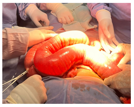
Figure 1: Sectioning of the transverse colon and release of the colonic framework.
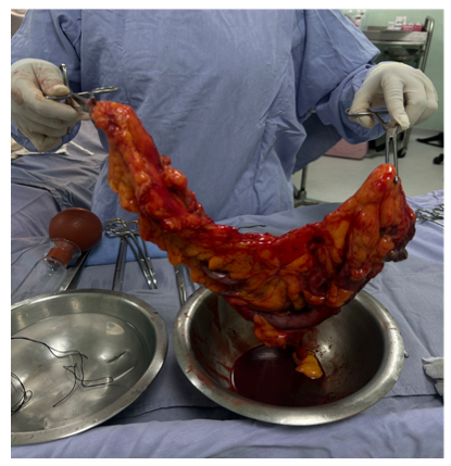
Figure 2: Stenosing tumor of 2 cm in the descending colon with an implant in the distal omentum of 1 cm.
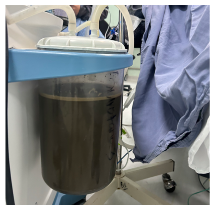
Figure 3: 3000 ml of intestinal contents were drained.
Clinical discussion
Malignant large-bowel obstruction (MLBO) is both a mechanical emergency and an oncologic event. Colorectal cancer remains the leading cause of large-bowel obstruction in adults and accounts for the majority of low intestinal occlusions; between 15% and 30% of colorectal cancers present as an emergency, and obstruction is the most frequent of those presentations [3]. Approximately 10–29% of patients with colon cancer will develop malignant bowel obstruction at some point in the disease course. More than three-quarters of obstructing tumours lie at or distal to the transverse colon, where the lumen is narrower; the sigmoid is the most common site, and tumours of the descending colon and splenic flexure also have a high obstructive potential. This topography matches the present case: a stenosing 2-cm mass in the descending colon, 50 cm from the anal verge on colonoscopy [4].
The pathophysiology explains the 20-day, subacute course and the systemic inflammatory response observed on admission. Intraluminal occlusion by the primary tumour interrupts propulsion, with progressive accumulation of gas, unabsorbed fluid and secretions proximal to the stenosis. Raised intramural pressure, mural oedema, splanchnic vasodilatation and hypersecretion—mediated in part by vasoactive intestinal peptide and other enteric neurotransmitters—create a self-perpetuating cycle of distension. Bacterial overgrowth and translocation then produce fever and tachycardia even before frank perforation. The 3,000 mL of intestinal contents drained at laparotomy illustrate this closed-loop physiology. Large-bowel obstruction typically evolves more slowly than small-bowel obstruction; a history measured in days to weeks, rather than hours, should therefore raise suspicion for a left-sided colorectal neoplasm rather than adhesive disease.
Diagnosis starts with the classic tetrad of colicky pain, abdominal distension, obstipation and vomiting, supported by laboratory markers of dehydration and systemic inflammatory response. Contrast-enhanced computed tomography is the imaging modality of choice: it confirms the transition point, estimates the likelihood of ischaemia or perforation, and contributes to oncologic staging. Colonoscopy, as performed here, remains useful when the patient is stable enough, because it localises the lesion, allows biopsy and can, in selected centres, serve as a platform for stent placement. It does not, however, replace cross-sectional staging.
Management of obstructing left-sided colon cancer must solve two problems at once: decompression of a potentially life-threatening mechanical obstruction and an oncologically adequate resection in a patient who may already harbour regional or peritoneal disease. The World Society of Emergency Surgery (WSES) recommends resection with primary anastomosis as the preferred option in uncomplicated left-sided malignant obstruction when patient and surgeon factors are favourable, and reserves Hartmann’s procedure for high-risk patients (perforation, faecal peritonitis, profound instability or poor physiological reserve) [5]. Total or subtotal colectomy is not superior to segmental resection in the absence of caecal ischaemia, perforation or a synchronous right-sided tumour, and it worsens bowel function. European and American endoscopic societies further recommend that self-expandable metallic stents (SEMS) be discussed, in experienced centres and after CT exclusion of perforation, as a bridge to elective surgery in potentially curable left-sided obstruction; stenting increases the rate of primary anastomosis, reduces permanent stoma and lowers short-term morbidity compared with emergency resection, although perforation of the stent tract may compromise oncologic outcomes [6,7]. In unresectable or widely metastatic disease, SEMS is the preferred palliative intervention when technically feasible.
In this 58-year-old patient without major comorbidity, with a single stenosing lesion, no free perforation and a proximal colon that remained viable after decompression, left hemicolectomy with end-to-side anastomosis was a guideline-concordant one-stage strategy. The early restoration of flatus and stool and discharge on postoperative day 5 support the safety of that choice in this individual. Nonetheless, fever of 39 °C and tachycardia of 120 bpm at presentation are markers of physiological stress; in a frailer patient the same findings would have favoured Hartmann’s procedure or a decompressive stoma followed by delayed oncologic resection [8,9].
Two findings must temper an exclusively “successful mechanical” reading of the case. First, the 1-cm implant in the distal omentum raises the possibility of peritoneal dissemination (M1c in the TNM framework) and converts what might have been a locally advanced primary into potentially stage IV disease [10]. Isolated omental deposits do not preclude resection of the primary when the obstruction is complete, but they mandate complete staging (thoraco-abdomino-pelvic CT, serum CEA, mismatch-repair status) and early referral to medical oncology. Bowel obstruction itself is a high-risk feature in stage II colon cancer and is among the criteria that support adjuvant chemotherapy after complete resection. Second, histopathology was still pending at the time of writing. Without grade, T and N categories, margin status, lymph-node yield (≥12 nodes) and molecular profile, neither prognosis nor adjuvant strategy can be defined. These data should be incorporated before publication [11,12].
A multidisciplinary pathway—emergency surgeon, endoscopist, radiologist, pathologist and oncologist—is therefore not an accessory but the standard of care. Conservative measures (nasogastric decompression, fluid and electrolyte resuscitation, analgesia and antibiotics when translocation is suspected) remain the first hours of treatment; they do not replace source control when obstruction is complete, symptoms persist, or warning signs of ischaemia or peritonitis appear [13,14].
This report is limited by the incomplete imaging and pathology record and by the short in-hospital follow-up. It does, however, illustrate that a one-stage oncologic resection with primary anastomosis can restore intestinal continuity promptly in a selected patient with left-sided obstructing colon cancer, provided that peritoneal findings and the definitive histological stage subsequently guide adjuvant and surveillance decisions.
Methods
This case report has been reported in line with the SCARE Criteria (11).
Conclusion
Obstructing colorectal carcinoma is the principal cause of large-bowel obstruction and often declares itself over days rather than hours. In left-sided disease, treatment must decompress the colon and respect oncologic principles at the same time. When there is no perforation, the proximal bowel is viable and the patient retains adequate physiological reserve, segmental resection with primary anastomosis—as performed here—is an accepted one-stage option. Stenting as a bridge to elective surgery and Hartmann’s procedure remain valid alternatives according to local expertise and individual risk. The presence of an omental implant and the still-pending pathology underline that relief of obstruction is only the first step: complete staging, molecular characterisation and multidisciplinary follow-up determine whether the operation was merely palliative decompression or the beginning of curative-intent therapy. Conservative care is appropriate for incomplete obstruction without alarm features; complete obstruction, systemic inflammatory response, intraperitoneal fluid, recent surgery, perforation or peritonitis require prompt source control
Sources of funding
This study did not receive any specific support from funding agencies, in the public, commercial or non-profit sector.
Conflict of interest
The authors declare that they have no conflict of interest in the writing of this manuscript.
Ethical considerations
Confidentiality of data: The authors state that they have followed their workplace protocols on the publication of patient data.
Protection of people and animals: The authors state that no experiments have been carried out on humans or animals for this research.
Right to privacy and informed consent
The authors declare that no patient data appears in this article.
Authors contribution
Gustavo Eduardo Sanchez-Martinez: Patient Care, Clinical case, writing. Diana Sofía Juárez-Mendoza: Writing, translation.
References
- Obstrucción intestinal. Medicina Integral.
- Gil Romea I, Moreno Mirallas MJ, Deus Fombellida J, et al. Obstrucción intestinal. Medicina Integral 38 (2001): 52-56.
- Ten Broek R, Krielen P, Di Saverio S, et al. Bologna Guidelines for Diagnosis and Management of Adhesive Small Bowel Obstruction (ASBO): 2017 Update of the Evidence-Based Guidelines from the World Society of Emergency Surgery ASBO Working Group. World Journal of Emergency Surgery 13 (2018): 1-13.
- Cárdenas J, Agamez C, Parra S. Obstrucción intestinal maligna. Revisión de tema. Revista Colombiana de Cancerología 17 (2013): 77-85.
- Ripamonti C, Mercadante S. Pathophysiology and Management of Malignant Bowel Obstruction. In: Doyle D, Hanks G, Cherny N, Keneth C, eds. Oxford Textbook of Palliative Medicine. 4th ed. Oxford University Press, Oxford (2010).
- Tuca A, Martínez E, Güell E, et al. Malignant Bowel Obstruction. Medicina Clínica 135 (2010): 375-381.
- Fraile Alonso I, Aranzana Gómez A, Blanco Bravo A. Obstrucción intestinal. In: Julián Jiménez A, ed. Manual de Protocolos y Actuación en Urgencias. 4th ed. (2016): 517-521.
- Pisano M, Zorcolo L, Merli C, et al. 2017 WSES Guidelines on Colon and Rectal Cancer Emergencies: Obstruction and Perforation. World Journal of Emergency Surgery 13 (2018): 36.
- Tuca A, Guell E, Martinez-Losada E, et al. Malignant Bowel Obstruction in Advanced Cancer Patients: Epidemiology, Management, and Factors Influencing Spontaneous Resolution. Cancer Management and Research 4 (2012): 159-169.
- van Hooft JE, Veld JV, Arnold D, et al. Self-Expandable Metal Stents for Obstructing Colonic and Extracolonic Cancer: European Society of Gastrointestinal Endoscopy (ESGE) Guideline – Update 2020. Endoscopy 52 (2020): 389-407.
- Sohrabi C, Mathew G, Maria N, et al. The SCARE 2023 Guideline: Updating Consensus Surgical CAse REport (SCARE) Guidelines. International Journal of Surgery 109 (2023): 1136-1140.
- Cazelles A, et al. Obstructed Colon Cancer: Review and Perspectives. Journal of Visceral Surgery (2025).
- Ripamonti CI, Easson AM, Gerber H. Management of Malignant Bowel Obstruction. European Journal of Cancer 44 (2008): 1105-1115.
- Schwartzberg DM, Valente MA. Surgical Dilemmas Associated with Malignant Large Bowel Obstructions. Clinics in Colon and Rectal Surgery 35 (2022): 197-203.