Angela Chen, Talia Fradkin, Charles Carpenter, Alexander Fethiere, Diego Avila Lessa Garcia*
University of Florida, Gainesville, Florida
Corresponding Author: Diego Avila Lessa Garcia, University of Florida, Gainesville, Florida
Received: 21 September 2026; Accepted: 28 September 2026; Published: 16 October 2026
Diffuse-type tenosynovial giant cell tumor (dt-TGCT), historically termed pigmented villonodular synovitis (PVNS), is an uncommon synovial neoplasm characterized by locally aggressive proliferation and hemosiderin deposition. Although the knee is the most frequently involved joint, the elbow is an uncommon site in which the tight joint capsule may facilitate pressure-related osseous erosion. We report the case of a 65-year-old man with more than 10 years of progressive left elbow pain and swelling and a slowly enlarging periarticular mass. Initial radiographs demonstrated chronic, well-corticated erosions involving both sides of the elbow joint with relative preservation of the joint space. MRI demonstrated a bulky, lobulated, circumferential synovial mass with predominantly low signal on proton-density fat-suppressed and T2-weighted sequences, associated with erosions of the distal humerus and proximal radioulnar structures. The combination of diffuse synovial proliferation marked low signal, and bicompartmental erosive change strongly suggested dt-TGCT/PVNS. CT further characterized cortical involvement and was used to guide biopsy, which confirmed the diagnosis. The patient was subsequently treated with vimseltinib, with early clinical improvement in pain, swelling, and range of motion. This case emphasizes that a chronic elbow mass with pressure-type erosion should not be assumed to represent degenerative or post-traumatic disease. Recognition of the characteristic MRI appearance, awareness of the erosive pattern in a tight-capsule joint, and appropriate tissue diagnosis are central to accurate diagnosis and management.
Diffuse-type tenosynovial giant cell tumor; Pigmented villonodular synovitis; Elbow; Magnetic resonance imaging; Hemosiderin; Synovial proliferation; Bone erosion; CSF1R inhibitor
Diffuse-type tenosynovial giant cell tumor articles; Pigmented villonodular synovitis articles; Elbow articles; Magnetic resonance imaging articles; Hemosiderin articles; Synovial proliferation articles; Bone erosion articles; CSF1R inhibitor articles
Introduction
Tenosynovial giant cell tumor (TGCT) is a rare benign neoplasm of synovial-lined structures, including joints, bursae, and tendon sheaths. The current classification distinguishes localized-type TGCT from diffuse-type TGCT, with the latter corresponding to the entity historically called pigmented villonodular synovitis (PVNS) when it involves a joint diffusely [1,2]. This terminology reflects an important conceptual shift: what was historically regarded as a reactive synovial process is now recognized as a clonal neoplastic disorder in which a relatively small population of neoplastic cells drives recruitment of a much larger population of CSF1R-expressing macrophages [3,4]. Clinically, dt-TGCT usually presents chronic pain, swelling, stiffness, recurrent effusions, and progressive limitation of motion. The disease is most encountered in the knee, whereas elbow involvement is distinctly uncommon [5-8]. Because symptoms may evolve slowly over years, patients can undergo prolonged conservative management or repeated evaluation for more common causes of chronic monoarticular symptoms. The disease nevertheless has the potential to produce substantial local morbidity, including pressure erosions, cartilage damage, restricted motion, recurrent disease, and, in advanced cases, progressive joint dysfunction [6,9,10]. Imaging plays a central role. Radiographs may demonstrate soft-tissue fullness and pressure-type erosions, but the findings can be subtle or nonspecific. MRI is the principal imaging modality for evaluating the extent and distribution of disease and typically demonstrates lobulated synovial proliferation with low signal intensity related to hemosiderin on T1- and T2-weighted sequences [11-14]. Gradient-echo or other susceptibility-sensitive sequences can demonstrate blooming, although blooming is not invariably present and its assessment is subject to interobserver variability [14]. The elbow is of radiologic interest because its relatively constrained capsule may allow extensive synovial proliferation to exert pressure on both sides of the joint, producing erosions despite relatively preserved joint space. We present a case of extensive dt-TGCT/PVNS of the elbow in a 65-year-old man with more than a decade of symptoms. The case illustrates the complementary roles of radiography, MRI, CT, and image-guided biopsy and highlights the evolving treatment landscape for symptomatic diffuse disease.
Case Presentation
A 65-year-old man presented for evaluation of a known left elbow mass in the setting of more than 10 years of progressive pain and swelling. The patient reported that the elbow enlargement had developed gradually and had been evaluated with outside imaging several years earlier. Thethat time, the lesion was reportedly considered benign, and the patient was managed conservatively. The prolonged clinical course was characterized by persistent enlargement and progressive local symptoms rather than an acute inflammatory presentation. He subsequently returned for reassessment because of concern for a possible pathologic fracture. He denied antecedent trauma and had no fever, weight loss, or other constitutional symptoms. On examination, there was diffuse swelling of the left elbow without overlying skin changes. The soft tissues involved were soft and compressible. Motor strength and distal pulses were preserved, and sensation was intact in the axillary, median, ulnar, and radial nerve distributions. Active elbow range of motion was limited to approximately 30–100 degrees. Complete blood count and comprehensive metabolic panel were within normal limits. CT was subsequently performed to further delineate cortical involvement, and the biopsy was performed under CT guidance.
Imaging Findings
Three radiographic views of the left elbow demonstrated circumferential soft-tissue fullness with geographic lucent lesions and erosive remodeling of the distal humerus. Additional well-defined lucencies with thin sclerotic margins involved the radial head and proximal ulna, producing a bicompartmental pattern with erosions on both sides of the joint (Figures 1 and 2). The osseous changes were chronic appearing, without an aggressive periosteal reaction or destructive permeative pattern. Mineralization was otherwise preserved, with no convincing chondroid or osteoid matrix. The elbow joint space remained relatively maintained despite the extensive periarticular abnormality.
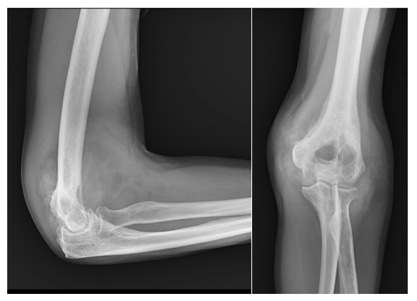
Figure 1: AP and lateral radiographs of the left elbow demonstrating circumferential soft-tissue fullness with geographic lucent lesions and bony erosion of the distal humerus.
MRI demonstrated a bulky, lobulated synovial proliferation distending from the elbow joint and extending through the coronoid, radial, and olecranon fossae. The lesion had a frond-like and nodular morphology and surrounded the joint circumferentially, involving both anterior and posterior compartments (Figures 3 and 4). Multiple nodular components were markedly hypointense on proton-density fat-suppressed and T2-weighted sequences, consistent with hemosiderin-rich synovial tissue. Interspersed regions of relatively higher signal likely reflected cellular or edematous synovium and associated joint fluid, resulting in a heterogeneous mixed-signal appearance. The MRI distribution favored diffuse-type rather than localized disease. The lesion was large and multinodular, with infiltrative margins and extensive articular and periarticular involvement rather than a single circumscribed nodule. Internal low-signal tissue was prominent and centrally distributed, while a discrete peripheral capsule was not identified. These features are consistent with the morphologic distinctions described between localized and diffuse TGCT [2,15]. Gradient-echo images were included and demonstrated marked susceptibility-related signal loss within the lesion. Although blooming is a classic imaging feature of TGCT, its conspicuity can vary, and assessment may be affected by sequence parameters and lesion distribution [13].
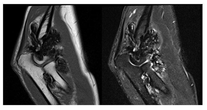
Figure 2: Sagittal MRI of the left elbow. (A) T1-weighted and (B) intermediate-weighted fat-suppressed (IW-FS), (C) gradient-echo, and (D) T1-weighted post-contrast images demonstrate the extensive lobulated synovial proliferation with predominantly low signal related to hemosiderin deposition.
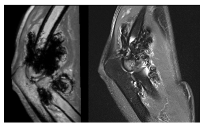
Figure 3: Sagittal MRI of the left elbow. (A) gradient-echo, and (B) T1-weighted post-contrast images demonstrate the blooming artifact with heterogeneous saturation on T1-weighted post-contrast image related to iron deposition.
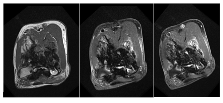
Figure 4: Axial MRI of the left elbow using the same sequence order as Figure 3. (A) T1-weighted, (B) intermediate-weighted fat-suppressed (IW-FS), and (C)) T1-weighted post-contrast images demonstrate circumferential involvement of the elbow joint and extensive nodular synovial proliferation.
The osseous abnormalities were most conspicuous along the distal humerus and proximal radioulnar structures and remained relatively well corticated. This pattern, in the setting of preserved joint space and a tight-capsule joint, favored chronic pressure erosion from synovial proliferation rather than a primary aggressive osseous neoplasm. The absence of matrix mineralization also made synovial chondromatosis less likely.
CT and Image-Guided Biopsy
CT of the left elbow was obtained during image-guided biopsy. Compared with radiography, CT more clearly depicted the extent of cortical irregularity and osseous loss along the lateral distal humerus and demonstrated the bulky circumferential soft-tissue component. The CT examination also allowed direct visualization of the biopsy trajectory and its relationship to the lesion. CT-guided core biopsy of the mass confirmed diffuse-type tenosynovial giant cell tumor, consistent with the historical diagnosis of pigmented villonodular synovitis.
Fracture Work-Up and Management
No acute fracture line was identified. Instead, imaging demonstrated chronic erosive bone loss involving the distal humerus and lateral epicondyle. The absence of an acute fracture was important because the patient's presentation was driven in part by concern for pathologic fracture in the setting of extensive chronic osseous remodeling. Given the diffuse extent of disease and the morbidity that can accompany extensive surgery in a complex joint, the patient was started on vimseltinib, an oral CSF1R inhibitor. At approximately 1.5 months of follow-up, the patient demonstrated clinical improvement in swelling, pain, and range of motion. Longer-term clinical and imaging follow-up was not available for this report.
Discussion
Although historically described as pigmented villonodular synovitis and sometimes regarded as a reactive synovial disorder, TGCT is now recognized as a clonal neoplastic process. Molecular studies have demonstrated recurrent rearrangements involving CSF1, including the classic COL6A3-CSF1 fusion in a subset of cases, with overexpression of CSF1 producing a microenvironment dominated by recruited CSF1R-positive macrophages [3,4,16]. This biology explains both the characteristic histologic composition and the development of targeted therapies directed at the CSF1/CSF1R axis. The clinical course in the present patient illustrates the indolent but locally destructive nature of dt-TGCT. More than a decade of pain, swelling, and progressive enlargement occurred without constitutional symptoms or an aggressive systemic presentation. Such a prolonged course should not be interpreted as evidence that the disease is clinically insignificant. Diffuse TGCT can progressively impair function and quality of life, and prospective studies have shown meaningful effects on physical function, stiffness, work, and daily activities [9,17,18]. Population-based data also indicate that diffuse disease is uncommon, with an estimated incidence substantially lower than localized TGCT [5,6]. Elbow involvement is particularly uncommon and is therefore a potential source of diagnostic delay. Published elbow cases have described patients presenting with pain, swelling, restricted motion, large masses, and, in some instances, neurologic compression. The literature remains limited largely to case reports and small series, but the available evidence emphasizes that elbow TGCT can behave as an extensive locally aggressive process and may involve both intra-articular and extra-articular compartments [7,8,19,20]. The current case is notable for the unusually long symptomatic interval and the extensive bicompartmental erosive remodeling despite preservation of joint space.
Radiographs remain useful as an initial examination, particularly when the presentation includes a palpable mass or concern for fracture. However, radiographic findings can be subtle, and normal radiographs do not exclude TGCT [11,12]. When present, osseous erosions are usually pressure-related and are more likely to occur in anatomically constrained joints. The elbow, wrist, ankle, and hip have relatively tight joint capsules compared with the knee, and this anatomic relationship may facilitate erosion of adjacent bone as proliferative synovium expands [12,13]. In this patient, the bilateral distribution of erosions across the elbow joint was an important clue that the process was centered in the synovium rather than arising primarily from bone. MRI is the key modality for characterizing the lesion. The combination of lobulated synovial proliferation, low signal on T1- and T2-weighted sequences, and susceptibility related to hemosiderin is highly characteristic. Nevertheless, the classic MRI signs should be interpreted as a pattern rather than as an absolute rule. Crim et al. demonstrated that blooming is not universal, particularly in localized lesions, and that assessment of blooming has substantial interobserver variability [14]. The absence of a dedicated gradient-echo sequence in the current case therefore represents a technical limitation, but it did not prevent recognition of the characteristic low-signal synovial tissue on conventional sequences. The differential diagnosis of a T2-hypointense intra-articular mass is broad and includes synovial chondromatosis, synovial hemangioma or intra-articular venous malformation, hemosiderotic synovitis, amyloid arthropathy, rheumatoid pannus, chronic crystal deposition, and selected fibrosing lesions [13,21]. In this case, the absence of mineralized nodules argued against synovial chondromatosis, while the marked low signal, diffuse synovial distribution, pressure erosion, and strongly favored dt-TGCT. Because imaging appearances may overlap and because the lesion was extensive, tissue confirmation remained appropriate.
CT has a complementary role. It is not usually the primary modality for diagnosing TGCT, but it can be particularly helpful for defining cortical integrity and osseous remodeling when radiographs are equivocal. In this patient, CT better demonstrated the cortical changes than radiography, and the biopsy was performed under CT guidance. This illustrates a practical multimodality sequence: radiography establishes the initial pattern, MRI maps the synovial disease and its extent, and CT can provide additional osseous detail while facilitating image-guided tissue diagnosis.
Management of dt-TGCT has evolved substantially. Surgical synovectomy remains an important treatment for resectable disease, but diffuse lesions can be difficult to eradicate completely, and recurrence remains a major consideration [1,2,10,15]. Elbow-specific literature is sparse; a review of published elbow cases found that most patients underwent open excision with or without synovectomy, while arthroscopic approaches have also been described [19]. The choice of surgery must balance disease control against the potential for postoperative stiffness, neurovascular injury, cartilage damage, and functional loss. Radiotherapy has historically been used selectively for residual or recurrent diseases, particularly when further surgery produces substantial morbidity. Long-term institutional series suggest that carefully selected patients with extensive or recurrent TGCT can achieve durable local control after surgery and radiotherapy, although this approach is generally reserved for situations in which other strategies are inadequate [19]. The increasing availability of systemic CSF1R inhibitors has further changed the treatment paradigm. Pexidartinib was the first CSF1R-targeted therapy to demonstrate substantial activity in advanced TGCT in a randomized phase 3 trial, but its use is limited by clinically important hepatotoxicity and the need for careful monitoring [20]. Vimseltinib is a more recent selective CSF1R inhibitor. In the phase 3 MOTION trial, vimseltinib produced a significantly higher objective response rate than placebo and improved symptoms and function in patients with symptomatic TGCT that was not amenable to surgery [21]. The early symptomatic improvement observed in our patient is consistent with the therapeutic rationale of CSF1R inhibition, although this single case cannot establish treatment efficacy, and the short follow-up limits assessment of radiologic response or durability. From an imaging perspective, the present case also illustrates why terminology matters. The historical term PVNS remains widely used and is familiar to radiologists, whereas dt-TGCT is the preferred contemporary term for diffuse disease. Using both terms at first mention can facilitate communication with clinicians and connect the current classification with the extensive historical literature. For radiologists, the most useful practical message is to recognize the combination of diffuse synovial proliferation, low T1/T2 signal related to hemosiderin, and pressure erosions in a chronic monoarticular process.
Conclusion
This case demonstrates the characteristic multimodality appearance of diffuse-type tenosynovial giant cell tumor of the elbow in an adult patient with an unusually prolonged clinical course. More than 10 years of pain and swelling culminated in evaluation for possible pathologic fracture, but imaging showed chronic bicompartmental erosive remodeling rather than an acute fracture. Radiographs established the chronic pressure-erosion pattern, MRI demonstrated the extensive circumferential synovial proliferation and hemosiderin-related low signal, and CT provided complementary assessment of cortical involvement and facilitated image-guided biopsy. The combination of a chronic enlarging periarticular mass preserved joint space, pressure-type erosions on both sides of a tight-capsule joint, and predominantly low T1/T2 signal should raise strong consideration of dt-TGCT/PVNS. Although MRI can be highly characteristic, histologic confirmation remains important when the imaging presentation is extensive or atypical. Recognition of this entity is particularly important in the elbow, where the uncommon location and nonspecific chronic symptoms may contribute to delayed diagnosis. Finally, the management of diffuse disease is evolving from a predominantly surgical paradigm toward individualized multidisciplinary care that may incorporate targeted CSF1R inhibition when surgery is not feasible or would result in substantial morbidity.
References
- Stacchiotti S, Dürr HR, Schaefer IM, et al. Best clinical management of tenosynovial giant cell tumour (TGCT): a consensus paper from the community of experts. Cancer Treat Rev 112 (2023): 102491.
- Gouin F, Noailles T. Localized and diffuse forms of tenosynovial giant cell tumor (formerly giant cell tumor of the tendon sheath and pigmented villonodular synovitis). Orthop Traumatol Surg Res 103 (2017): S91-S97.
- West RB, Rubin BP, Miller MA, et al. A landscape effect in tenosynovial giant-cell tumor from activation of CSF1 expression by a translocation in a minority of tumor cells. Proc Natl Acad Sci U S A 103 (2006): 690-695.
- Mastboom MJL, Verspoor FGM, Verschoor AJ, et al. Tenosynovial giant cell tumor: incidence, prevalence, patient characteristics, and recurrence. A registry-based cohort study in Denmark. J Rheumatol 44 (2017): 829-835.
- van der Heijden L, Gibbons CLMH, Dijkstra PDS, et al. Higher incidence rates than previously known in tenosynovial giant cell tumors. Acta Orthop 88 (2017): 688-694.
- DiCaprio MR, Damron TA, Stadnick M, et al. Pigmented villonodular synovitis of the elbow: a case report and literature review. J Hand Surg Am 24 (1999): 386-391.
- Koto K, Murata H, Sakabe T, et al. Magnetic resonance imaging and thallium-201 scintigraphy for the diagnosis of localized pigmented villonodular synovitis arising from the elbow: a case report and review of the literature. Exp Ther Med 5 (2013): 1277-1280.
- Mastboom MJL, Verspoor FGM, Verschoor AJ, et al. The patient perspective on the impact of tenosynovial giant cell tumors on daily living: crowdsourcing study on physical function and quality of life. JMIR Musculoskelet Disord 1 (2018): e9325.
- Verspoor FGM, Mastboom MJL, Hannink G, van der Graaf WTA, van de Sande MAJ, Schreuder HWB. The effect of surgery in tenosynovial giant cell tumours as measured by patient-reported outcomes on quality of life and joint function. Bone Joint J 101-B (2019): 272-280.
- Jelinek JS, Kransdorf MJ, Utz JA, et al. Imaging of pigmented villonodular synovitis with emphasis on MR imaging. AJR Am J Roentgenol 152 (1989): 337-342.
- Cheng XG, You YH, Liu W, et al. MRI features of pigmented villonodular synovitis (PVNS). Clin Rheumatol 23 (2004): 31-34.
- Murphey MD, Rhee JH, Lewis RB, et al. Pigmented villonodular synovitis: radiologic-pathologic correlation. Radiographics 28 (2008): 1493-1518.
- Crim J, Dyroff SL, Stensby JD, et al. Limited usefulness of classic MR findings in the diagnosis of tenosynovial giant cell tumor. Skeletal Radiol 50 (2021): 959-967.
- Palmerini E, Staals EL, Maki RG, et al. Diffuse-type tenosynovial giant cell tumour: current treatment concepts and future perspectives. Eur J Cancer 63 (2016): 34-40.
- Righi A, Gambarotti M, Sbaraglia M, et al. Detection of CSF1 rearrangements deleting the 3' UTR in tenosynovial giant cell tumors. Genes Chromosomes Cancer 58 (2019): 653-660.
- Mastboom MJL, Verspoor FGM, Verschoor AJ, et al. The diffuse-type tenosynovial giant cell tumor patient journey: a prospective multicenter study. Orphanet J Rare Dis 16 (2021): 133.
- Al Farii H, Zhou S, Turcotte R. The surgical outcome and recurrence rate of tenosynovial giant cell tumor in the elbow: a literature review. J Shoulder Elbow Surg 28 (2019): 1835-1840.
- Kohyama K, Sugiura H, Yamada K, et al. Posterior interosseous nerve palsy secondary to pigmented villonodular synovitis of the elbow: case report and review of literature. Orthop Traumatol Surg Res 99 (2013): 247-251.
- Griffin AM, Ferguson PC, Catton CN, et al. Long-term outcome of the treatment of high-risk tenosynovial giant cell tumor/pigmented villonodular synovitis with radiotherapy and surgery. Cancer 118 (2012): 4901-4909.
- Tap WD, Gelderblom H, Palmerini E, et al. Pexidartinib versus placebo for advanced tenosynovial giant cell tumour (ENLIVEN): a randomised phase 3 trial. Lancet 394 (2019): 478-487.
- Gelderblom H, Bhadri V, Stacchiotti S, et al. Vimseltinib versus placebo for tenosynovial giant cell tumour (MOTION): a multicentre, randomised, double-blind, phase 3 trial. Lancet 403 (2024): 2709-2719.