Marcelo Di gregorio1*, Manhal Kalaji2, Marie Cécile Nolleveaux3, Francis Lorge1, Michaël Dupont4
1Urology, CHU UCL Namur site-Godinne, Belgium
2Surgery Resident UCL, Urology, UCL Saint-Luc, Belgium
3Anatomopathology, CHU UCL Namur site Godinne, Belgium
4Radiology, CHU UCL Namur site Godinne, Belgium
*Corresponding Author: Dr. Marcelo Di gregorio, Urology, CHU UCL Namur site-Godinne, Belgium
Received: 20 Sep 2019; Accepted: 14 October 2019; Published: 03 December 2019
Background: Cystic nephroma It is a benign lesion that can mimic a malignant tumor of the kidney.
Case presentation: Here we report a case of multilocular cystic nephroma with unusual localization, largely invading the pyelocaliceal cavities.
Conclusions: The importance of its accurate diagnosis is to avoid a radical surgery proposing a partial surgery to preserve a vital organ.
Cystic nephroma; Cystic kidney tumor
Abbreviations:
CN-Cystic nephroma; CT-Computed tomography; MRI-Magnetic resonance imaging
1. Introduction
Cystic nephroma—also known as cystoadenoma or benign nephroblatoma in adults—is a benign cystic kidney tumor that can mimic renal carcinoma [1]. It is a non-hereditary pathology that shows bimodal distribution, most commonly occurring in children under 10 years old, and among adults of 45–60 years of age (mainly women) [2]. Clinically, some patients may complain of back or abdominal pain, while others remain entirely asymptomatic. The etiology is unknown, and diagnosis requires anatomo-pathologic confirmation based on immunohistochemical characteristics [3].
2. Case Presentation
During routine post-gastric bypass follow-up, a 46-year-old woman underwent a scheduled non-contrast-enhanced abdominal CT scan, which revealed a voluminous 15-cm left renal heterogeneous mass. The patient had no relevant personal or family history. She didn’t have any renal complaint. Clinical examination was unremarkable, with normal blood pressure, and normal renal function (creatinine: 0.75 mg/dl; glomerular filtration: 88.48 ml/min). Contrast-enhanced MRI showed enhancement of the solid part of the tumor (Bosniak 4 lesions) (Figure 1A-1C). Following this fortuitous discovery, the patient was referred to urology consultation. A surgical management was decided, and a total left nephrectomy was performed (due to suspicion of a malignant tumor). Histopathology excluded renal carcinoma, and revealed a cystic nephroma (Figure 2A, 2B).
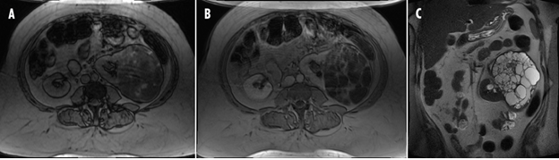
Figure 1A-C: Contrast-enhanced MRI showed enhancement of the solid part of the tumor (Bosniak 4 lesions).
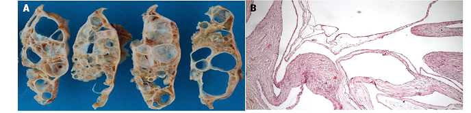
Figure 2A&B: Histopathology excluded renal carcinoma, and revealed a cystic nephroma.
3. Discussion and Conclusions
The cystic nephroma, also known as cystoadenoma or benign nephroblastoma in adults, is a benign cystic kidney tumor. The problem of this rare condition of the adults remains its diagnosis in order to propose conservative surgery (partial nephrectomy). Indeed, the cystic cancer of kidney constitutes the main differential diagnosis for which an enlarged and total nephrectomy is “the gold standard.” (cystic cancer of the kidney). Cystic nephroma is one of the benign renal tumors, which can mimic renal carcinoma. Diagnosis needs anatomo-pathologic confirmation. It remains challenging to diagnose this rare condition before surgery, which would support conservative surgery (partial nephrectomy) [4] rather than total nephrectomy, which is the gold standard treatment for cystic cancer of the kidney [5, 6].
Ethics Approval and Consent to Participate
Not applicable
Consent for Publication
Written informed consent was obtained from the patient for publications of this case reports and any accompanying images.
Availability of Data and Material
Data are available at the CHU UCL Namur.
Competing Interests
The authors declare that they have no competing interests.
Funding
No funding was obtained for this study.
Authors' Contributions
Marcelo Di Gregorio conceived, coordinated and designed of study. Took care of the patient in the emergency service and performed the surgery.
Francis Lorge: helped to draft the manuscript and took care of the patient in the emergency service and performed the surgery.
Manhal Kalaji: helped to write the manuscript
Michaël Dupont, Radiologist (CHU UCL Namur site Godinne), performed the radiology examinations to discover the lesion.
Marie Cécile Nollevaux, anatomopathologist (CHU UCL Namur site Godinne) performed pathological analysis revealed the malignant NSGCT.
All authors read and approved the final manuscript
Acknowledgements
The authors acknowledge Axel Baily for picture edition and Edith Botek for reading support.
References
- Castillo OA, Boyle ET, Kramer SA. Multilocular cysts of kidney: a study of 29 patients and review of literature. Urology 37 (1999): 156-162.
- Thibeau JF, Sourtzis S, Lufuma EM, et al. Néphrome kystique multiloculaire de l’adulte. Présentation inhabituelle et revue de littérature. Progrès en urologie 11 (2001): 507-511.
- Raspollini MR, Castiglione F, Martignoni G, et al. Multiple and bilateral kidney tumors with clear cells of three different histotypes: A case report with clinicopathologic and molecular study. Apmis 124 (2016): 619-623.
- Kural AR, Öbek C, Özbay G, et al. Multilocular cystic nephroma: an unusual localization. Urology 52 (1998): 897-899.
- Billiet I, Van Poppel H, Baert L. Actual approach to cystic nephroma. European urology 14 (1988): 280-286.
- Granja MF, O'brien AT, Trujillo S, et al. Multilocular cystic nephroma: a systematic literature review of the radiologic and clinical findings. American Journal of Roentgenology 205 (2015): 1188-1193.