Ramakrishna Narra1*, Radha Bhavani Varikuntla2, Jukuri Naga Narasimha Raju3
1Professor of Radiology and Neuroradiology, Katuri Medical College, Katuri Nagar, chinakondrupadu, NH 16, Guntur, Andhra Pradesh 522019, India
2Deprtment of Radiology, Katuri Medical College, Guntur, Andhra Pradesh, India
3Department of Radiology, Katuri Medical College and Hospital, Guntur, Andhra Pradesh, India.
*Corresponding Author: Narra Ramakrishna, Associate Professor of Radiology and Neuroradiology, Katuri Medical College, Katuri Nagar, hinakondrupadu, NH 16, Guntur, Andhra Pradesh 522019, India.
Received: 08 February 2022; Accepted: 04 March 2022; Published: 22 March 2022
Severe acute respiratory syndrome coronavirus 2 (SARS-CoV-2) is the causative agent of the global COVID 19 pandemic. COVID 19 presents with varying imaging features along the course of the disease. The lower sensitivity of reverse transcriptase-PCR assay and other rapid detection tests lead the CT scan as the ancillary mode for early detection and management of suspected COVID-19 patients. The first published article on imaging findings of COVID-19 was in January 2020, stating bilateral peripheral Ground Glass Opacities (GGOs) as the predominant feature in most cases. This imaging pattern-based review gives a brief outline of different CT imaging features of COVID 19 and the CO-RADS grading along with the CT severity score index helps the radiologists for better conveying the disease burden to the concerned clinicians in making an accurate diagnosis and for adopting appropriate therapy protocols.
Covid-19; Corads; Ct Severity Score Index
1. Introduction
CT is sensitive in identifying the lung involvement in the early stages of Corona viral disease. It is used for the screening and diagnosing the clinically suspicious COVID-19 patients [1, 2]. Though chest CT scan may at times be normal [3]. This pictorial essay aims to overview the various spectrum of HRCT patterns in COVID-19 pneumonia.
1.1 Technique f Hrct chest protocol
Patients referred to the radiology department for High Resolution Chest Tomography (HRCT) in suspicion of COVID should undergo non-contrast chest CT by using a low-radiation-dose protocol to lower radiation burden [4]. Low-radiation-dose HRCT images are obtained by using lower KV settings and iterative reconstructions for decreasing the noise [5]. HRCT images are acquired in a single inspiratory breath-hold. Expiratory phase CT increases the radiation dose, and studies have shown no extra benefit in evaluating suspected COVID-19 cases [4, 5].
1.2 Ct patterns
Multifocal subpleural GGOs are the most common HRCT pattern. Other features include crazy paving patterns and segmental/ subsegmental pulmonary consolidations [6].
1.3 Ground glass opacity
Peripheral and subpleural multifocal GGOs are the initial CT manifestation of COVID-19. GGOs together with patchy areas of consolidation would suggest an organizing pneumonia pattern [7].
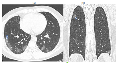
figure 1a,b: A 28 years male presenting with fever and cough and RT-PCR confirmed COVID 19. Non contrast axial (1a) and coronal (1b) chest HRCT images performed 4 days after the onset of symptoms showing bilateral multifocal patchy subpleuralGOs (arrow, double headed arrow) predominantly in posterior segments of lower lobes with CTSSI of 8/25.
GGO pattern associated in cases of: Infectious diseases like atypical pneumonias, Interstitial lung disease, Pulmonary edema, Diffuse alveolar hemorrhage, Bronchogenic carcinoma.
1.4 Crazy paving pattern
GGOs with superadded interlobular septal thickening and intralobular lines is another common finding of initial COVID-19 disease, bearing resemblance to paving stones [8].
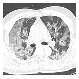
figure 2: A 34 years male presenting with history of fever, dry cough and dyspnea with RT-PCR confirmed COVID 19 disease. Non contrast axial HRCT image performed 7 days after onset of symptoms showing diffuse GGOs with interlobular interstitial septal thickening (arrow) forming typical geographic crazy paving pattern.
Crazy paving pattern associated with: Pulmonary alveolar proteinosis, Acute interstitial pneumonias, Acute respiratory distress syndrome
1.5 Halo sign
Is a rare finding in COVID-19, seen as a focal nodule surrounded by ground-glass haziness [8].
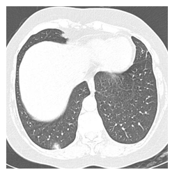
figure 3: A 48 years old male presenting with fever, cough with RT-PCR confirmed COVID 19. Non contrast axial chest HRCT image after 9 days of symptom onset shows few ill-defined nodules (arrow) and one of them showing surrounding ground glass haze suggestive of Halo is most commonly seen in: Angioinvasive aspergillosis.
1.6. Dandelion sign
The "dandelion sign" on the CT image includes thickened blood vessels and interlobular septal thickening within the focal stripe of GGO [9].
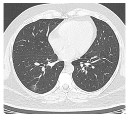
figure 4: A 37 years old female presenting with fever and RT-PCR confirmed COVID 19. Non contrast axial chest HRCT image after 4 days of onset of symptoms showing focal vascular thickening (arrow) within the area of ground glass opacity giving the appearance of a Dandelion fruit.
1.7 Pomegranate sign
It is an uncommon feature characterized by an increase in the sub-segmental extent of ground-glass opacity with significant interlobular septal thickening and associated spotty alveolar haemorrhage, giving an imbricated pomegranate appearance [9].
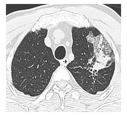
figure 5: A 42 years old female with cough for 7 days with RT-PCR confirmed COVID-19 disease. Non contrast axial chest HRCT performed after 5 days of onset of symptoms showing focal GGO with interlobular septal thickening and spotty hemorrhage resembling a pomegranate appearance.
1.8 Subpleural curvilinear line
It manifests as a thin, less than 3 mm peripheral curvilinear opacity, along the viscera pleura [10].
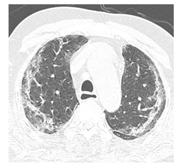
figure 6: A 22 years old female with cough and RT-PCR confirmed COVID -19. None contrast axial chest HRCT showing curvilinear fibro reticular opacity appearing as typical subpleural lines (arrow) in bilateral lungs.
Subpleural line Discerned in: Dependent atelectasis, Pulmonary edema, fibrosis secondary to varied etiologies.
2. Consolidation
Subpleural or peribronchovascular, patchy or segmental multifocal consolidation is usually present in COVID-19 patients and is a pointer for the worsening course of disease [10]. Consolidation frequently seen in patients with renal failure, possibly due to secondary bacterial infection. Consolidation is uncommon in the initial stages of COVID-19, but it establishes in course of disease progression. In the peak stage of the disease, the extent of pulmonary consolidation becomes more and involves diffuse lung parenchyma [11].
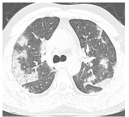
figure 7: A 48 years old male with fever, cough and dyspnea with RT-PCR confirmed COVID-19 disease. Non contrast axial chest HRCT performed 8 days after the symptom onset showing multifocal peripheral patchy areas of consolidation (arrow) in bilateral lung fields. Consolidation visualised in cases of other pneumonias, Pulmonary edema secondary to heart failure, Pulmonary hemorrhage Bronchoalveolar carcinoma, Alveolar proteinosis.
2.1. Lymphadenopathy
Hilar and mediastinal lymphadenopathy is considered as an uncommon finding and can be associated with severe pneumonia or bacterial superinfection [11].
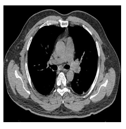
figure 8: A 25 old female with fever and RT-PCR confirmed COVID – 19 diseases. Non contrast axial chest HRCT performed after 10 days after onset of symptoms showing enlarged prevascular (arrow) and hilar lymph nodes. Lymphadenopathy seen secondary to Reactive to Infectious aetiology, Neoplastic, Granulomatous diseases, Occupational lung diseases
2.2. Air bronchogram
Seen as gas-filled bronchi surrounded by high attenuating airless lungs. It can be associated with bronchiolectasis [12].
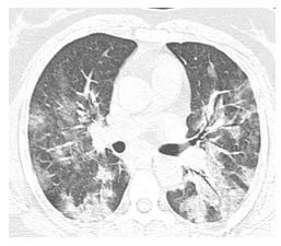
figure 9: A 44 years old female with fever, cough, dyspnea and RT-PCR confirmed COVID-19 pneumonia. Non contrast axial chest HRCT performed after 1 week of symptom onset showing multifocal areas of patchy consolidation with gas filled air bronchogram (arrow). Air bronchogram seen with Pulmonary edema Non obstructive atelectasis, Neoplasms, Pulmonary infarct and Pulmonary hemorrhage.
2.3. The air bubble sign
Is a small lucent air-containing space, due to the alveolar sac or bronchiolar pathological expansion or by the absorption process of consolidation [13].
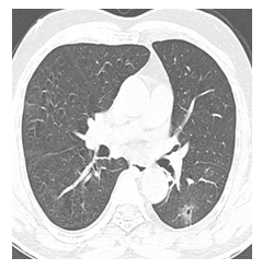
figure 10: A 20 years old asymptomatic male with RT-PCR confirmed COVID-19 diseases. None contrast axial chest HRCT showing a small air containing lucent space within the previous consolidation focus. It is encountered in some of the cases of complicated perforated pulmonary hydatid cyst.
2.4. Vascular Dilation or Thickening
The dilatation of pulmonary vessels within the vicinity of GGO or consolidation. Pulmonary vascular enlargement (PVE) may be secondary to inflammatory infiltration of the vascular wall. It is significantly associated with COVID-19 [13].
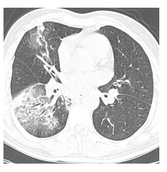
figure 11: A 38 years old male with fever since a week with RT-PCR confirmed COVID -19 diseases. Non contrast axial chest HRCT showing enlarged pulmonary vessels within the region of consolidation. It could be seen secondary to primary pulmonary diseases like severe COPD or congestive heart failure, connective tissue disorders.
2.5. Nodule
Spontaneously subsiding SPN may be associated with organizing pneumonia secondary to COVID-19 infection [14].
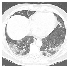
figure 12: A 52 years old male with fever, cough and RT-PCR confirmed COVID - 19 diseases. None contrast axial chest HRCT showing a small sub centimetric nodule in the GGO. Nodule has a wide spectrum of aetiologies like infectious, hypersensitivity pneumonitis, granulomatous, small air way diseases, interstitial diseases, neoplasms.
2.6. Reversed Halo Sign (Atoll sign)
It has been reported in many COVID-19 cases. It is seen as a central focal area of GGO surrounded by complete ring-like consolidation [15].
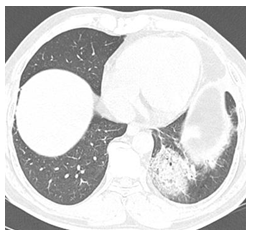
figure 13: A 43 years old female with fever, cough and dyspnea for 8 days and RT-PCR confirmed COVID – 19 disease. Non contrast axial chest HRCT showing ground glass opacity surrounded by rim of consolidation. Atoll sign seen in cases of crypto-genic organising pneumonia, invasive pulmonary fungal infections like Mucor mycosis and aspergillosis, granulomatous diseases, pulmonary infarction due to venous thromboembolism.
2.7. The spider web sign [16]
It is a focal peripheral triangular GGO with interstitial septal thickening, simulating that of spider web in the corner.
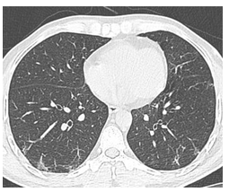
figure 14: A 33 years old asymptomatic male with RT-PCR confirmed COVID -19 diseases. Non contrast axial HRCT chest showing focal subpleural angular ground glass opacity like that of a spider web.
2.8. Bronchiectasis with thickened wall
Is not commonly in COVID-19 pneumonia. They are more frequent in late-stage and in critically ill patients [17].
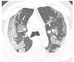
figure 15: A 37 years old male with fever for 5 days and RT-PCR confirmed COVID -19 diseases. Non contrast axial chest HRCT showing focal dilated bronchiole with thickened wall. It is seen secondary to varying aetiologies like congenital, impaired host defences, post infectious, allergic, autoimmune, post obstructive cases.
2.9. Pleural changes
Pleural involvement is generally seen in the late stage of the COVID-19 as pleural effusion or focal pleural thickening [17]. Among the two, pleural thickening is more prevalent. pleural effusion can be secondary to intricating co-morbid condition or suggest a worse prognosis in COVID-19 [17].
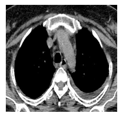
figure 16: A 54 years old female with fever for 4 days with RT-PCR confirmed COVID-19 diseases. Non contrast axial chest HRCT performed 10 days after the onset of symptoms showing left sided pleural thickening. Pleural thickening usually follows the recurrent inflammation, pneumothorax, empyema and sometimes seen in cases of collagen vascular diseases, post exposure occupational/ inhalational lung diseases, primary malignant pleural disease and pleural metastasis.
2.10. Cavity sign
The late stage of the covid 19 disease may be associated with cavities due to focal haemorrhage and necrosis of the lung parenchyma after drainage through bronchioles [18].
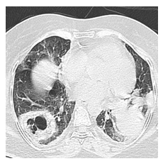
figure 17: A 42 years old female with cough for 10 days and RT- PCR confirmed COVID – 19 disease. Non contrast axial chest HRCT performed after the onset of symptoms showing a thick walled subpleural cavity in the right lower lobe. Pulmonary cavity can be congenital or may result secondary to infection, infarct, trauma, granulomas, cavitating lung carcinomas.
2.11. Reticular pattern
It presents as sub-pleural coarse or fine linear opacities due to intralobular and interlobular septal thickening. It is relatively late but is also the common HRCT manifestation of COVID-19 [19]. Reticular pattern is associated with pulmonary oedema, infection and post infectious scar related changes, inhalational & interstitial lung diseases, granulomatous conditions, collagen vascular disorders, drug related changes and pulmonary neoplasms.
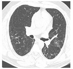
figure 18: A 26 years old asymptomatic male with RT-PCR confirmed COVID-19 diseases. Non contrast axial chest HRCT showing fine lacy network like reticular opacities noted in the subpleural regions of bilateral lower lobes.
2.12. fibrosis
fibrous lesions are seen as sequelae to the chronic healing process of pulmonary inflammation in COVID-19 suggesting the disease stabilization. It may proceed to pulmonary interstitial fibrosing disease changes [20].
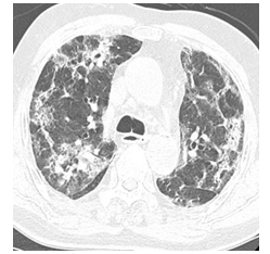
figure 19: A 38 years old female with dry cough for 10 days with RT- PCR confirmed COVID – 19 disease 2 weeks ago. Non contrast axial chest HRCT performed 3 weeks after the onset of symptoms showing multiple linear fibrotic bands in bilateral lung parenchyma. fibrosis results as an end stage disease process following a wide range of conditions like acute insult to lung, inhalational lung disease, radiation induced, autoimmune, granulomatous conditions, connective tissue disorders and drug related.
3. Temporal Evolution of Hrct Lung Abnormalities
CT findings correspond to that of the inflammatory lung injuries, COVID-19 has varying manifestations at different stages of the disease. In the initial stages of the disease, unifocal or multifocal GGOs are seen which increase further along the course of the disease. GGO is usually seen in the initial mild pulmonary infection [21]. In the late stage of COVID-19, GGO is usually associated with consolidation, crazy paving, and pleural effusion [21]. Pulmonary consolidation in the COVID-19 is usually considered a sign of worsening disease course.
Four stages of COVID-19 have been described in chest CT:
- Early-stage (up to 5 days after the onset of symptoms), normal CT or subtle GGOs;
- Progressive stage (6 to 8 days after the onset of symptoms), On CT-widespread GGOs, crazy-paving appearance;
- Peak stage (9 to 13 days after the onset of symptoms), on CT- predominantly consolidation is seen; and
- Late-stage (14 days after the onset of symptoms), Gradual resolution of consolidation and GGOs, parenchymal fibrous bands, architectural distortion, and traction bronchiectasis starts appearing [21].
Depending on the severity of the disease, temporal changes of imaging features and extent of lung involvement vary in different cases [21].
3.1. Corads
The Dutch radiological society developed a categorical CT scheme CORADS-to assess the suspicion for pulmonary involvement of COVID-19 based on features seen at unenhanced chest CT [22].
|
CO-RADS grade |
Degree of Suspicion |
HRCT features |
|
1 |
Very low |
Normal/non-infectious |
|
2 |
Low |
Consistent with infections other than COVID-19 |
|
3 |
Indeterminate |
Similar features of COVID-19 & other diseases |
|
4 |
High |
Features suspicious of COVID-19 |
|
5 |
Very high |
Typical COVID-19 features |
|
6 |
RT- PCR positive |
Table 1: CO-RADS grading demonstrating the degree of suspicion of COVID-19 disease.
3.2. Ct Severity Score
Lung involvement due to COVID-19 is assessed using the CT severity score index. five lung lobes are given a score from 1 to 5 depending on the extent of lung involvement.
|
The score of each lobe |
Percentage of involvement |
|
1 |
< 5% of lobar involvement |
|
2 |
5 to 25% of lobar involvement |
|
3 |
26–50% of lobar involvement |
|
4 |
51–75% of lobar involvement |
|
5 |
> 75% of lobar involvement |
Table 2: CT SEVERITY SCORE demonstrating the severity of COVID – 19 disease.
The summation of each lobe score gives the total score (out of 25) [18].
Another system for grading of lung involvement:
none (0%) = score 0,
minimal (1% to 25%) = score 1,
mild (26% to 50%) = score 2,
moderate (51 to 75%) = score 3, and
severe (76 to 100%) = score 4.
The total score of lung involvement is obtained by the sum of the individual score of each lobe (0-20) [18].
4. Conclusion
In conclusion, with increasing COVID-19 pandemic cases, chest CT is a rapid tool for screening suspicious cases and assessing the extent of involvement of COVID 19 cases. Familiarity with imaging patterns of COVID 19 and further conveying the same results with referring physicians through structured reporting systems like CO-RADS with corresponding CT severity score, as represented in this pictorial review article, the radiologist can play a vital role in the management of this global outbreak.
References
- Memish ZA, Al-Tawfiq JA, Assiri A. Middle east respiratory syndrome coronavirus disease in children Pediatr. Infect Dis J 33 (2014): 904-906.
- Rao TA, Paul N, Chung T, et al. Value of CT in assessing probable severe acute respiratory syndrome Am J Roentgenol 181 (2003): 317-319.
- Furkan U, Recep S. Chest CT features of the novel coronavirus disease (COVID-19) Turk J Med Sci 50 (2020): 664-678.
- Rodrigues JCL, Hare SS, Edey A, et al. An update on COVID-19 for the radiologist - A British Society of Thoracic Imaging statement Clin Radiol Epub 75 (2020): 323-325.
- Agostini A, floridi C, Borgheresi A, et al. Proposal of a low-dose, long-pitch, dual-source chest CT protocol on third-generation dual-source CT using a tin filter for spectral shaping at 100 kVp for CoronaVirus Disease 2019 (COVID-19) patients: a feasibility study La Radiologia Medica 125 (2020): 365-373.
- Kim H. Outbreak of novel coronavirus (COVID-19): What is the role of radiologists Eur Radiol 30 (2020): 3266-3267.
- Kanne JP. Chest CT findings in 2019 Novel Coronavirus (2019-nCoV) Infections from Wuhan, China: Key Points for the Radiologist Radiology 295 (2020):16-17.
- Sana Salehi, Aidin Abedi, Sudheer Balakrishnan, et al. Coronavirus Disease 2019 (COVID-19): A Systematic Review of Imaging findings in 919 Patients. Am J of Roentgenol 215 (2020): 87-93.
- Jin J, Gao Dh, Mo X, et al. Analysis of 4 imaging features in patients with COVID-19 BMC Med Imaging 20 (2020): 84.
- Ye Z, Zhang Y, Wang Y, et al. Chest CT manifestations of new coronavirus disease 2019 (COVID-19): a pictorial review Eur Radiol 30 (2020): 4381-4389.
- Song F, Shi N, Shan F, et al. Emerging 2019 novel coronavirus (2019-nCoV) pneumonia Radiology 295 (2020): 210-217.
- Khazaei M, Mahdavi A, Mahboubi-Fooladi Z, et al. Chest CT Imaging Spectrum of Coronavirus Disease 2019 (COVID-19) Pneumonia: A Pictorial Essay Iran J Radiol 17 (2020): 103166.
- Wang J, Xu Z, Wang J, et al. CT characteristics of patients infected with 2019 novel coronavirus: association with clinical type, Clinical radiology 75 (2020): 408-414.
- Pogatchnik BP, Swenson KE, Sharifi H, et al. Radiology-pathology Correlation in Recovered COVID-19, Demonstrating Organizing Pneumonia Am J Respir Crit Care Med 202 (2020): 598-599.
- Bernheim A, Mei X, Huang M, et al. Chest CT findings in Coronavirus Disease-19 (COVID-19): Relationship to Duration of Infection Radiolog 295 (2020): 200463.
- Lei P, Fan B, Mao J, et al. The progression of computed tomographic (CT) images in patients with coronavirus disease (COVID-19) pneumonia: Running title: The CT progression of COVID-19 pneumonia The Journal of infects 80 (2020): 30-31.
- Shi H, Han X, Jiang N, et al. Radiological findings from 81 patients with COVID-19 pneumonia in Wuhan, China: a descriptive study Lancet Infect Dis 20 (2020): 425-434.
- Xu Z, Shi L, Wang Y, et al. Pathological findings of COVID-19 associated with acute respiratory distress syndrome Lancet Respir Med 8 (2020): 420-422.
- Wang Y, Dong C, Hu Y, et al. Temporal Changes of CT findings in 90 Patients with COVID-19 Pneumonia: A Longitudinal Study Radiolog 296 (2020): 55-64.
- Pan Y, Guan H, Zhou S, et al. Initial CT findings and temporal changes in patients with the novel coronavirus pneumonia (2019-nCoV): a study of 63 patients in Wuhan, China Eur Radiol 30 (2020): 3306-3309.
- Feng P, Ye T, Sun P, et al. Time Course of Lung Changes at Chest CT during Recovery from Coronavirus Disease 2019 (COVID-19) Radiolog 295 (2020): 715-721.
- Prokop M, Van Everdingen W, Van Rees Vellinga T. CO-RADS: a categorical CT assessment scheme for patients suspected of having COVID-19 definition and evaluation Radiolog 296 (2020): 97-104.