Antonio Pierro*
Radiology Department, Gemelli Molise Hospital, Campobasso, Italy
*Corresponding Author: Dr. Antonio Pierro, Radiology Department, Gemelli Molise Hospital, Campobasso, Italy
Received: 20 November 2020; Accepted: 30 November 2020; Published: 04 December 2020
Bifid tail of pancreas; Fish tail pancreas; Branching of the main pancreatic duct
1. Main Point
This paper would like to present one of the possible causes of recurrent acute pancreatitis, namely the bifid pancreas. This anatomical variant is best investigated only with the abdominal MRI, including MRCP (MR cholangiopancreatography).
2. Clinical Image
Bifid tail of pancreas (fish tail pancreas) or pancreas bifidum is extremely rare congenital malformation. This anatomical variant is characterized by an abnormal branching of the main pancreatic duct. This anomaly is generally not associated with abdominal pain or pancreatic disease. Most cases are asymptomatic, detected incidentally. Nevertheless, pancreas bifidum may cause acute pancreatitis [1]. Herein, we report the case of a 65-year-old female patient with a previous recurrent episode of acute pancreatitis. Patient had none clinical predisposing factors for acute pancreatitis. Abdominal MRI, including MRCP (MR cholangiopancreatography), was performed to better evaluation of the biliary tract and pancreatic parenchyma. MRCP (Figure 1) showed duplication of the major duct in the tail of the pancreas. Axial T2-weighted image (Figure 2) showed ventral and dorsal pancreatic buds with the typical fish tail appearance.
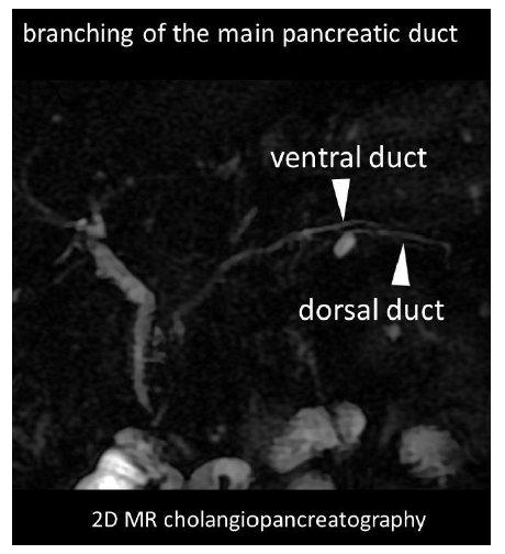
Figure 1: 2D MR cholangiopancreatography.
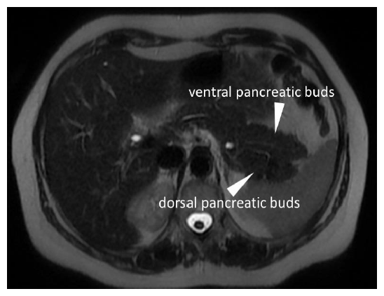
Figure 2: Axial T2-weighted image showing ventral and dorsal pancreatic buds with the typical fish tail appearance.
In the absence of clinical predisposing factors (alcohol or smoking history), in cases of acute pancreatitis, it is advisable to perform an abdominal MRI, including MRCP to rule out anatomical abnormalities as predisposing factors. Our case corroborates what reported in very few literature cases [2] of association between acute pancreatitis and bifid pancreas, besides displaying the typical MRI findings.
Conflict of Interest
The authors declare that they have no conflict of interest.
References
- APA Kahramanoglu Aksoy Evrim, Koklu Seyfettin, Öztürk Ömer, et al. An Unusual Cause of Recurrent Acute Pancreatitis. Pancreas 45 (2016): e11-12.
- Koyasu S, Isoda H, Nakase H, et al. Bifid tail of the pancreas with localized acute pancreatitis. Magn Reson Med Sci 12 (2013): 315-318.