André Léké1*, Maurice Biendo2
1Soins Intensifs de Néonatologie et Médecine Néonatale, CHU Amiens Picardie, F-80054 Amiens, France
2Laboratoire Péritox UMR 101 CURS-UPJV, F-80054 Amiens, France
*Corresponding author: André Léké, Soins Intensifs de Néonatologie et Médecine Néonatale, CHU Amiens Picardie, F-80054 Amiens, France.
Received: 01 September 2026; Accepted: 07 September 2026; Published: 13 September 2026
Background: The course of fetal growth relies on how much nutrition is accessible. This supply hinges on how well the placenta can move vital molecules such as fatty acids, glucose, and amino acids delivered via placental transfer.
Objective: The aim of this study is to show how placental programming influences newborn weight in pregnancies that are normal as well as those affected by obesity. It also considers how obesity in pregnancy is linked to a greater chance of atypical fetal size, including unusually small or unusually large infants.
Methods: A PRISMA-guided systematic review was carried out, using an extensive search of several databases like PubMed, ScienceDirect, and ResearchGate. Sources from 2020 through 2026 were used for this work.
Results: Fetal tissues contain docosahexaenoic acid and arachidonic acid during pregnancy. Some proteins, like FATPs, FAT/CD36, and FABPs, move fatty acids by crossing placental membranes. Liver X receptors, peroxisome proliferator-activated receptors, and sterol regulatory element-binding proteins are transcription factors involved in placental gene regulation. Maternal obesity causes lipotoxicity and inflammation in the placenta, which can affect angiogenic signaling and endothelial metabolism. Gestationalore GLUT1 in babies that were large for their gestational age (LGA) than in babies that were small for their gestational age (SGA). SGA was linked to lower SNAT2, and LGA-obese mothers had lower SNAT4. FATP1 was lower in SGA yet higher in LGA.
Conclusion: This review assembles evidence that maternal obesity impairs placental vascular performance, emphasizing the interplay between angiogenic mediators and endothelial metabolic activity to produce dysfunction.
Long-chain polyunsaturated fatty acid; Fatty acid binding/transport proteins; Nuclear transcription factors; Placental glucose transporters; Placental amino acid transporters; Placental dysfunction.
Long-chain polyunsaturated fatty acid articles; Fatty acid binding/transport proteins articles; Nuclear transcription factors articles; Placental glucose transporters articles; Placental amino acid transporters articles; Placental dysfunction articles
Introduction
Pregnancy represents a crucial phase of physiological transformation for both the mother and the fetus. As gestational age advances, the energy requirements rise to meet the increasing nutritional demands of fetal development. Pregnancy involves a natural state of maternal insulin resistance, and the growing gap in glucose levels between mother and fetus as gestation progresses helps the fetus absorb more macronutrients. Fetal development depends strongly on how much nourishment is accessible, and this supply is enabled by transport activity across the placenta. This indicates that shifts in how nutrients are moved through the placenta directly affect fetal growth. For appropriate growth and maturation ‘’in utero”, the fetus requires an adequate delivery of vital nutrients provided via the placenta. A vital biological role of the placenta is to determine how nutrition-focused approaches affect results for the pregnant woman, the developing fetus, and the infant. As pregnancy progresses, the placenta develops alongside the embryo and subsequently the fetus. It promotes growth by allowing delivery of nutrients including fats, glucose, amino acids, vitamins, and minerals, while also removing waste and aiding protection of the fetus against harmful influences. The placenta can adapt to buffer brief changes in the mother’s nutrient state, whether low or high, allowing the fetus to develop within a stable nutritional environment [1].
Notably, the placenta is itself an organ still forming, and it depends on sufficient nutrition to grow and work as it should. When resources are limited, proper placental development and performance can be disturbed. Diet-focused interventions can raise the supply of nutrients to the developing placenta, which may then affect placental gene activity, blood-vessel formation, and nutrient transfer [2]. Placental pathologies are linked to disruptions in maternal nutrient status that affect how the placenta senses, processes, and transports nutrients such as fats, glucose, and amino acids to the fetus. In the short term, a functioning placenta can also benefit the mother by releasing hormones that adjust her physiology to prepare her for pregnancy and lactation. Consequently, dietary interventions designed to enhance outcomes for both the mother and the infant may be accompanied by alterations in the placenta [3]. Within the placenta, nutrients and other substrates are handled, and the production pattern of particular placental transport proteins seems aligned with fetal needs [4]. When substances do not pass through membranes easily, the placenta uses both diffusion-driven movement and transport that requires energy. The amounts of fatty acids (FAs), glucose, and amino acids delivered to the fetus depend on regulation of placental enzymes, receptors, and transporters. To support healthy infant growth, essential nutrients must be provided by the mother and transported through the placenta. In the final trimester, Docosahexaenoic acid (DHA) is viewed as a vital nutrient for the developing fetal nervous system [5-9]. The mother’s liver is able to produce DHA starting from α-linolenic acid (ALA) through desaturase and elongate. In contrast, neither the placenta nor the fetus can carry out this synthesis. As a result, fetal DHA levels rely on maternal DHA status together with placental processing and transport of this fatty acid. During pregnancy, DHA and arachidonic acid (ARA) move from maternal circulation to the fetus through the placenta. Glucose serves as the primary fuel for both the developing fetus and the placenta. Proteins in the glucose transporter (GLUT) group help shuttle glucose across the placenta by allowing it to pass down its concentration slope [10]. During pregnancy, the placenta serves as a vital organ that facilitates the transfer of essential amino acids to the developing fetus. The higher concentration of amino acids in fetal blood plasma compared to the mother’s suggests a net movement of these molecules across the placenta. Transporting amino acids through this organ is a multifaceted process influenced by numerous interdependent factors, such as amino acid concentrations in the maternal, placental, and fetal compartments, the blood flow between mother and fetus, and the characteristics of the transport proteins responsible for carrying amino acids [11]. The aim of this study is to show how placental programming influences newborn weight in pregnancies that are normal as well as those affected by obesity. It also considers how obesity in pregnancy is linked to a greater chance of atypical fetal size, including unusually small or unusually large infants
Methods
Database search strategy
Following the PRISMA guidelines, the team conducted this review. To identify relevant studies addressing the influence of obesity, FAs, glucose, and amino acids on pregnancy and on preterm versus term infants, the researchers carried out a broad search of online databases. They derived the findings from articles listed in PubMed, ScienceDirect, and ResearchGate. A date restriction was applied to include papers published from 2020 through 2026. To run the web-based database search, the authors used selected MeSH headings along with connected search words. They combined phrases covering the human placenta (HP), obesity associated with placental dysfunction, gestation, defects in placental blood vessels, and placental transfer of FAs, glucose, and amino acids. The selected references were saved using citation-management software.
PICOS framework
Population
The study included pregnant women with or without obesity, as well as infants born to mothers who are healthy, obese, or have gestational diabetes mellitus (GDM). In this study, the age of healthy pregnant women ranged from 18 to 40 years, while those with GDM were between 23 and 40 years old. Furthermore, while the weights of the participating healthy pregnant women ranged from 49 to 100 kg, those with GDM weighed between 60 and 118 kg. Additionally, children and adolescents aged 6 to 18 years who were estimated to be overweight or obese were enrolled in the study.
Intervention
Microscopically, the placentas of obese pregnant women were examined for abnormalities. It is estimated that women with GDM or type 2 diabetes mellitus (T2DM) are affected by pregnancy-related diabetes. The height of pregnant women in this study was measured to calculate their body mass index (BMI) using the formula: BMI = weight (kg)/height (m2). GDM was diagnosed based on the criteria established by the International Association of Diabetes and Pregnancy Study Groups.
Comparison
Because the global maternal obesity rate and the pooled prevalence of overweight and obesity during pregnancy were estimated using a random-effects meta-analytic approach, a direct comparison is not feasible. Furthermore, the systematic review with meta-analysis conducted during pregnancy did not compare maternal BMI categories to evaluate their association with adverse fetal and maternal outcomes. However, a prospective case-control study compared the microscopic placental pathology of obese pregnant women without diabetes to that of lean pregnant women. This study revealed that obesity is associated with a higher risk of maternal and fetal vascular malperfusion, inflammatory placental lesions, and villitis in both the full cohort and those with uncomplicated pregnancies compared to the control group.
Outcomes
In individuals with T2DM, GLUT1 protein expression increased in the bone marrow, while GLUT4 levels decreased. In both T2DM and normoglycemic placentas, higher microvillous plasma membrane (MVM) FATP6 levels were positively correlated with birth weight. Basal plasma membrane (BM) FATP6 levels were found to be significantly increased in patients with T2DM. SNAT1 levels were elevated at the maternal-fetal interface in placentas of women with T2DM and correlated with birth weight, while SNAT2 levels also increased. In T2DM, MVM LAT1 expression was associated with neonatal fat mass and birth weight. Placenta-related disorders encompassing primary outcomes such as placenta previa, accreta, increta, abruption, retained placenta, and postpartum hemorrhage. Other secondary outcomes evaluated included ectopic pregnancy, gestational hypertension, preeclampsia (PE), intrauterine growth restriction (IUGR) and diabetes mellitus.
Study design
We included studies that reported obstetric and neonatal outcomes for all pregnant women hospitalized between 2020 and 2026 who experienced pregnancy complications or had fetuses with growth appropriate for gestational age.
Inclusion and exclusion criteria
Only publications written in English-language were eligible for the review, and primary research papers were included as well. Items left out were repeated records, articles not in English, content intended for the wrong readership, sources that did not match the topic, private correspondence, editorials expressing opinions, unpublished writing, papers still being submitted, and commentary articles. Figure 1 presents the summary via the PRISMA flow diagram.
Results
The query produced 3724 results overall. However, among these records, 3024 entries appeared, including repeated files, publications dated before 2020, sources not aligned with the review objectives, and articles lacking an obtainable full text. Consequently, 800 articles proceeded to full-text review, with 703 meeting the criteria. For the eligibility check, 3724 indexed full-text papers were evaluated in PubMed (n=2432), ScienceDirect (n=759), and ResearchGate (n=533). Furthermore, about an extra quarter was discarded because 703 items were excluded for several causes and did not satisfy even one criterion. Consequently, 96 papers were left for assessment (Figure 1). At the beginning, eight items were identified as absent from indexing and were subsequently removed automatically by the database.
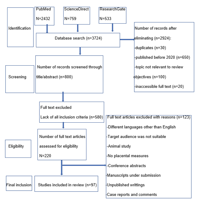
Figure 1: The process that was examined in this review is depicted in the flow chart.
Nutrient Transfer through the Placenta during Pregnancy in Women
Hemochorial placenta: structure and functions
HP has a villous hemochorial type, meaning that once maternal blood enters the terminal compartment, the maternal vessel lining is absent. Serving as a protective barrier, this organ safeguards the fetus and is made up of both maternal and fetal parts. Its composition consists of the decidua plus the embryonic trophoblast, together with numerous folded outgrowths known as chorionic villi. The placenta chiefly exists to form a tangible connection between the uterine wall and the fetus, making it possible to build a maternal-fetal exchange interface. The umbilical cord links the fetus with the placenta and supports blood interchange between mother and fetus. Within it are three vessels: one vein delivering blood into the fetus and two arteries carrying blood away toward the placenta. Transplacental ion channel transport involves (i) passive diffusion (O2, CO2, H2O, and steroids), (ii) carrier-mediated diffusion of ions and organic solutes (glucose and amino acids), (iii) ATP-dependent active transport of ions, (iv) endocytosis that either leads to molecular break down directly, or proceeds without breakdown via receptor-assisted uptake [12]. At full term in people, the placental barrier is made up of 7 main strata: (1) endothelium on the maternal side, (2) maternal connective tissue, (3) epithelium of the uterus, (4) trophoblast tissue, (4a) syncytiotrophoblast layer, (4b) cytotrophoblast layer, (5) connective tissue on the fetal side, (6) fetal endothelium, and (7) a trophoblast giant cell [12]. The placenta is a distinctive organ that exists only briefly and serves as both a structural and biological link between the growing embryo and the uterine lining. As pregnancy progresses, it supplies the embryo and fetus with necessary water and nutrients. Also, metabolic wastes produced by the fetus, including urea, uric acid, and creatinine, need elimination through the mother’s bloodstream and are cleared by transport across the placenta. By serving in respiration, the placenta allows the fetus to receive oxygen and expel carbon dioxide. Maternal blood rich in O2 and umbilical arterial blood, a blend of arterial and venous blood with low O2, exchange gases with one another [13]. During pregnancy, the placenta contributes several physiological activities, including producing hormones needed to sustain gestation, protecting the fetus from environmental threats, and creating immune tolerance between mother and fetus [14]. The syncytiotrophoblast constitutes the main obstacle limiting nutrient passage across the placenta. The MVM is oriented toward maternal blood flow, whereas the BM is oriented toward fetal blood flow. Placenta-related pregnancy complications, including (i) pregnancy-induced hypertension, (ii) abnormal implantation locations (placenta previa, placenta abruption, placenta accreta, increta, and percreta), (iii) insufficient placentation with PE, (iv) trophoblast-related disease (hydatiform mole and choriocarcinoma), and (v) additional outcomes (spontaneous abortion, preterm birth, fetal growth restriction-FGR) [15,16].
Hormones in pregnancy
Maternal circulation receives steroid hormones such as progesterone and estrogen, alongside protein hormones including human placental lactogen (hPL), human chorionic gonadotropin (hCG), adrenocorticotropic hormone (ACTH), CRH, gonadotropin releasing hormone (GnRH), and human growth hormone variant (hGH-V). It also receives other decidual proteins like calcitonin, relaxin, inhibins, activins, atrial natriuretic peptide, hypothalamic-like releasing, inhibiting hormones, thyrotropin releasing hormones (TRH), parathyroid hormone-related protein (PTH-rP), somatostatin, growth hormone-releasing hormone (GHRH), α-fetoprotein, prolactin, and relaxin. Control of the stress reaction is coordinated by the anterior pituitary through the CRH generated in the hypothalamus’s paraventricular nucleus [17]. Adrenaline, norepinephrine, acetyl choline, and glucocorticoids like cortisol are released by the adrenal medulla. Two outputs occur: ACTH formation [18] and cortisol secretion. Once cortisol circulates in the blood, it triggers feedback inhibition in the hypothalamus and pituitary glands, which lowers CRH and ACTH concentrations [19-21]. During gestation, CRH acts on the placenta, the myometrium’s smooth muscle, and the fetus’s pituitary-adrenal axis. By driving progesterone and estrogen production, it also contributes to the onset and continuation of labor. Elevated estrogen leads to the appearance of oxytocin receptors on the uterine muscle tissue. Cervical ripening involves estradiol, which increases glycosaminoglycan synthesis. Pregnancy is sustained by progesterone, which limits uterine contractility and diminishes estrogen’s influence [22,23]. During pregnancy, hormones such as progesterone, estrogen, hCG, and placental lactogenic hormone (also called chorionic somatotrophin hormone) enter the maternal circulation [24-26]. Placental activity is reduced by progesterone and nitric acid, whereas glucocorticoids, interleukin1 (IL1), noradrenaline, and acetylcholine increase it.
Nutrient transport in human placenta
Figure 2 illustrates the transfer of FAs, glucose, and amino acids across the placenta. For a successful pregnancy, the placenta must effectively uptake nutrients, metabolize them, and transfer them to the fetus to support the growth and development of both the placenta and the fetus. Active transport systems in the MVM and BM of the placental syncytiotropho blast facilitate the transfer of placental nutrients.
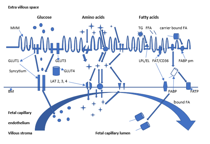
Figure 2: Nutrient transport across the placenta.
Note: The position of essential proteins responsible for the movement of macronutrients (glucose, amino acids, and FAs) at the MVM and BM. The SCTB serves as the main barrier for transferring nutrients from the mother to the developing fetus. Maternal blood accumulates in the intervillous space surrounding the developing fetus. The SCTB'BM is directed toward the fetal circulation. System L transporters (LAT1, LAT2, 3, 4) and exchangers facilitate the movement of amino acids from the BM to the fetal capillary. GLUT1 is the main transporter responsible for moving glucose across the MVM and BM to the syncytium. LPL and el break down maternal TG into FFA that can cross the MVM through FATPs, FAT/CD36, and FABPpm. FFAs are able to move through the cytosol using FABPs and through the bm using FATPs and FAT/CD36.
The composition of fatty acids
Maternal-to-fetal movement of fatty acids across the placenta.
During the first part of gestation and throughout breastfeeding, stores of FAs build up to meet the nutrient needs of the pregnant woman and the developing fetus. This period is referred to as the maternal catabolic and fetal anabolic phase. For the fetus to grow and mature properly, FAs are required. They are also used by the organism as an energy substrate [27]. Higher levels of maternal triglycerides (TGs), along with dyslipidemia and hyperleptinemia are associated with adverse outcomes in GDM. The movement of FAs from maternal blood to the fetus is supported by non-esterified fatty acids (NEFAs) and glycerol. In typical conditions, placental NEFAs provide the key nutrients needed for fetal development. By contrast, when pregnancy is complicated by GDM and obesity, excess lipid exposure can occur and this increases the expression of placental FA transporters. In GDM, the placental FA transport system is more complex and more susceptible to alterations in placental influences and in abnormal pregnancies tied to GDM-related metabolic changes. In maternal blood, lipoproteins (LPs) transport TGs that include essential as well as non-essential FAs. These lipids are taken up by the placenta and then supplied to the developing fetus. Before reaching the fetus as NEFAs, maternal TGs need to be broken down by lipoprotein lipases (LPL) and endothelial lipase (EL) so they can passa across the placental MVM and BM. NEFAs generation from maternal TGs likely relies on the activity of LPL and EL. LPL is abundant in the MVM, while EL is located in the capillary endothelial cell (EC) membranes of the HP. Within the HP, multiple transport and binding locations exist in the cytoplasm as well as within membranes. Fatty acid transport protein (FATP) has a mass of 63-70 kDa, fatty acid translocase/cluster of differentiation 36 (FAT/CD36) is 88 kDa, FA binding protein (FABP) is 40 kDa [28,29], major facilitator superfamily domain-containing protein 2A (MFSD2A) functions as a sodium-dependent lysophosphatidylcholine (LPC) symporter with a molecular mass of 50-60 kDa [30,31] and free fatty acid receptors (FFARs) (Figure 3) [29].
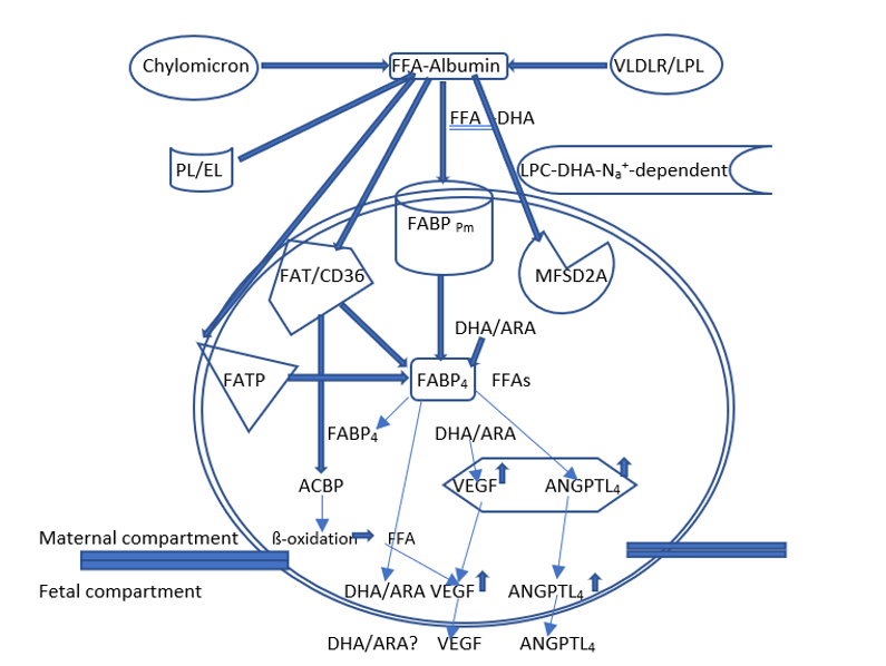
Figure 3: Schematic representation of placental fatty acid.
Note: The extended-chain fatty acids in this panel have an impact on the growth of cells and the development of new blood vessels in the placental trophoblast during the initial trimester. This also indicates that growth factors that aid in the development of blood vessels, such as ANGPTL4 and FABP4, play a role in the growth of the placenta. Cellular proteins and their function in facilitating the movement of maternal FAs across the human placental trophoblast. The transport of all FAs molecules between the mother and the fetus is made possible by the presence of FAT/CD36 and FATP on both sides of the bipolar placental cells. In the placenta, MFSD2A aids in cell fusion and forms syncytiotrophoblasts. MFSD2A carries DHA as an esterified single-tail phospholipid, lysophosphatidylcholine-DHA (LPC-DHA). By stimulating angiogenesis, DHA can assist in the early development of the placenta. DHA's unique capability to express VEGF mRNA sets it apart from other growth factors and cytokines. All LCFAs induce the secretion of ANGPTL4, while DHA promotes the expression and secretion of VEGF. FAs also activate intracellular FABP4 and FABP3, which are known to directly influence angiogenesis. Angiopoietin -like protein 4 (ANGPTL4); vascular endothelium growth factor (VEGF), fatty acid-binding proteins (FABPs); major facilitator superfamily-domain2A (MFSD2A).
The FATP group consists of six distinct isoforms, with five of these (FATP1-4, 6) encoded by the SLC27A gene family. Expression of everyone has been reported in the HP. Within this set, the highest mRNA levels are seen for FATP2, FATP4, and FATP6. FATP1 protein can be found in both the BM and the MVM. In placental tissue, mRNA for FATP1 along with FATP4 is detected and is associated with DHA concentrations in maternal blood and in placental phospholipids (PLs). These findings indicate that FATP4 plays an essential role in moving long-chain polyunsaturated FAs (LC-PUFAs) from the placenta to the developing fetus [27,28,30-32]. In the placenta, FATPs serve as primary transport proteins for LC-PUFAs such as DHA and ARA, and these lipids are required for fetal development and maturation of the brain. Using trophoblast cells obtained from term placentas, hypoxia exposure alters FATP1, FATP2, and FATP4 transcript levels via signaling that involves peroxisome proliferator-activated receptors-ϒ (PPARϒ) acting in concert with retinoid X receptor (RXR). Under these conditions, FATP4 transcripts declined, yet the protein level remained stable [30]. Elevated LC-PUFA levels in fetal blood may be linked to how these FAs are associated with specific transport molecules, including FATP4, which could directly affect uptake of DHA and ARA. An explicit link was found connecting DHA concentrations in plasma taken from the placenta during pregnancy with how strongly the FATP-1 and FATP-4 genes were expressed. From these findings, FATP-4 appears to contribute to the selective transfer of DHA across placental tissue. Proteins in the FATP family are able to carry free fatty acids (FFAs) across cell barriers and subsequently convert them into acyl-coA via esterification, which simplifies processing within the cell. A FABP protein that spans the membrane resides in trophoblast plasma membranes. It is made up of two parts: FABPpm and the placental FA-binding-plasma membrane protein (p-FABPpm). Data suggest that FABPpm enables one-way movement of LC-PUFA from the maternal side to the fetal side. In the HP, p-FABPpm appears exclusively in the syncytiotrophoblast MVM. Inside the syncytiotrophoblast cytosol, FABPs transport FA molecules toward specific intracellular destinations. This route can involve esterification, ß-oxidation, or passage onward to the fetus. The FABP family includes five members consisting of FABP1, FABP3, FABP4, FABP5, and FABP7, which are encoded by the genes FABP1, 3, 4, 5, and 7 genes. In the HP, these proteins are restricted to the syncytiotrophoblast MVM [30]. Trophoblast cells produce FABP4 and FABP7 [32]. From HP membranes, researchers extracted FAT/CD36, which attaches to LC-PUFAs [31, 32]. Acting in several roles, this protein associates with multiple ligands, such as oxidized low-density lipoprotein (LDL), and FFAs. As Figure 3 shows, long-chain FAs in this set affect trophoblast proliferation and the formation of new blood vessels. Across the syncytiotrophoblast MVM, maternal FAs move either by simple diffusion or by carrier-supported transfer involving FAT/CD36, FATPs, and the plasma-membrane FA-binding protein (FABPm). Placental transporter expression permits NEFA to travel from the placenta to the fetus by way of the syncytiotrophoblast BM. During 2022, Xintong’s team [33] carried out a retrospective analysis of GDM that relied on first-trimester body-composition metrics spanning multiple BMI groupings. When compared with the non-GDM cohort, participants with GDM had markedly higher measures of total body water, protein and mineral quantities, bone mineral content. Looking at pre-gestation BMI categories, among women with BMI below 24 kg/m2, 10. 9% received a GDM diagnosis, while among those with BMI at or above 24 kg/m2, 25.7% likewise diagnosed with GDM. Relative to typical controls, the same trend is characterized by reduced expression of FATP1 and FATP4 alongside increased levels of FAT/CD36 and FATP6 levels. The investigators found that indicators associated with body makeup had separate associations with the onset of GDM in individuals whose pre-pregnancy BMI was under 24 kg/m2 and in those whose BMI prior to pregnancy was 24 kg/m2 or higher. Before pregnancy, BMI corresponded more closely with total fat amount, visceral adiposity category, and the waist-to-hip ratio. In the HP, MFSD2A [28,30,31] carries fetal DHA as a Lysophospholipid (Lyso-PL). Based on what is presented, MFSD2A is viewed as a specialized carrier for Lyso-PL in both the brain and the placenta. In addition, MFSD2A permits DHA to reach the brain and contributes to preserving blood-brain barrier (BBB) integrity. The structural firmness of the BBB relies on the lipid environment that MFSD2A supplies to ECs within the central nervous system (CNS). Studies have examined MFSD2A levels under different pregnancy states, and lower expression has been reported in severe PE and GDM (Figure 3) [30]. G protein-coupled receptors (GPCRs) constitute a family of receptor proteins [31,33]. Researchers have recognized four FFARs: FFAR1 (GPR40), FFAR2 (GPR43), FFAR3 (GPR41), and FFAR4 (GPR120) [34]. In humans, the genes coding for FFAR1, FFAR2, and FFAR3 are positioned close together on the long arm of chromosome 19. Within that same chromosomal area, the human GPR42 gene produces multiple proteins whose architectures are similar to FFAR3. Located on the long arm of chromosome 10 is the FFAR1 gene, which determines human characteristics. Wherever FFAs may be found outside the cell, in the cytosol, or within the nuclear matrix. FABPpm, FAT/CD36, and FATPs mediate the transport and interaction of FFAs. In addition, FFAs may activate FFARs on the cell membrane by initially attaching to cytosolic FABPs and subsequently being guided into cellular compartments or metabolic pathways [30]. Also, the placenta contains Caveolae, which are protein-based formations that take part in FA transport. They are membrane in-foldings shaped like small tubes and enriched with cholesterol. As integral membrane proteins, Caveolae fall under the caveolin group and are generally around 21-24 kDa in size. In placental tissue from humans, there are three isoforms: caveolin-1 (cav-1), cav-2, and cav-3 [35]. Inside cells, cav-1 is found in Caveolae on membranes, within the Golgi apparatus, and in transport vesicles that derive from the trans-Golgi network. When considering cav-1 abundance, ECs show high expression, while cytotrophoblasts and syncytiotrophoblasts exhibit lower expression. Cav-2, is mainly located in endothelium, adipocytes, and fibrous tissue. Cav-3 is present only in skeletal muscle. In addition to forming Caveolae, cav-2 and cav-3 can also affect cav-1 expression and its metabolic processing [36-38]. By aiding cellular movement toward the membrane, Caveolae may influence FAT/CD36-mediated FA uptake. In certain tumors, PPARϒ boosts the functions of cav-1 and cav-2 [35-37]. Within a trophoblast, LPL function can be reduced by TGs together with non-essential FAs. This happens despite LPL’s primary role in hydrolyzing TGs transported by chylomicrons, very low-density lipoprotein (VLDL), and high-density lipoprotein (HDL) particles. EL has been shown in placental ECs and also in the syncytiotrophoblast. Movement of lipids from placenta to fetus may be aided by PLs as well as by TGs. Additionally, human placental tissue contains receptors for VLDL, LDL, and HDL. Through passive diffusion involving certain FA carriers, placental tissue draws FFAs from maternal circulation and transfers them through the placenta to the fetus. Since the mother’s circulating FFA reserve is limited, FAs need to be released from maternal LPs, so the placenta can absorb and deliver them to the fetus. In the placenta, LPL functions to break down TGs in maternal blood into FFAs that placental cells can take in and use.
LC-PUFAs are transferred from the mother to the fetus
The fetus is able to produce some monounsaturated and saturated FAs de novo by using glucose. However, essential FA such as ARA and DHA, have to be provided by the mother’s diet. Throughout pregnancy, the placenta transports ARA and DHA in an active and selective manner from maternal blood to the developing fetus [14]. The greater concentrations of ARA and DHA in fetal blood are attributed to transport that is stronger and more selective than what occurs for linoleic acid (LA) and ALA. Beginning at the 22nd week of amenorrhea and continuing until delivery, the fetus depends on the placenta for nutrient acquisition. The LA, ALA, ARA, DHA, and eicosapentaenoic acid (EPA) present in breast milk originate from the mother’s diet via lipomobilization and from maternal fat reserves that are partially established before and during gestation. DHA in red blood cells (RBC-DHA) reflects both DHA consumption and levels within tissues. In pregnancy, the mother’s DHA drops and ARA, along with other key FAs are mainly directly to the fetus through the placenta [8]. At birth, DHA in cord blood (CB-DHA) is frequently greater than the mother’s RBC-DHA, and this is called biomagnification. In contrast, when an infant’s CB-DHA matches or falls below maternal RBC-DHA, it is labeled bio-attenuation [8].
Esterified fatty acids from the mother's blood.
The esterified FAs (EFAs) from the mother's circulation to the fetus is essential for ensuring normal fetal growth and development. ELs are capable of sequestering both FFAs and NEFA that are released from the mother’s LPs. The results show that NEFA levels rise in the blood of both the mother and the fetus as delivery advances. Elevated maternal cholesterol levels during pregnancy can impact the developing fetus. Following their release from maternal LPLs by placental LPL and EL, they are subsequently taken uptake within the placental tissue. In Brenes-Martin and colleagues’ 2022 [38] work, they assessed how maternal visceral fat thickness relates to the likelihood of unfavorable pregnancy outcomes. They found that pregnant women with complications tended to have higher amounts of visceral fat. The findings indicated that the most common adverse pregnancy outcomes (APO) appearing during pregnancy were atypical fetal growth,13.6% (SGA, IUGR); gestational diabetes (GD), 9.9%, and pregnancy-related hypertensive conditions, 5.2%; with preterm birth prior to 37 weeks at 3.9% next; then membrane rupture before 37 weeks at 1.6%; and stillbirth at 1%. Significant differences were identified between the no-complication group and the APO group for pre-pregnancy weight and BMI, along with gestational age at delivery. Concerning biochemical measures, the APO group had elevated TGs (p=0.001), LDL cholesterol (p=0.02), and C-reactive protein (CRP) levels (p=0.04), while showing reduced soluble fms-like tyrosine kinase-1 (sFlt-1) similar to fms (p=0.005) and a lower sFlt-1/PlGF (placental growth factor) (p=0.03). With respect to ultrasound measures, the APO group demonstrated greater visceral fat thickness (VFT), p=0.001, and greater subcutaneous fat thickness (SFT), p=0.03, compared with women who had no pregnancy complications. Maternal visceral and SFT were significantly associated with insulin (r=0.37, p<0.001; r=0.30, p<0.001, respectively), and with the homeostasis model assessment insulin resistant (HOMA-IR) index (r=0.33, p<0.001; r=0.34, p<0.001, respectively), and they were also linked with CRP (r=0.47, p=0.001; r=0.28, p<0.001, respectively). BMI showed correlations as well (r=0.47; p<0.001; r=0.65; p<0.001). VFT in the mother also correlated significantly with TGs (r=0.24; p=0.02). Maternal erythrocytes may build up lyso-PL by incorporating them into their membranes. Through these cells, Lyso-PL can reach the uterus. Phospholipase type II (PLA2), located mainly in the placenta and choriodecidua, is a lipase that helps FFAs from maternal plasma LP. A triacylglycerol (TAG) hydrolase is located within the placental syncytiotrophoblast layer. In placental tissue, HDL and LDL are able to bind to LP receptors [39,40].
Placental lipid transport and carrier proteins
Prostaglandins (PGs) associate with FABP-1, and this protein, through protein-to-protein coupling, transports the ligands to PPAR. Once FAs reach the placenta, some are broken down into mitochondria to provide energy, while the remainder becomes part of placental tissue. Throughout gestation, the placenta serves as the principal source of PGs in uterine tissue. Within the placenta, the fetal-placental unit generates numerous eicosanoids that function as ligands for paracrine and lipoxin (LX) E molecules. These PGs are produced from HP that originates from ARA via cyclooxygenase (COX)-1/2, along with particular PG isomerase/synthase enzymes [41]. In HP, PGD2 is transformed into PGJ2 and hence acts as a ligand for PPARϒ. Through the lipoxygenase (LOX) pathway, which converts ARA and LA, they generate oxygenated lipid mediators along with 9- and 13-hydroxyoctadecadienoic acid (HODE), and hepoxilin, that act as endogenous ligands to spark off PPARϒ. Those lipid-activated transcription factors then adjust genes involved in inflammation, lipid metabolism, and cell differentiation [14].
Nuclear receptors during pregnancy
In an uncomplicated pregnancy, progesterone and estrogen, together with the nuclear receptors, including PPAR, liver X receptor (LXR), farnesoid X receptor (FXR), pregnane X receptor (PXR), retinoic acid receptors (RARs), and RXR contribute substantially to the stepwise metabolic changes that support the continued survival of both mother and fetus [42,43]. Throughout the reproductive cycle and during pregnancy, PPAR signaling helps the uterus carry out functions such as making steroids, producing cytokines, and angiogenesis. When these pathways are switched on, they can promote the synthesis and secretion of hormones, including gonadotrophins, which help maintain gestation and support fetal development. There are three variants of PPAR: α, β/δ, and ϒ. These receptors take part in key early gestational processes including embryo attachment, placental development, and maturation of trophoblast cells [14,44]. Reports describe PPARα in placental choriocarcinoma cell lines and also in term amnion, choriodecidua, placental syncytial cells, and cytotrophoblast cells. Evidence indicates that it helps establish the fetal skin barrier, limits cell growth, and lowers secretion of hCGß [14]. PPARß/δ has been identified in human trophoblast cells, and this receptor is required for fetal growth and progression. It aids implantation and decidualization, stimulates cell division, and increases nutrient absorption as well as nutrient specialization [14]. In HPs, PPARϒ is expressed at high levels, most notably within trophoblasts, villous syncytiotrophoblasts, and cytotrophoblasts. It is necessary for building the placenta, enabling trophoblast invasion, and driving the shift from cytotrophoblasts to syncytiotrophoblasts [14]. There are two variants of LXR: α and ß, and both pair up with RXRs. LXRα has been detected in placental samples taken from women whose pregnancies were normal. Also, LXRα influences LP metabolism by modulating enzymes including LPL, cholesterol ester transfer protein, and PL transfer protein. The gene for LXRß is found in many tissues. Its role is crucial in restraining how invasive human trophoblast cells occur. Sterol regulatory element-binding proteins (SREBPs) constitute a specialized class of transcriptional regulators. Researchers have identified three members within the SREBP family across various mammalian species. The human SREBP-1a and SREBP-1c isoforms are derived from a single gene, SREBPF-1, located on chromosome 17p11.2, whereas SREBP-2 is encoded by a distinct gene, SREBPF-2 found on chromosome 22q13.3 [14]. SREBP-1 stimulates genes essential for FA biosynthesis, whereas SREBP-2 regulates those involved in cholesterol biosynthesis. Evidence suggests that SREBPs can directly activate numerous genes, including 3-hydroxy-3-methylglutaryl-coA synthase1, 3-hydroxy-3-methylglutaryl-coA reductase, and FAs, which play roles in the synthesis and uptake of cholesterol, TAG, and PL [14]. SREBP-1c regulates genes critical for de novo lipogenesis and serves as a key modulator of the placenta’s dynamic pH balance. Once polyunsaturated fatty acids (PUFAs) trigger the transcription of carrier proteins and activate mitochondrial peroxisomal ß-oxidation, the catabolic degradation pathway is initiated.
How fatty acid pathways contribute to fetal and placental development
To ensure proper fetal development and readiness for birth, the mother must supply the LC-PUFA DHA. During the last weeks of pregnancy and following birth, DHA transfers from the mother to the infant, while simultaneously increasing significantly within the mother’s fat reserves [14]. Since fetal fat tissue grows rapidly, the majority of fat accumulation occurs during this broader mobilization period. During pregnancy, a mother’s blood levels of TAG, PLs, and NEFAs rise. In the placenta, NEFAs may either be re-esterified and stored as TAG or oxidized within trophoblast cells. Lipolysis in women becomes more pronounced during the third trimester. Maternal blood TAG rises as a result of both increased hepatic production and dietary intake. During this same late stage, placental lipase activity increases, thereby restricting the storage of TAGs. In the placenta, LPL hydrolyzes TAG within LDL and in VLDL produced by the liver, but it does not act on TAG found in chylomicrons. How placental fats are processed could significantly impact pregnancy outcomes and the baby’s health.
The human placental organ's sugar
Glucose transfer through the placenta
Glucose is a vital nutrient required for normal development and maturation. This fuel supplies the majority of the energy required by both the fetus and the placenta [40,41]. Because the fetus generates very little glucose independently, it depends entirely on glucose provided by the mother. Glucose is transferred across the placenta via facilitated diffusion, a process that relies on specific glucose carrier proteins. Glucose diffuses solely down its concentration gradient. When maternal blood glucose levels exceed those of the fetus, additional glucose is transported to the fetus. Due to the presence of many transporters, the syncytiotrophoblast can absorb glucose rapidly. GLUT proteins are present on both the maternal-facing and fetal-facing membranes of the syncytiotrophoblast, including the MVM and the BM. The MVM is characterized by a high glucose-transfer capacity, which maintains a concentration gradient between the syncytiotrophoblast cytosol and the fetal capillary. To achieve a net delivery of glucose to the fetus, this difference is essential, and it also sufficiently satisfies the placenta's own glucose requirements. Compared with the MVM, the BM has far fewer GLUTs. Lower GLUTs across the BM complement the disparate surface areas of the BM compared with the placental membrane. Glucose processing involves three categories of protein solute carriers: sodium-coupled symport systems (SGLT, SLC5), facilitative carriers (GLUT, SLC2), and SWEET transporters (SLC50) [41, 45]. Even though those last two groups belong to the larger GLUT-related family, they are absent from the HP. The GLUT family consists of fourteen proteins that are divided into three distinct classes. In class, I have the standard GLUT 1-4 along with GLUT14, which is a duplicated form of GLUT3. Class II includes the GLUT5, 7, 9, and 11 forms, while class III contains the GLUT 6, 8, 10, and 12 variants [45]. Within the HP, reports describe seven GLUT forms; GLUT1, 3, 4, 8-10 and 12 [40,45,46] (Table 1). The GLUT1 (SLC2A) isoform, about 55 kDa, is recognized as being synthesized by the placenta, encompassing the syncytiotrophoblast, cytotrophoblast, EC, and villous stroma components [41]. GLUT1 binds glucose with strong affinity and also operates on various other substrates, including galactose, mannose, glucosamine, and DHA. Studies indicate that diabetes in the mother correlates with elevated GLUT1 amounts in breast milk. This points to a greater capacity of the BM to transport glucose. The GLUT1 gene has been located within a region of human chromosome 1p35-p31.3. Glucose transfer from mother to fetus occurs mainly through placental GLUT1, which is present at high levels on both the syncytiotrophoblasts MVM and BM [47]. This transporter is likewise found on the cell of surface extravillous cytotrophoblasts as well as within the placental endothelial lining [48]. Yet, additional studies indicate that the amount or activity of GLUT1 varies over gestation, spanning the first to the third trimester [49]. Rising maternal age was linked to higher placental GLUT1 mRNA, whereas increasing placental weight corresponded to lower levels. Also, placental GLUT1 protein correlated positively with pre-gestational maternal BMI and umbilical artery glucose, yet it did not correlate with insulin levels in umbilical blood [49]. When GLUT1 is expressed more strongly, glucose uptake rises at the syncytial BM, but the MVM remains unchanged. Studies assessing GLUT1 expression have yielded mixed outcomes, since certain reports find no difference between placentas from GDM cases and those from pregnancies without diabetes. At term, placenta tissues show GLUT3 (SLC2A3) with a mass of 49 kDa within placental membrane layers (MVM and BM) as well as intraplacental endothelial cells [47]. On chromosome 12, the SLC2A3 gene maps to a band that is located in the region of chromosome p13.3. During the first trimester, GLUT3 plays a key role in maintaining sufficient glucose levels in fetal tissues. Because GLUT3 binds glucose with high affinity, it can also transport several other substrates, including galactose, fructose, mannose, xylose, and DHA. Findings from Huang’s team and Feng’s group, referenced in Guadix et al. 2023 [49], described GLUT3 presence in HPs that were altered by GDM. Based on these findings, GLUT3 abundance could markedly influence placental glucose transport in GDM through effects on hyperglycemia. During the first trimester, HPs additionally produce GLUT4 (SLC2A4), another GLUT-family transporter (48 kDa), and it is most prominent at the syncytiotrophoblast MVM [50]. Unlike many others, GLUT4 operates as a carrier regulated by insulin and is mainly located in adipose tissue plus skeletal and cardiac muscle [45]. Researchers have mapped the SLC2A4 gene to a segment on human chromosome 17p13. GLUT4 binds glucose with high affinity and can also utilize glucosamine and DHA as substrates. At term, HP samples show GLUT4 localization alongside insulin receptors [51]. When GDM complicates pregnancy, GLUT4 amounts drop, shaped by components of insulin signaling and by sex hormone-binding globulin. In contrast, in cases of insulin-requiring GDM, the quantity of placental GLUT4 protein correlated positively with neonatal birth weight. Earlier results suggest that influences linked to GDM, not solely insulin, could shift levels of GLUT4. In particular, research in obese gestations without GDM found that maternal insulin boosts glucose transfer through the placenta by promoting relocation of GLUT4 toward the fetal-facing of the syncytiotrophoblast BM [49]. Evidence suggests GLUT8 (SLC2A8) is present in extravillous trophoblasts and, at term, within both syncytio-and cytotrophoblast cells. Similar to GLUT4, GLUT8 relies on insulin. GLUT8 mediates the insulin-stimulated uptake of glucose into the blastocyst. Alongside fructose and galactose, glucose functions as the main fuel used for GLUT8. Within syncytiotrophoblasts, GLUT8 has additionally been observed in the cytosol, as well as in endosomes, lysosomes, and the endoplasmic reticulum membrane [45]. The SLC2A8 gene has been mapped to a region on human chromosome 9q33.3. Research indicates that the 59 kDa GLUT9 (SLC2A9) variant operates in urate movement, and it is also able to transport hexose, since it binds fructose strongly. GLUT9 consists mainly of two subtypes, GLUT9a and GLUT9b, and these show distinct distribution patterns in polarized cells. On the apical membrane, GLUT9b acts, while GLUT9a is found on the basal side [45, a 49]. The SLC2A9 gene resides on chromosome 4 within the p15.3-p16 interval. Within HPs, both GLUT9 isoforms are detected in the syncytiotrophoblast. Relative to healthy control groups, their amounts were greatly increased in placentas from pregnancies affected by GD as well as pre-gestational diabetes (PGD). Guadix and coauthors [49] reported that Stanirowski et al. showed that, even though the two GLUT9 isoforms display distinct expression profiles. Trophoblast investigations verified a marked rise in total GLUT9 abundance in placental tissue from patients with insulin-dependent diabetes, GDM, and PGD. Taken together, the results imply that GLUT9 could contribute to the later-life consequences linked with fetal macrosomia in pregnancies affected by GDM. Although Information regarding the specific placental location and function of GLUT10 (SLC2A10) remains scarce, research across various organs and species suggests that GLUT10 may be localized intracellularly, potentially within the endoplasmic reticulum or mitochondria. Like GLUT1, GLUT10 facilitates the transport of dehydroascorbic acid into the endoplasmic reticulum or mitochondria, and its absence can result in protein folding defects and/or increased oxidative stress [45]. GLUT10 exhibits a strong preference for binding D-glucose and D-galactose, whereas it shows minimal affinity for fructose [49]. Furthermore, the GLUT10 gene has been localized to a region on human chromosome 20q12-13.1 associated with type 2 diabetes mellitus (T2DM), suggesting that GLUT10 may be involved in glucose metabolism and the pathogenesis of T2DM. GLU10 mRNA has been detected in the placenta, liver, pancreas, and kidneys, as well as in human adipose tissue [45]. GLUT12 (SLC2A12) was initially reported as a distinct insulin-responsive variant associated with the GLUT4 carrier. When stimulated by insulin, estradiol, dihydrotestosterone, or epidermal growth factor (EGF), It likewise shifts to the cell surface membrane. However, unlike GLUT4, its transport profile is not the same, since GLUT12 is considered a primary transporter together with GLUT1. The chief substrate for GLUT12 is glucose, and it can also take up galactose and fructose. In placental at normal term, GLUT12 has been detected, while in syncytiotrophoblast during the first trimester it appeared predominantly in the cytoplasm. Under both conditions, it is also found in villous cytotrophoblast and extravillous trophoblast. The GLUT12 gene is located within the human chromosomal region 6q23.2. With progression of pregnancy, placental glucose absorption increases in efficiency, and the mechanisms controlling glucose movement are different in the first and the third trimester (Table 1) [45].
|
Protein name |
Gene Localization |
Localization |
Transported substrates |
Ref |
|
GLUT 1; solute carrier family 2, member 1; Class I |
SLC2A1 1p35-p31.3 |
Placental membranes: syncytiotrophoblast MVM, BM, cytotrophoblast cell, fetal endothelium, plasma membrane of the villous, |
Glucose transporter (50-55 kDa); galactose, mannose, glucosamine transporter |
[40, 45, 49] |
|
GLUT 3; solute carrier family 2, member 3; Class I |
SLC2A3 12p13.3
|
Placental membranes: syncytiotrophoblast MVM, BM, placental endothelial cells |
Glucose transporter (49 kDa); galactose transporter |
[40, 45, 49] |
|
GLUT 4; solute carrier family 2, member 4; Class I |
SLC2A4 17p13
|
Placental membranes: syncytiotrophoblast BM, MVM |
Glucose transporter (48 kDa), dehydroascorbic acid transporter, glucosamine transporter |
[40, 45, 49] |
|
GLUT 8; solute carrier family 2, member 8; Class III |
SLC2A8 9p33.3 |
Placental membranes: syncytiotrophoblast MVM BM |
Glucose transporter is insulin dependent. |
[45, 49] |
|
GLUT 9; solute carrier family 2, member 9; Class II GLUT 9a and GLUT 9b |
SLC2A9 4p15.3-p16 |
Placental membranes: syncytiotrophoblast MVM, BM; GLUT 9b is found localized apically, while GLUT 9a is found on basolateral membranes; |
Glucose transporter (59 kDa), Urate transporter, hexose transporter, and fructose transporter |
[40, 45, 49] |
|
GLUT 10; solute carrier family 2, member 10; Class III isoform |
SLC2A10 Localization: Chromosome 20q12-13.1 |
Placental membranes: |
Glucose transporter, dehydroascorbic acid transporter. Strong affinity for D-glucose and D-galactose, but not for fructose |
[45, 49] |
|
GLUT 12; solute carrier family 2, member 12; Class III |
SLC2A12 6q23.2 |
Placental membranes: basal membrane of the syncytio totrophoblast and extra-villous trophoblast cells |
Glucose transporter, Galactose transporter, and fructose transporter |
[40, 45, 49] |
Table 1: Glucose transporter in the human placenta.
Note. Glucose is the primary nutrient required for fetal growth and development. Glucose transport across the placenta to the fetus is mediated by a family of facilitated diffusion transporters, named GLUTs, encoded by the solute carrier family 2 and facilitated glucose transporters (SLC2A) family of genes. Seven GLUT isoforms have been reported to be expressed in human placenta at different sites of the syncytiotrophoblast membranes.
Predominantly pronounced; less pronunced; similar level in placental cells; MVM: microvillous plasma membrane; BM: basal plasma membrane.
In 2023, Aldahmash and colleagues [10] carried out a study indicating that placentas from pregnant women with GD had markedly elevated amounts of GLUT1 and GLUT3 compared with placentas from healthy pregnant women. From these findings, the researchers inferred that GDM is associated with more frequent apoptosis in the placental villi and changes in the protein expression levels of GLUT1 and GLUT3 in the placenta of women affected by GD.
Hormone effectors and substrates
Multiple reports examine how circulating factors influence the expression of the GLUT family within the HP. These factors include insulin, insulin-like growth factor 1 (IGF-1), adiponectin, resist in, leptin, tumor necrosis factor-α (TNF-α), hepatocyte growth factor (HGF), glucocorticoids, and CRH. They regulate the expression of the GLUT family in the HP [46]. Insulin, IGF-1, adiponectin, and resist in are among the earliest identified factors, generally considered contributing to fetoplacental metabolic activity and development, with alterations in their levels associated with changes in fetal and placental mass. Some studies suggest that insulin-driven increases in glucose transport at term may conflict with the known locations of the insulin receptor. However, the observation of elevated GLUT levels in first-trimester villous tissue is consistent with the presence of GLUT4 and the insulin receptor in the syncytiotrophoblast. The observed changes in placental GLUT1 levels correspond to the combined effects of IGF-1-driven increases in glucose transport and variations in fetal and/or placental weight. Adiponectin partially modulates placental function by downregulating the expression of nutrient transport proteins, a mechanism that aligns with the observed inverse relationship between maternal blood adiponectin concentrations and newborn birth weight. In humans, resistin is secreted by circulating blood cells and macrophages, and it plays roles beyond insulin resistance, including involvement in cardiovascular and inflammatory conditions. It also modulates GLUT1 expression. In BeWo cells, researchers assessed the combined effects of leptin and TNF-α, finding that both induced a concentration-dependent increase in GLUT1 expression. Due to its significant role in regulating glucose absorption and metabolic processes within pancreatic ß-cells, intestinal epithelial cells, adipocytes, and skeletal muscle cells, HGF is considered a key metabolic regulator alongside other established factors. In placental explant preparations, HGF enhances glucose uptake via a mechanism that can be blocked by cytochalasin ß, which is consistent with the observed increase in GLUT1 expression. Like resist in, HGF appears to regulate glucose transporter expression through a pathway mediated by extracellular signal-regulated kinase 1/2 (ERK1/2). When the effect of glucocorticoids on GLUT expression in cultured term primary trophoblast cells derived from term placenta was investigated, it was found that increasing doses led to a reduction in both GLUT1 and GLUT3 at the mRNA and protein levels. In contrast, exposure of human placental endothelial cells to glucocorticoids resulted in increased levels of both GLUT1 and GLUT3 mRNA and protein. The placental growth hormone (PGH) is derived from a growth hormone (GH) variant gene situated on chromosome 17, specifically within the q22-q24 region. Within the HP, PGH is synthesized in both the syncytiotrophoblast and extravillous cytotrophoblast layers. It is also present in fetal-related compartments, such as the umbilical cord circulation and amniotic fluid. PGH is considered to play a central role in regulating maternal IGF-1. Furthermore, it stimulates glucose production, fat mobilization, and tissue growth in the mother, while also influencing fetal size, placental development, and the mother’s physiological adaptation to pregnancy. While most of these effects are mediated by the modulation of maternal IGF-1 levels, activity within extravillous trophoblast cells suggests a more direct role in placental growth. Since both GH and PGH receptors are present in the syncytiotrophoblast, this hormone facilitates placental maturation and function via autocrine signaling. In villous tissue from a full-term placenta, GH enhances glucose transfer into the tissue. However, in first-trimester villi, GH leaves glucose entry unchanged. The action of CRH is mediated through two distinct corticotropin-releasing hormone receptors, CRH-R1 and CRH-R2. In placentas affected by IUGR, CRH-R1 expression is downregulated, while in trophoblast cells, CRH-R1 positively regulates GLUT1 expression. Placenta-derived CRH regulates glucose transport within the placenta via autocrine or paracrine signaling pathways [52]. Steroid hormones can also alter the activity or expression of GLUTs in the placenta during late pregnancy. In contrast, estrogens and progesterone were found to reduce the rate of glucose uptake by MVM vesicles. The placenta contains various signaling networks, including the mechanistic target of the Rapamycin (mTOR) pathway, which regulates nutrient transport needs [53]. This mTOR pathway governs cellular growth and metabolic status by sensing extracellular nutrients, growth factors, and signals via two distinct complexes: mTOR Complex 1 (mTORC1) and mTOR Complex 2 (mTORC2) [50]. In 2023, Shi and colleagues [53], reported that varying glucose levels influence the function and growth of bovine placental trophoblast cells (BPTCs), with effects observed in GLUT-related indices, ATP levels, and mTOR-associated signaling pathways. Additionally, inhibiting mTOR activity led to a decline in GLUT4 mRNA production and a reduction in protein levels within the mTOR cascade following glucose addition. Overall, the data suggest that in BPTCs, glucose may influence its own transport by modulating the mTOR pathway, providing fundamental insights into how glucose transport is regulated in these.
The human amino acid pattern of the human placenta
Normal development and maturation of the fetus require amino acids to move from the placenta into fetal circulation. How efficiently this occurs is determined largely by the placenta’s capacity to carry out that movement. Moving amino acids through the placenta relies on energy-requiring mechanisms that work against a concentration difference, using accumulative carriers and exchange systems located in the MVM and BM.
Movement of amino acids through the placenta
Various amino acid transporter systems in the placenta are displayed in Table 2. For this one-way movement to occur, more than 20 different amino acid transporter proteins must work together. They are positioned on both the maternal-side and fetal-side plasma membranes of the placental epithelium. Their role is to bring amino acids in from the mother and pass them on to the fetus. Every placental amino acid transporter falls within the solute carrier (SLC) superfamily. The diverse network of carrier mechanisms can be sorted into 7 groups: A, ASC, X-AG, L, y+, y+L, and T, that together allow amino acids the placenta. Among these, two systems have been studied in depth [54]. System A operates with sodium and brings in neutral, non-essential neutral amino acids, whereas System L functions without sodium and brings in essential amino acids within the placenta. In addition, these transporters can be grouped by function into accumulative, exchange, or facilitated categories. In HP, three distinct isoforms of the system A transporter, including the sodium-coupled neutral amino acid transporters, also called the serotonin N-acetyltransferase family (SNAT1, SNAT2, and SNAT4), are encoded by the SLC38A gene (known as SLC38A1, SLC38A2, and SLC38A4) [51,55]. SNAT1, which contributes most to system A activity, influences the performance of this system in the placenta. During the first trimester, SNAT4 is more abundant than it is at term placenta. The system A transporter carries many neutral non-essential amino acids, such as alanine, glutamine, and serine [55]. Concerning placental amino acid transporter expression in GDM, certain reports describe decreased system A transporter expression in the MVM of placentas from GDM-complicated pregnancies. This reduction could be due to fewer amino acid transporters being present. In contrast, another study found that placental amino acid uptake is higher in human pregnancies complicated by GDM. It has been suggested that leptin helps control placental amino acid transport by increasing system A transporter activity in long-term placentas. The presence of accumulative transporters, primarily systems A and X-AG, involving SLC1 and SLC7, make it easier for certain amino acids to enter the syncytiotrophoblasts. Through amino acid exchange transporters, amino acid movement across the membrane promotes the entry of additional amino acids from outside the cell. SLC16A10 and SLC43 are transporter proteins located in the BM of syncytiotrophoblasts. When these specialized transporters act on the syncytiotrophoblast surface, they help clear specific amino acid from the fetal bloodstream. An exchanger is an agent able to convert those amino acids into other amino acid forms. In this way, the developing fetus receives the full set of required amino acids. System A function occurs in both the syncytiotrophoblast and the BM, yet it is most abundant at the MVM. By driving system A, intracellular levels of amino acids such as glycine become high, and system L then swaps these for essential amino acids available outside the cell. As a result, System A together with L jointly ensure the availability of both indispensable and dispensable amino acids. Data suggest that placental amino acid carriers perform effectively only when supported by adequate hormones and nutrients. Growth-related signals, including Insulin, IGF-1, IGF-2, IL-6, TNF-α, leptin, and adiponectin, enhance System A by engaging mTOR, which increases transport capacity and thereby helps sustain faster fetal growth [54]. It is proposed that IL-6 modulates system A through the signal transducer and activator of transcription (STAT)-3, and investigations using villous tissue have verified that STAT3 plays a major role in controlling amino acid movement across the placenta.
When leptin attaches to its receptor (LepR), it is abundantly present in the syncytiotrophoblast MVM. It triggers signaling through Janus kinase 2 (JAK2) and STAT3. In human placental villous explants, leptin boosts the System A transporter by altering gene transcription via stimulation of the JAK2/STAT3 route. Using that same model, blocking STAT3 or JAK2 diminishes the activity of the System A amino acid transporter [54]. Placental system A activity has been shown to fall in response to influences such as hypoxia, IL-1ß, angiotensin II (AngII), urocortin, and CRH [54]. Maternal diet, smoking habits, and vitamin D consumption may affect how certain placental amino acid transporters are produced. Regulation of amino acid transporter expression in the placenta might also be shaped by epigenetic influences. The serine/threoline kinase mTOR is particularly important in regulating cell growth and tumor progression. mTOR forms two complexes, mTORC1 and mTORC2 [50, 56]. Recently, greater focus has been placed on links between amino acid transporters, especially those that carry leu, and the placental mTOR signaling network, since mTOR responds to nutritional inputs like amino acids without relying on GHs. Disrupted mTOR signaling has been linked to numerous conditions, including metabolic disorders, neurodegeneration, cancer, growth abnormalities, and aging, underscoring the pathway's critical role in maintaining cellular and physiological homeostasis. The body’s overall metabolic state likely modulates the regulation of amino acid transport across the placenta. In addition to regulating transporter activity and protein synthesis, the mTORC1 proteinase kinase cascade likely plays a pivotal role in controlling placental amino acid exchange. However, evidence indicates that the mTOR signaling pathway, specifically mTORC1, regulates system A and L activity by reducing the plasma membrane trafficking of SNAT2, while placental mTOR activity evidenced by decreased levels of downstream signaling molecule phosphorylated ribosomal S6 kinase 1 is diminished in FGR [57]. When intracellular leucine is present, Sestrine 2 attaches to it, and this interaction triggers activation of the GATOR2 complex together with the mTORC1 signaling route. In IUGR, lowered mTORC1 activity changes system A and system L elements are transported across cell membranes. Through modifying particular placental amino acid carriers, mTORC1 controls fetal development. When oxygen availability is low, reactive oxygen species (ROS) rise and trigger oxidative stress, a process that could explain how hypoxia influences amino acid transfer in the placenta. Oxidative stress at the cellular level occurs once ROS generation outpaces enzymatic and nonenzymatic antioxidant defenses that normally keep ROS in a healthy range. Multiple findings indicate that heightened oxidative stress plays a role in pregnancy disorders including miscarriage, PE, and IUGR [58]. It has also been shown that greater oxidative stress during fetal development raises the fetus’ likelihood of later brain impairment and cardiovascular disease [59]. Under hypoxic conditions, SNAT1 and SNAT2 gene activity is reduced, and System A amino acid uptake is suppressed in cultured primary human trophoblast cells. Overall, the results indicate that both hypoxia and oxidative stress act to restrain amino acid transport across the placenta. System L, also called the large neutral amino acid transporter (LAT) system, is a sodium-independent exchanger. It carries essential neutral amino acids such as L-leucine and L-phenylalanine across the placenta [49]. System L (SLC7) is a heterodimer, consisting of a light chain, including isoforms LAT1(SLC7A5) or LAT2 (SLC7A8). Both isoforms have been found to be expressed in trophoblast cells of the HP, and they are located in MVM and BM of the syncytiotrophoblast [49,54,60]. In contrast, the SLC43 family comprising LAT3 (SLC43A1) and LAT4 (SLC43A2) are monomeric and function as facilitated amino acid transporters [60]. LAT3 is poorly expressed in the HP, whereas LAT4 is highly expressed. Reports indicate that increased abundance of both System A and System L in GDM accelerates fetal growth, leading to macrosomia and subsequently affecting placental performance. Leptin is essential for both cell division and protein synthesis. In human trophoblastic cells, leptin stimulates amino acid synthesis by activating the mitogen-activated protein kinase (MAPK) and phosphatidylinositol-3-kinase (PI3K) signaling pathways. Beside system y+ (SLC7A1), which carries cationic amino acids at the MVM, the syncytiotrophoblast includes additional transport proteins. Moreover, the main BM route, indicated as y+L, is also found at the MVM. Operation of system y+ occurs without sodium. Accumulative transporter members of the SLC1 and SLC38 groups are present on the BM and contribute to placental uptake of amino acids from the fetus. Within the fetal BM, highly active SLC1 transporters move glutamate as well as aspartate. These SLC1 proteins help bring fetal glutamate into the placenta. Because fetal amino acid concentrations generally exceed maternal levels, an active placental transport process is thought to exist. Amino acid transporters are essential for taking up small non-essential amino acids, including alanine, glycine, and serine. With this transport arrangement, sodium enters the cell too, enabling amino acids uptake even when extracellular levels are lower than intracellular ones. System ASC (referred to as SLC7A10 or Asc-1) is the light chain of the heterodimer neutral amino acid transporter, and is a sodium-independent antiporter distributed throughout the CNS. Asc-1 is present in both astrocytes and neurons, and displays high affinity for neutral amino acids and uses diverse substrates, such as L-serine, L-alanine, L-cysteine along with glycine and D-serine [61]. Evidence suggests it resides in the BM area, although activity has also been noted at the MVM of the syncytiotrophoblast [57]. Glutamine deamination in the fetal liver yields glutamate, a vital nitrogen source and precursor of ϒ-amino butyric acid (GABA), a key inhibitory neurotransmitter. Glutamate is transported across the MVM and BM membranes of the syncytiotrophoblast via high-affinity excitatory amino acid transporters (EAATs; system X-AG), where it is subsequently converted into glutamine within the placenta. Glutamine serves as a substrate for the amino acid transporter systems A (isoforms SNAT1, 2, and 4), N (SNAT5); L (LAT1 and LAT2); Y+L and ASC. However, the transfer of glutamine across the MVM and BM of the syncytiotrophoblast occurs via the system Y+L (Table 2) [57].
|
Protein name |
Gene |
Transport system |
Localization in the syncy- tiotrophoblast |
Predominant substrates |
Ref |
|
Na+-dependent systems |
|||||
|
SNAT1 SNAT2 SNAT4 |
SLC38A1 SLC38A2 SLC38A4 |
A A A |
MVM, BM MVM, BM MVM, BM |
Q, A, N, C, H, S A, N, C, Q, G, H, M, P, S A, N, C, G, S, T |
[49, 54] |
|
ASCT1 ASCT2 |
SLC1A4 SLC1A5 |
ASC ASC |
MVM, BM MVM, BM |
A, S, C, T A, C, Q, S, T, N |
[54, 57] |
|
EAAT3 EAAT2 EAAT1 EAAT4 |
SLC1A1 SLC1A2 SLC1A3 SLC1A6 |
X-AG X-AG X-AG X-AG |
MVM, BM MVM, BM MVM, BM MVM, BM |
Glu, Asp, Cys Glu, Asp Glu, Asp D, E |
[57] |
|
Na+-independent systems |
|||||
|
LAT1 LAT2 LAT3 LAT4 |
SLC7A5 SLC7A8 SLC43A1 SLC43A2 |
L L L L |
MVM, BM MVM, BM MVM, BM MVM, BM |
F, Y, W, M, V, I, L, H, BCH L, A, S, T, C, F, Y, W, BCH, N, H, I, M, V, Q, G |
[49, 54, 60] |
|
Asc1 |
SLC7A10 |
asc |
MVM, BM |
L-serine, L-alanine, L- cysteine,D-serine, glycine |
[61] |
|
CAT1 CAT2B CAT3 CAT4 Y+LAT1 Y+LAT2 |
SLC7A1 SLC7A2 SLC7A3P SLCA4
SLC7A7 SLC7A6 |
Y+ Y+ Y+ Y+
Y+L Y+L |
MVM, BM MVM, BM MVM, BM MVM, BM MVM, BM MVM, BM |
R, H, K R, H, K, M, AL |
[57] [57] |
Table 2: Amino acid transporter systems in the human placenta.
Note. Na+-dependant systems: A, ASC,asc, X-AG; Na+independant systems: L, Y+L, Y+; A (L-Alanine, R (L-arginine); N (L-asparagine); D (L-aspartate; C (L-cysteine), E (L-glutamate); Q (L-glutamine); G (glycine); H (L-histidine); I (L-isoleucine); K (L-lysine); M (L-methionine); F (L-phenylalanine); P (L-proline; S (L-serine); T (L-threoline); W (L-tryptophan); Y (L-tyrosine); V (L-valine); BCH (2-aminobicyclo-(2,2,1)-heptane-2-carboxylic acid; EAAT, excitatory amino acid transporter.
Placenta-related pregnancy complications
During the periconceptional window, running from 14 weeks before conception until at least 10 weeks afterward, the placenta undergoes its most vital growth, serving as the link between the mother, the embryo, and the fetus. These unfavorable, short-term health effects are also tied to a higher likelihood of non-communicable diseases in later life. A suggested mechanism behind these connections involves epigenetic processes, in which how the placenta forms and functions is a key contributor [62].
Overweight and obesity
Preconception of obesity is linked to numerous pregnancy and birth complications, including GDM, PE, IUGR, gestational hypertension (GsH), placenta previa, placental abruption, placenta accreta/increta, retained placenta, ectopic pregnancy, birth defects, stillbirths, prolonged labor, miscarriage, fetal macrosomia, preterm delivery, labor induction, postpartum hemorrhage, significantly worsened maternal and neonatal pregnancy outcomes, and a higher rate cesarean [62-66]. It has been suggested that obesity affects placental function not only by exacerbating inflammation but also by altering placental development. Obesity also impacts angiogenesis, potentially causing abnormal placental circulation or epigenetic changes that can influence fetal development and increase the risk of macrosomia, SGA status, low birth weight, or IUGR. The World Health Organization (WHO) [67] scheme groups adults older than 20 years as underweight (BMI<18.5kg/m2), normal weight (BMI 18.5-24.9 kg/m2), overweight (BMI 25-29.9 kg/m2), obesity category I (BMI 30-34.9 kg/m2), obesity category II (BMI 35-39.9 kg/m2), and obesity category III (BMI>40 kg/m2) [68]. On the other hand, four phenotypes of obesity have been described based on body fat composition and distribution: (1) normal weight obese; (2) metabolically obese normal weight; (3) metabolically healthy obese; (4) metabolically unhealthy obese. Kent et al. employed a random-effects meta-analytic approach. A 2024 study [63] estimated that the global maternal obesity rate stands at 16.3%. The pooled prevalence of the combined measure of overweight and obesity during pregnancy was 43.8%. The highest prevalence was observed in North America (obesity at 18.7% and overweight/obesity at 47. 0%), whereas the lowest was reported in Asia (obesity at 10.8% and overweight/obesity at 28.5%). Kelly and colleagues, 2022 study [64] reported that the prevalence of obesity has increased markedly around the world. In France, approximately 15% of women are obese. In both the UK and the USA, over 30% of women of reproductive age are obese. A systematic review with meta-analysis by Vats et al. 2021 [69] evaluated how maternal BMI categories during pregnancy relate to unfavorable fetal and neonatal negative outcomes. The findings suggest maternal obesity has effects in two directions on fetal growth, increasing the risk of both LGA and macrosomia while also elevating the risk of low birth weight and FGR [70,71]. Lewandowska et al. 2021 [70] analyzed body measures (BMI, waist circumference [WC], and waist-to-hip ratio [WHR]) along with physical activity (PA) levels in 76 women postmenopause. In their sample, 13.1% were in the normal weight range, 43.4% were classified as overweight, and 43.4% were classified as obese. Most participants reported PA exceeding 600 MET-min/week (78.9%) and fell into the overweight or obese groups (86.8%). WC above 80 cm, reflecting higher metabolic-syndrome risk, was found in 85.8% of those studied. A study including 413 participants [72] found that children ages 6-12 had the lowest obesity rate (11.2%), while teens ages 16-18 showed the greatest combined overweight-and-obesity level (59.0%), pointing to a steady increase in excess body weight as age rises. Data from the Kingdom of Saudi Arabia indicate that childhood overweight prevalence ranges from approximately 5% to 29%, while obesity estimates span from 3.8% to 49.7% [72]. Between 1990 and 2022, the prevalence of overweight in children and adolescents aged 6 to 18 years rose from approximately 8% to about 20%. Pooled analyses estimate that the obesity prevalence among children and adolescents is approximately 8.5% [Zhang et al. as cited by Alqahtani et al. 2025] [72]. In a prospective case-control study conducted by Beneventi and colleagues (2023) [71], placental microscopic pathology was assessed in obese pregnant women without diabetes compared to lean participants. This study found that, in both the full cohort and among those with uncomplicated pregnancies, obesity was associated with a higher likelihood of maternal and fetal vascular malperfusion, placental lesions indicative of maternal and fetal inflammation, and villitis compared to the control group. In the group uncomplicated gestations, and once confounding factors were accounted for, BMI in the first trimester showed a significant relationship with total maternal vascular malperfusion, total fetal vascular malperfusion, maternal inflammatory lesions, and the fetal inflammatory response [71].
Consequences of obesity on the placental transporter system
Obesity-related changes within placental blood vessels drive shifts in the placenta’s ability to move nutrients. Garcia-Santillan and colleagues (2022) [73] assessed placental nutrient transporter protein levels in term infants classified as SGA, appropriate (AGA), or LGA, born to healthy mothers or to mothers with obesity (LGA-OB). These authors examined links with maternal FAs, placental TGs, and newborn outcomes. The outcomes below were reported: GLUT1 showed increased levels in LGA, decreased levels in SGA, and a positive association with placental weight (PW). SNAT2 was reduced in SGA, whereas SNAT4 was reduced in LGA-OB. FATP1 was decreased in SGA but elevated in LGA. The PW/birth anthropometry (BA) ratio was observed to be inversely correlated with the SNAT4 level, whereas the FATP1 level was positively correlated. Placental TG levels were elevated in LGA and LGA-OB and were associated with pre-pregnancy BMI, maternal insulin, and BA. Maternal DHA was elevated in SGA, particularly in male placenta, and showed negative relationships with maternal TGs, PW, cord glucose, and abdominal perimeter. Palmitic acid (PMA) showed a positive relationship with FATP4 and cord insulin, LA showed a negative relationship with PMA and maternal cholesterol, and ARA showed an inverse relationship with maternal TG and direct relationship with FATP4. A study of ECs from term placentas described lowered expression of FABP3, FABP4, and FABP5. In addition, investigations using placental homogenates have shown reduced levels of FAT/CD36, FATP1, FATP4, and GLUT1, along with decreased expression of FABP1 and FABP3. These transporter and binding proteins support substrate uptake for endothelial metabolism and modulate nutrient passage into the fetoplacental circulation [74].
Obesity alters placental angiogenic mediators
Since the placenta regulates the exchange between mother and fetus, it is believed to play a central mediating role in these outcomes. Neither obesity nor GDM affected the expression of endocrine and growth factor genes in the placenta. However, in the placentas of obese women, leptin gene expression was reduced, TNFα levels were increased in syncytiotrophoblasts, and IL-6 levels were decreased in stromal and fetal vessels. With GDM, placental TNFα protein abundance and TNFα levels in the mother’s bloodstream are both reduced. Distinct changes in placental structure were linked to maternal obesity and, though less strongly, to GDM. Maternal blood pressure, gestational weight gain, and the newborn’s ponderal index were also affected by obesity and/or GDM [75]. In the setting of maternal obesity, the placenta shows lipotoxicity effects, inflammatory activity, and several cellular stress pathways, which could impair angiogenic signaling and endothelial metabolic function, thereby disrupting vascular formation [76]. How these disruptions manifest both in kind and severity varies with the extent of the mother’s fat mass, her metabolic and inflammatory state, and any associated comorbid conditions, leading to a range of placental ‘’insults’’ across pregnancies complicated by obesity [76]. The placenta contains a complex network of blood vessels that requires robust angiogenesis. In typical placental development, multiple cytokines participate, such as vascular endothelial growth factor A (VEGFA), Ang I, and Ang II, along with PPAR-ϒ coactivator-1α (PGC-1α) [76]. Studies indicate that when PGC-1α becomes more active, VEGFA output increases. As VEGFA climbs, the secretion of additional factors that promote vascular development typically rises too. The creation of new vessels is enhanced, helping these vessels form and work properly. In pregnant women experiencing oxidative stress, ROS rise sharply and injure vascular endothelial cells (VECs). Meanwhile, inflammatory mediators such as TNF-α increase in these women, impairing normal placental function and resulting in abnormal pregnancy. Peng and co-workers (2022) [77] examined how lowering the Sirtuin1(SIRT1)/ PGC-1α axis suppresses placental blood-vessel growth. The SIRT1, recognized as a PGC-1α known to be a NAD± -dependent deacetylase, serves as a regular of PGC-1α by controlling its activity via deacetylation. The researchers assessed SIRT1/PGC-1α and VEGFA expression in human umbilical vein ECs (HUVECs) exposed to high-fat diet conditions. Following PMA exposure, HUVECs showed reduced protein amounts of SIRT1, PGC-1α, and VEGFA [77]. Likewise, the mRNA abundances of SIRT1, PGC-1α, and VEGFA dropped markedly. Together, these findings imply that high-fat stress suppresses the SIRT1/PGC-1α signaling route in HUVECs. When PMA-treated HUVECs were given the SIRT1 activator SRT1720, levels of SIRT1, PGC-1α, and VEGFA rose significantly [77]. The team also tested the SIRT1 blocker EX-527 and found that expression within the SIRT1/PGC-1α pathway was further reduced and VEGFA was additionally suppressed. Overall, the data indicate that SRT1720 lessened high-fat-stress-driven suppression of SIRT1/PGC-1α and VEGFA, whereas EX-527 intensified inhibition of SIRT1/PGC-1α and VEGFA [77]. These works indicate that obesity in the mother weakens placental angiogenesis. It further offers experimental support that stimulating SIRT1/PGC-1α signaling enhances angiogenesis in vitro.
Metabolism of VECs in a physiologically normal state
Exposure of VECs to blood oxygen triggers oxidative phosphorylation (OXPHOS) generating ATP for energy. Nevertheless, since VECs possess relatively few mitochondria, glycolysis remains the primary source of their energy production. In contrast, glucose-derived pyruvate moves into the mitochondria to facilitate glucose breakdown via the tricarboxylic acid cycle (TCA). This process generates ATP, though the yield is minimal, accounting for less than 1% [66]. During aerobic glycolysis in VECs, four advantages result: decreased ROS from OXPHOS; superior delivery of O2 to perivascular cells; an increased adaptability to hypoxia; and a production of lactate that stimulates angiogenesis [Eelen et al., cited by Peng and colleagues (2021)] [66].
Another advantage of glycolysis is its capacity to divert glucose into alternative metabolic routes, such as the hexosamine biosynthesis pathway (HBP), pentose phosphate pathway (PPP), and polyol pathway (PP), thereby supporting the synthesis of biomacromolecules [De Bock et al. cited by Peng et al. (2021)] [66]. In addition to glucose, FAs are another energy source for VECs. FAs can be converted into acetyl-CoA, and the latter can be used to generate reducing power and produce ATP in the mitochondria. However, the VEC mitochondria serve as a signaling mitochondrial calcium signaling, ROS generation from the electron transport chain and nitric oxide (NO) production catalyzed by nitric oxide synthase (eNOS). Therefore, FAO occurring in the mitochondria may not contribute substantially to total ATP production in VECs [Groschner et al. cited by Peng and colleagues (2021)] [66]. The metabolism of amino acids in VECs has been studied only for arginine and glutamine. NO, a crucial modulator of VEC function, is generated from arginine by endothelial eNOS. The conversion of glutamine into glucosamine inhibits the activity of the oxidative PPP (oxPPP), thereby leading to the reduction of NADPH and inhibiting the production of endothelial NO [Wu et al. cited by Peng and colleagues (2021)] [66]. In summary, the main energy sources of VECs are derived from glucose glycolysis, whereas fatty acid oxidation (FAO) and glutamine oxidation are generally thought to supplement the TCA cycle via OXPHOS. When the rate of glycolysis decreases, the energy provided by the oxidative metabolism of glucose, FAs, and amino acids might alternatively increase for supporting the VEC activity.
Metabolism of VECs in pathological conditions
Changes in the metabolism of VECs
VECs can be activated by a variety of factors, such as lipopolysaccharide (LPS), IL-1, and TNF-α. Upon activation, VEC metabolism becomes dysregulated, marked by increased glycolysis and upregulated expression and activity of fatty acid synthase (FAS). These factors promote VEC proliferation, migration, and inflammation, leading to cellular dysfunction and vascular disorders [Li et al. cited by Peng et al. (2021)] [66]. During tumor angiogenesis, VECs migrate into the anoxic microenvironment. The energy that powers this process is mainly derived from the anaerobic metabolism of VEC. As a result, migrated VECs depend much more heavily on glycolysis compared to silenced VECs. Glycolysis typically occurs in the perinuclear cytoplasm of VECs. However, as these cells begin to move, glycolysis takes place in their lamellipodia and filopodia to produce the ATP necessary for rapid migration. As the rate-limiting enzyme of the PPP, overexpression of glucose-6-phosphate dehydrogenase (G6PD) enhances VEC migration by increasing NO and NADPH levels [Pan and co-workers cited by Peng and colleagues (2021)] [66]. Conversely, elevated glucosamine levels facilitate protein glycosylation while impeding the migration of VECs [Li et al. cited by Peng and colleagues (2021)] [66]. Inflammatory VECs are characterized primarily by a metabolic shift toward increased glycolysis. Research has shown that mechanical low shear stress activates the nuclear factor-κB (NF-κB) pathway in cultured VECs, thereby triggering hypoxia-inducible factor 1α (HIF-1α). HIF-1α promotes the production of inflammatory factors in VECs by upregulating glycolytic regulators such as hexokinase 2 (HK2), ENO2, GLUT1, and 6-phosphofructo-2-kinase/fructose-2,6-biphosphatase 3 (PFKFB3), as well as the glycolysis marker extracellular acidification rate (ECAR) [66,78]. FA synthesis, transport, and FAO all contribute to VEC proliferation. In hypoxic human pulmonary artery endothelial cells (HPAECs), FAS expression and activity increase, and inhibiting FAS reduces their proliferation, a process associated with FATP and FABP levels. When FABP4 was inhibited, VECs proliferated less. In addition, lowering levels of carnitine palmitoyltransferase 1A (CPT1A), an essential factor in FA metabolism, or eliminating CPT1A likewise proved to curb VEC proliferation. Together, these results indicate that FA metabolism plays an important role in controlling VEC proliferation [66, 78]. The expansion of VECs is affected by glutamine metabolism as well. In particular, preventing glutaminase 1 (GLS1) activity, either by genetic deletion or by pharmacological intervention, reduces VEC growth. Asparagine synthetase (ASNS) drives the production of asparagine using nitrogen and aspartic acid [Huang et al. cited by Peng et al. (2021)] [66].
Changes in obesity-related endothelial cell metabolism
VECs can, in fact, produce and secrete multiple endothelial-origin vasoactive mediators that control perfusion, coagulation, and the balance of fibrinolysis [78-80]. Initial responses to damage in the endothelium can set off activation in VECs. However, if this activation becomes too intense, metabolic programs are disrupted, VEC performance declines, and angiogenesis is altered. When VECs are activated and then become impaired, it elevates the activity of enzymes involved in glycolysis, which in turn initiates pro-inflammatory signaling that promotes vascular harm, including atherosclerosis [78]. In contrast, lowering the function of glycolysis-related enzymes may stop the process, causing intermediates to build up and potentially be processed via other metabolic routes. Diabetic angiopathy can result from uncoupled eNOS, the generation of advanced glycation end (AGE) products, and the ensuing creation of multiple oxidative compounds. Moreover, alterations in VECs metabolism have been linked to the development of hypertension and pulmonary arterial hypertension. In the end, during tumor-associated neovascularization, VECs often show pronounced reliance on glycolysis while still maintaining working mitochondria [66]. Yang and Ji (2025) [81], explained that angiogenesis and shifts in metabolism a lot in tumors. By building new vessels, tumors receive more oxygen and nutrients, which supports their growth and, like, helps them spread. Meanwhile, tumors also rework how they use metabolism so they can adapt and, like, generate more energy. These processes are really key not only for tumors turning malignant as they develop, but also for noncancerous ones such as infantile hemangioma (IH) and Kaposiform hemangioendothelioma (KHE). Because of that, angiogenesis and cellular energy use started to look like a pretty hopeful direction for tumor therapy.
Pregnancy-related placental lipotoxicity in obese women
When pregnancy occurs in women with obesity, fat depots in the hips and thighs may max out and/or storage may redistribute to the belly, which can drive FA spillover and harm from lipid overload. This fat-driven lipotoxicity then causes malfunction in the mother’s endothelial layer, restrains trophoblast invasion, disrupts placental metabolism and function, and raises the likelihood of unfavorable pregnancy results. Too much lipid load, along with oxidative pressure, triggers endothelial dysfunction. Because this lipotoxicity may interfere with the maternal vascular lining and also with placental function, it may connect maternal obesity with poor pregnancy outcomes, such as miscarriage and PE. Childhood obesity may develop through the joint effect of excessive or modified fat intake, an unfavorable prenatal metabolic setting, and changes in placental gene expression, inflammation, and metabolic function. During late gestation, placental tissue from mothers with obesity displays lower mitochondrial FA ß-oxidation together with accumulated lipids. This produces a lipotoxic environment that weakens how well the placenta works. In 2022, Rasool and colleagues [82] suggested that, from early pregnancy onward, obesity in women is linked to impaired placental FA handling. In samples from mothers with obesity, levels of transcripts tied to FAO and to esterification were both decreased. In placentas from the first trimester, relative to women with lower body weight, those with obesity had markedly lower gene expression related to mitochondrial pathways (CPT2, PRKAA1) and peroxisomal oxidation (ACOX1), lipases that break down triglyceride (LPL, LIPG), the FA transporter protein (MFSDA), processes of lipid esterification (ACACA, DGAT1), and lipid storage mechanisms (PLIN2) [82]. In placental tissue collected at delivery from women with obesity, the FAO-related gene profile is lower, with the central controller PPARα also decreased. This occurs together with reduced mitochondrial abundance and diminished Acylcarnitine amounts, and these combined findings point to a weaker FAO ability. When lipid-handling pathways shift, the delivery of fetal FAs is altered, underscoring how central they are to lipotoxicity and fetal development. In placentas of women with obesity, ACOX1 protein levels rose markedly (p=0.0017) and this pattern did not depend on gestational age (GA). The results imply that starting around week seven, maternal obesity and hyperlipidemia modify how the placenta processes FAs.
Gestational diabetes mellitus and placental dysfunction
Infants born to mothers with GDM are at a higher risk of being LGA due to increased fetal fat accumulation. The transport of nutrients across the placenta determines fetal supply and consequently influences fetal growth [83]. Globally, pregnancy-related diabetes is estimated to affect 14% of women, with GDM accounting for over 90% of cases and T2DM representing approximately 8%. The increasing prevalence of T2DM reflects the surge in obesity and the transition from T2DM [84]. Castillo-Castrejon and colleagues, 2021 [83] investigated the impact of T2DM on insulin signaling, nutrient transporters, and neonatal adiposity. In the bone marrow of T2DM patients, GLUT1 protein expression increased significantly (p=0.001), whereas GLUT4 levels decreased (p=0.006). Elevated MVM FATP6 levels (p=0.02) were positively associated with birth weight in both T2DM and normoglycemic placental samples (r=0.65, p=0.02). T2DM was associated with a significant increase in BM FATP6 expression (p=0.01). In T2DM placental MVM, SNAT1 levels increased (p=0.05) and correlated with birth weight (r=0.84, p=0.004), while SNAT2 also showed an increase (p=0.01). However, no significant differences in System A transporter activity were observed among the groups. Elevated MVM LAT1 expression in T2DM (p=0.01) was associated with birth weight (r=0.59, p=0.04) and neonatal fat mass (r=0, p=0.06). This study found that in pregnancies complicated by T2DM, elevated placental expression of glucose, amino acid, and FA transporters may drive increased fetal growth and neonatal adiposity [83]. Worldwide, GD affects about 14% of women, with obesity and advanced maternal age serving as the main risk factors that trigger insulin resistance and disrupt glucose regulation at the cellular and molecular levels. GD is linked to both short-term and long-term health risks for both the mother and her baby. GD increases the likelihood of T2DM in mothers, while exposure during fetal development heightens the risk of obesity, glucose intolerance, and metabolic disorders in offspring later in life. The precise pathophysiological mechanisms behind GDM are not yet fully understood. Kisspeptin, a peptide encoded by the KISS1 gene, is mainly expressed in placental syncytiotrophoblasts during pregnancy. It acts as a crucial ligand for the kisspeptin-1 receptor (KISS1R), which is present in both villous and invasive extravillous cytotrophoblast cells [85,86]. Its crucial function in fertility arises from its regulation of hypothalamic GnRH secretion through the GPCR54 (Kiss1R) receptor. In addition to its hypothalamic function, the kisspeptin/Kiss1R system is also expressed in the placenta [86]. This suggests that the placenta is a major source of systemic kisspeptin. Evidence from studies shows that kisspeptin influences blastocyst attachment and implantation, trophoblast decidual transformation and movement, along with immune characteristics and uterine natural killer cell function [87,88]. Therefore, abnormal expression at the maternal-fetal boundary could play a role in placental pathologies. Disorders such as PE, GsH, miscarriage, GDs, and obesity have been reported to alter kisspeptin amounts in plasma and/or within the placenta. As a result, evaluating its pattern in plasma can be used to anticipate pregnancy outcome [85]. During the second and third trimesters of pregnancy, circulating kisspeptin levels in blood increase dramatically, coinciding with the period when insulin resistance reaches its peak. In a glucose-linked way, kisspeptins stimulate insulin secretion, and reduced amounts of these peptides in plasma are associated inversely with measures of insulin resistance. Moreover, kisspeptins are essential for regulating energy expenditure and maintaining glucose homeostasis. Although pregnancies affected by GDM show elevated placental expression of kisspeptin and KISS1R, the level of kisspeptin in the bloodstream is reduced [89].
Impaired placental function and preeclampsia
PE develops through several interacting influences and is defined by high blood pressure along with proteinuria [90]. Worldwide, it occurs in roughly 3-8% of pregnancies and remains a major cause behind increased maternal and fetal mortality. Newly reported population-level data show that hypertension during pregnancy makes up 14% of maternal deaths around the world, and PE is seen globally at an incidence of 4.6% [90]. When PE is grouped by when it starts, early-onset PE (EOP), refers to births that happen before 34 weeks of gestation (WG), whereas late-onset PE (LOP), applies to births occurring at 34 WG or beyond. The PE is seen as the riskiest complication of pregnancy, since it is linked to an enlarged placenta, premature detachment of the placenta, and the fetus’ death in utero [91]. PE linked to the placenta follows a two-phase process: initially, early gestation is disrupted when placental development goes awry, including shallow invasion and incomplete remodeling of the spiral vessels, resulting in impaired function. Subsequently, an injured placenta releases mediators into maternal circulation, and these trigger elevated blood pressure and damage to the maternal organs [92]. While researchers map pregnancy-related hypertension with greater detail at cellular and tissue levels, they emphasize signaling pathways involving VEGFA, RNA products that give rise to VEGFC, PlGF, Ang1, and Ang2, together with receptors like vascular endothelial growth factor receptor-3 (VEGFR-3/Flt-4=Fms-related tyrosine kinase 4) [76,90]. Studies have identified unusual changes in the placenta in PE, and a key placental characteristic believed to contribute to PE is its association with lowered blood flow through the placenta. In addition, immune system disruptions can either raise the likelihood of PE or arise due to oxidative damage within the placenta or heightened inflammatory processes. While a woman is pregnant, cholesterol functions as a major chemical compound. Serving as a precursor, it helps produce steroid hormones in the placenta, and as pregnancy advances, rising cholesterol levels promote the buildup of maternal fat reserves during about the first two-thirds of gestation, ensuring there is enough energy for both the mother and the growing fetus. It has been established that cholesterol is able to pass from the mother’s bloodstream into the fetus’ circulation through syncytiotrophoblast uptake of lipoprotein particles, including LDL and HDL [93]. When circumstances shift, mitochondria adjust by changing how they use oxygen and available nutrients so they can better support fetal growth and sustain placental vessel formation [91]. This process is controlled by many factors, including both pro-angiogenic and anti-angiogenic, such as soluble Fms-like tyrosine kinase-1, known as sFlt-1. Higher amounts of sFlt-1 in the bloodstream have been associated with multiple forms of PE presentation. Among the many outcomes related to elevated sFlt-1 is interference with normal mitochondrial activity [91]. Current findings on PE indicate that the preeclamptic phenotype results from the interplay of mitochondrial malfunction, the oxidative stress that follows, and the ensuing impairment of endothelial function [91]. In the HP, some adenosine triphosphate (ATP)-binding cassette (ABC) transporter subfamilies B, C, and G, such as MDR1/P-glycoprotein, MRPs, and BCRP. They help move endogenous substances and may shield the fetus from outside agents like prescribed drugs. Placental studies have also demonstrated transporter activity from ABCA, ABCB, ABCC and ABCG families [90]. Within the HP, ABCA1 plays a central role, and its function likely extends past managing cholesterol to involve phospholipids such as sphingomyelin, phosphatidylcholine, and phosphatidylserine. Findings indicate that levels of ABCA1 are associated with conditions tied to impaired placental formation, including PE. In this tissue, ABCA1 is abundantly present, located in villous cytotrophoblasts, at the syncytiotrophoblast membrane exterior, and inside placental ECs. It supports cholesterol and additional phospholipids and helps generate HDL particles [90]. Evidence further indicates that reduced ABC transporter activity is tied to inflammation together with oxidative and metabolic stress, factors that play a major role in the use of PE and GDM [90]. Following activation, LXRs promote expression of numerous mRNAs that help regulate cholesterol homeostasis. Researchers identify two variants of LXR: LXRA (nuclear receptor subfamily 1, group H, member 3, NR1H3) and LXRB (nuclear receptor subfamily 1, group H, member 2, NR1H2). Expression of LXRA is especially high in adipose tissue, liver, and macrophages, while LXRB is detected across all tissues that have been tested. These receptor types are triggered by oxysterol, meaning cholesterol that has been oxidatively modified [90]. According to Wolski and colleagues (2022) [90], elevated placental LXRA RNA levels combined with increased LXRA protein abundance are associated with a reduced likelihood of LOP. Dysregulated lipid metabolism mediated by these nuclear receptors may contribute to the pathogenesis of PE. Another study revealed that LXRα protein levels are significantly elevated in the placental tissue of individuals with (PE), and this increase may drive both the transcription and translation of its downstream effector, SREBP1-c, which is implicated in the onset and progression of PE. In women experiencing LOP, lower placental LXRB mRNA together with decreased LXRA protein suggests that disturbances in these nuclear receptors may aid LOP emergence by reshaping lipid metabolic activity. During gestation, LXR could be one of the factors that preserve cholesterol balance, and it may have a central role in PE through its impact on lipid handling in the bloodstream. The extent to which PE worsens is linked to numerous molecular and epigenetic mechanisms that shape placental development, remodel blood vessels, activate immune responses, and guide fetal programming [94,95]. Studies released between 2022 and 2025 indicate that serious complications affecting mothers and fetuses are often associated with repeated epigenetic alterations, including atypical DNA methylation patterns, shifts in histone states, changes in epitranscriptomic signatures, and disturbed microRNA circuitry [96]. These consequences include EOP, pronounced hypertension, biochemical abnormalities resembling HELLP (involving hemolysis, increased liver enzyme levels, and reduced platelet numbers), FGR, and a heightened inflammatory profile. Severe clinical presentations were described as occurring when several epigenetic disturbances co-occur and influence inflammatory signaling pathways. As an example, methylation and histone changes triggered by oxidative stress have been reported to increase the expression of pro-inflammatory mediators such as IL-6 and TNF-α. In addition, modifications in placental epitranscriptomic features, including N4-acetyl cytidine, have been connected to impaired mitochondrial and metabolic pathways that are required for early placentation [94,96].
Conclusions and Suggestions for the Future
Placental levels of certain nutrient carriers correlate with infant growth in the womb and with the mass of the placenta. Maternal conditions, including BMI before conception and circulating glucose, can modify how strongly these carriers are produced. More work is needed not only to pinpoint which nutrients are required for placental activity and fetal maturation, but also to clearify the controls behind these carriers, such as maternal micronutrients or placental energy-sensing pathways which may point to future therapeutic targets. Research must continue to improve our understanding and management of maternal-placental-fetal stress factors within the framework of the developmental origins of health and disease, with a specific focus on the first 1,000 days. This would allow the fetus to be spared, as much as possible, from an environment responsible for growth disorders that can have long-term consequences. Obesity and GDM each separately reshape the placenta’s hormone activity, immune signaling pattern, and physical form, which makes them major indicators of results for the pregnant person and the baby. Even so, more studies are required to clarify and define the ways GDM and obesity act together to affect outcomes in pregnancy. With that aim, existing studies may help guide the development of treatments aimed at the placenta that support wellbeing for both parent and child. In maternal obesity, the main way placental architecture and performance are disturbed is through lipotoxic injury, inflammatory activation, cellular stress, and shifts in the levels of nutrient transporter genes. These disruptions may alter outcomes by disturbing angiogenic pathways and EC metabolism function. To establish when placental alterations begin and how they evolve, longitudinal research is indispensable, particularly in the first part of gestation. In that context, obesity could function as an early contributing risk factor. In the end, exploring placental EC metabolism sheds light on the processes that drive vascular changes and helps define specific therapeutic targets. At the broadest level, expanding understanding of placental vascular pathophysiology in maternal obesity is necessary to identify early biomarkers and to steer focused interventions that strengthen placental performance and lead to better pregnancy outcomes.
Data Accessibility Declaration.
The datasets used in this study can be obtained from the corresponding author upon reasonable request.
Author Contributions :
Credit MB, and AL all played a role in the Conceptualization, Formal analysis, Methodology, Visualization, Supervision, Writing-original draft, Designed of the study, Analyzed and Interpreted the data. All authors have reviewed and agreed upon the final version of the submitted manuscript.
Conflict of Interest.
The authors state that they have no personal or financial interests that could influence the content of this manuscript.
Sources of Financial Support
No financial support was granted for this investigation.
Ethics Approval
In the region of the Ile de France 3, the local Personal Protocol Committee (PPC) that checks this Review determined that the study did not qualify as clinical research or experimental research with human subjects as defined in the Medical Research involving Human subjects. Consequently, the PPC’s approval was unnecessary.
References
- Thayer ZM, Rutherford J, Kuzawa CW. The maternal nutritional buffering model: an evolutiuonary framework for pregnancy nutritional intervention. Evol Med Public Health 21 (2020): 14-27.
- Huang Z, Huang S, Song T, et al. Placental angiogenesis in mammals: a review of the regulatory effects of signaling pathways and functional nutrients. Adv Nutr 12 (2021): 2415-2434.
- Bonnell V, White M, Connor K. Do nutritional interventions before or during pregnancy affect placental phenotype? Findings from a systematic review of human clinical trials. J Glog Health 14 (2024): 04240.
- Erichsen C, Heiser A, Haack N, et al. Increasing the understanding of nutrient transport capacity of the ovine placentome. Animals 14 (2024): 1294.
- Zamai N, Cortie CH, Jarvie EM, et al. In pregnancy, maternal HDL is specifically enriched in, and carries the highest proportion of, DHA in plasma. Prost Leukot Fatty Acids163 (2020): 102209.
- Basak S, Mallick R, Banerjee A, et al. Matternal supply of both arachidonic and docosahexaenoic acids is required for optimal neurodevelopment. Nutr 13 (2021): 2061.
- Joshi NP, Mane AR, Sahay AS, et al. Role of placental glucose transporters in determining fetal growth. Reprod Sci 29 (2022): 2744-2759.
- Gustafson KM, Christifano DN, Hoyer D, et al. Prenatal docosahexaenoic acid effect on maternal-infant DHA-equilibrium and fetal neurodevelopment: a randomized clinical trial. Pedr Res 92 (2022): 255-264.
- Bidne KL, Berry KZ, Dillon M, et al. Maternal docosahexaenoic acid supplementation alters maternal and fetal docosahexaenoic acid status and placenta phospholipids in pregnancies complicated by high body mass index. Nutr 16 (2024): 2934.
- Aldahmash W, Harrath AH, Aljerian K, et al. Expression of glucose transporters 1 and 3 in the placenta of pregnant women with gestational diabetes mellitus, Life 13 (2023): 993.
- Hussain T, Tan B, Murtaza G, et al. Role of dietary amino acids and nutrient sensing system in pregnancy associated disorders. Front Pharmacol 11(2020): 586979.
- Zhao Y, Pasanen M, Rysä J. Placental ion channels: potential target of chemical exposure. Biol Reprod 108 (2022): 41-51
- Salni BS, Morrison JL, Seed M. Gas exchange across the placenta. In Respiratory Disease in Pregnancy, from section 2, Chapter 5. Edited by S.E. Lapinsky and L.A. Plante. Publisher: Cambridge University Press (2020).
- Biendo M, Kongolo G, Bertin ED, et al. Is there a relationship between pregnancy and the levels of long-chain polyunsaturated fatty acids? Role of placental fatty acid binding/transporter proteins and nuclear transcription factors in this relationship: a review. Biomed Res Review 6 (2022): 1-15.
- Kong F, Fu Y, Shi H, et al. Placental abnormalities and placenta-related complications following in-vitro fertilization: based on national hospitalized data in China. Front Endocrinol 13 (2022): 924070.
- Somer S, Kabiri D, Yaeger LH, et al. Subsequent pregnancies after conservative placenta accreta management: recurrent accreta and preserved fertility, a systematic review and meta-analysis. J Clin Med 15 (2026): 1684.
- Rasiah NP, Loewen SP, Bains JS. Windos into stress: a glimpse at emerging roles for CRHPVN neurons. Physiol Rev 103 (2023): 1667-1691.
- Kaur J, Gandhi J, Sharma S. Physiology, Cortisol. In: StatPearls [Internet]. Treasure Island (FL): StatPearls Publishing; 2026 Jan. pp. 1-5, 2025 Dec 1. Bookshelf ID: NBK38239.
- Kassotaki DI, Valsamakis G, Mastorakos G, et al. Placental CRH as a signal of pregnancy adversity and impact on fetal neurodevelopment. Front Endocrinol 12 (2021): 714214.
- Chen HJ, Antonson AM, Rajasekera TA, et al. Prenatal stress causes intrauterine inflammation and serotonergic dysfunction, and long-term behavioral deficits through microbe- and CCL2-dependent mechanisms. Transl Psychiatry 10 (2020): 191.
- Nomura Y, Rompala G, Pritchett L, et al. Natural disaster stress during pregnancy is linked to reprogramming of the placenta transcriptome in relation to anxiety and stress hormones in young offspring. Mol Psychiatry 26 (2021): 6520-6530.
- Valsamakis G, Papatheodorou D, Chalarakis N, et al. Maternal chronic stress correlates with serum levels of cortisol, glucose and C-peptide in the fetus, and maternal non chronic stress with fetal growth. Psychoneuroendocrinol 114 (2020): 104591.
- Nolvi S, Merz EC, Kataja E-L, et al. Prenatal stress and the developing brain: postnatal environments promoting resilience. Biol Psychiatry 93 (2023): 942-952.
- Lautarescu A, Craig AM, Glover V. Prenatal stress: effects on fetal and child brain development. Int Rev Neurobiol 150 (2020): 17-40.
- Cao-Lei L, de Rooij SR, King S, et al. Prenatal stress and epigenetics, Neurosc. Biobehav Rev 117 (2020): 198-210.
- Herghelegiu CG, Veduta A, Stefan MF, et al. Hyperglycosylated-hCG: Its role in trophoblast invasion and intrauterine growth restriction. Cells 12 (2023): 1647.
- Duttaroy AK, Basak S. Maternal dietary fatty acids and their roles in human placental development. Prostaglandins Leukot Essent Fatty Acids 155 (2020): 102080.
- Duttaroy AK, Basak S. Maternal fatty acid metabolism in pregnancy and its consequences in the feto-placental development. Front Physiol 12 (2022): 787848.
- Mallick R, Basak S, Duttaroy AK. Fatty acids and evolving roles of their proteins in neurological, cardiovascular disorders and cancers. Prog Lipid Res 83 (2021): 101116.
- Origüela V, Ferrer-Aguilar P, Gazquez A, et al. Placental MFSD2A expression in fetal growth restriction and maternal and fetal DHA status, Placenta, 150 (2024): 31-38.
- Walter JD, Remm S, Seeger MA. Fatty acid transporter MFSD2A is a multifunctional gatekeeper in brain and placenta. Nat Struct Mol Biol 29 (2022): 504-506.
- Samovski D, Jacome-Sosa M, Abumrad NA. Fatty acid transport and signaling: mechanisms and physiological implications. Annu Rev Physiol 10 (2023): 317-337.
- Xintong L, Dongmei X, Li Z, et al. Correlation of body composition in early pregnancy on gestational diabetes mellitus under different body weights before pregnancy. Front Endocrinol 13 (2022): 916883.
- Frei R, Nordlohne J, Hüser U, et al. Allosteric targetting of the FFA2 receptor (GPR43) restores responsiveness of desensitized human neutrophils. J Leukoc Biol 109 (2021): 741-751.
- Luse MA, Jackson MG, Juskiewicz ZJ, et aI. Physiological functions of caveolae in endothelium. Curr Opin Physiol 35 (2023): 100701.
- Parton RG, McMahon KA, Wu Y. Caveolae: formation, dynamics, and function. Curr Opin Cell Biol 65 (2020): 8-16.
- Badaut J, Blochet C, Obenaus A, et al. Physiological and pathological roles of caveolins in the central nervous system. Trends Neurosci 47 (2024): 651-664.
- Brenes-Martin F, Melero-Jiménez V, Lopez-Guerrero MA, et al. First trimester evaluation of maternal visceral fat and its relationship with adverse pregnancy outcomes. Biol 12 (2023): 144.
- Herrera E, Ortega-Senovilla H. Dietary implications of polyunsaturated fatty acids during pregnancy and in neonates. Life 13 (2023): 1656.
- Sibiak R, Ozegowska K, Wender-Ozegowska E, et al. Fetomaternal expression of glucose transporters (GLUTs)-biochemical, cellular and clinical aspects. Nutr 14 (2022): 2025.
- Illsley NP, Baumann MU. Human placental glucose transport in fetoplacental growth and metabolism. Biochim Biophys Acta Mol Basis Dis 866 (2020): 165359.
- Manna LB, Williamson C. Nuclear receptor in pregnancy and outcomes: clinical perspective. Adv Exp Med Biol 1390 (2022): 3-19.
- Manna LB, Williamson C. Nuclear receptors, gestational metabolism and metabolic disorders. Mol Aspects Med 78 (2021): 100941.
- Wagner N, Wagner K-D. The role of PPARs in desease. Cells 9 (2020): 2367.
- Szablewski L. Human glucose transporters in health and selected neurodegenerative diseases. Int J Mol Sci 26 (2025): 7392.
- Chang Y-L, Chao A-S, Chang S-D, et al. Placental glucose transporter 1 and 3 gene expression in monochorionic twin pregnancies with selective fetal growth restriction. BMC Pregn chilbirth 21 (2021): 260.
- James-Allan LB, Teal S, Powell TL, et al. Changes in placental nutrient transporter protein expression and activity across gestation in normal and obese women. Reprod Sci 27 (2020): 1758-1769.
- Balachandiran M, Bobby Z, Dorairajan G, et al. Decreased maternal serum adiponectin and increased insulin-like Growth factor-1 levels along with increased placental glucose transporter-1 expression in gestational diabetes mellitus: possible role in fetal overgrowth. Placenta 104 (2021): 71-80.
- Guadix P, Corrales I, Vilariño-Garcia T, et al. Expression of nutrient transporters in placentas affected by gestational diabetes: role of leptin. Front Endocrinol 14 (2023): 1172831.
- Vaughan OR, Thompson F, Lorca RA, et al. Effect of high altitude on human placental amino acid transport. J Appl Physiol 128 (2020): 127-133.
- Dumolt JH, Powell TL, Jansson T. Placental function and the development of fetal overgrowth and fetal growth restriction. Obstet Gynecol Clin North Am 48 (2021): 247-266.
- Daida T, Shin B-C, Cepeda C, et al. Neurodevelopment is dependent on maternal diet: placenta and brain glucose transporters GLUT1 and GLUT3. Nutr 16 (2024): 2363.
- Shi L, Kang K, Wang Z, et al. Glucose regulates glucose transport and metabolism via mTOR signaling pathway in bovine placental trophoblast cells. Animals 14 (2024): 40.
- Shimada H, Powell TL, Jansson T. Regulation of placental amino acid transport in health and disease. Acta Physiol 240 (2024): e14157.
- Johns EC, Dernison PC, Reynolds RM. The impact of maternal obesity in Pregnancy on placental glucocorticoid and macronutrient transport and Biochem Biophys Acta Mol Basis Dis 1866 (2020): 165374.
- Wälchli M, Berneiser K, Mangia F, et al. Regulation of human mTOR complexes by DEPTOR. Elife 10 (2021): e70871.
- Mclntyre KR, Vincent KMM, Hayward CE, et al. Human placental uptake of glutamine and glutamate is reduced in fetal growth Scientific Rep 10 (2020): 16197.
- Joo EH, Kim YR, Kim N, et al. Effect of endogenic and exogenic oxidative stress triggers on adverse pregnancy outcomes: preeclampsia, fetal growth restriction, gestational diabetes mellitus and preterm birth. Int J Mol Sci 22 (2021): 10122.
- Camm EJ, Cross CM, Kane AD, et al. Maternal antioxidant treatment protects adult offspring against memory loss and hippocampal atrophy in a rodent model of development hypoxia. FASEB J 35 (2021): e21477.
- Owaydha WH, Ashton N, Verrey F, et al. Differential expression of system L amino acid transporter subtypes in rat placenta and yolk sac. Placenta 103 (2021): 188-198.
- Mikou A, Cabayé A, Goupil A, et al. Asc-1 transporter (SLC7A10): homology models and molecular dynamics insights into the first steps of the transport mechanism. Scient Rep 10 (2020): 3731.
- Wenzhi X, Xin X, Ping Z, et al. A meta-analysis of obstetric and neonatal outcomes in patients after treatment of hysteroscopic adhesiolysis. Front Endocrinol 14 (2023): 1126740.
- Kent L, McGirr M, Eastwood K-A. Global trends in prevalence of maternal overweight and obesity: a systematic review and meta-analysis of routinely collected data retrospective cohorts. Int J Popul Data Sci 9 (2024): 2401.
- Kelly AC, Powell TL, Jansson T. Placental function in maternal obesity. Clin Sci 134 (2020): 961-984.
- Ke J-F, Liu S, Ge R-L, et al. Associations of maternal pre-pregnancy BMI and gestational weight gain with the risks of adverse pregnancy outcomes in Chinese women with gestational diabetes mellitus. BMC Pregn Childbirth 23 (2023): 414.
- Peng H, Wang X, Du J, et al. Metabolic reprogramming of vascular endothelial cells: basic research and clinical applications. Front Cell develop Biol 9 (2021): 626047.
- World Health Organization Global Health Observatory, Prevalence of obesity among adults, BMI≥30, crude-estimates by WHO region. https://doi.org/apps.who.Int/gho/data/view.main.BMI30CREGv?lang=en (access 2023.08.25).
- Ramanlal I, James JL, Boss AL. The impact of obesity on placental vascular development and function. Mol Hum Reprod 23 (2026): gaag002.
- Vats H, Saxena R, Sachdeva MP, et al. Impact of maternal pre-pregnancy body mass index on maternal, fetal and neonatal adverse outcomes in the worldwide populations: a systematic review and meta-analysis. Obes Res Clin Prect 15 (2021): 536-545.
- Lewandowska J, Tomczak M, Wilk I, et al. Obesity and low levels of physical activity are associated with a decreased health-related quality of life in postmenopausal women: a wroclaw pilot study. Med Sci Pulse 15 (2021): 35-42.
- Beneventi F, Bellingeri C, De Maggio I, et al. Placental pathologic features in obesity. Placenta 144 (2023): 1-7.
- Alqahtani YA, Shati AA, Asiri AA, et al. Prevalence of obesity and overweight among children in Aseer Region, Saudi Arabia. J Clin Med 15 (2026): 76.
- Garcia-Santillan J-A, Lazo-de-la-Vega-Monroy M-L, Rodriguez-Aldaña G-C, et al. Placental nutrient transporters and maternal fatty acids in SGA, AGA, and LGA newborns from mothers with and without obesity. Front Cell Develop Biol 10 (2022): 822527.
- Nogues P, Dos Santos E, Couturier-Terrade A, et al. Maternal obesity influences Placental nutrient transport, inflammatory status, and morphology in human term placenta. J Clin Endocrinol Metab 106 (2021): e1880-e1896.
- Musa E, Salazar-Petres E, Arowolo A, et al. Obesity and gestational diabetes independently and collectively induce specific effects on placental structure, inflammation and endocrine function in a cohort of South African women. J Physiol 00 (2023): 1-20.
- Umapathy A, Chamley LW, James IL. Reconciling the distinct roles of angiogenic/anti-angiogenic factors in the placenta and maternal circulation of normal and pathological pregnancies. Angiogenesis 23 (2020): 105-117.
- Peng H, Wei M, Huang L, et al. Maternal obesity inhibits placental angiogenesis by down-regulating the SIRT/PGC-1α pathway. Ann Transl Med 10 (2022): 446.
- 78 Kane K, Edwards DJ, Chen J. The influence of endothelial metabolic reprogramming on the tumor microenvironment. Oncogene 44 (2025): 51-63.
- Pandian K, van Zonneveld AJ, Hams A, et al. Matabolic alterations of endothelial cells under transient and persistent hypoxia: study using a 3D microvessels-on-chip model. Tissue Barriers 13 (2025): e2431416.
- Jiang M, An X. 6-phosphofructo-2-kinase/fructose-2,6-biphosphatase 3: a potential target in the development of diabetes and its complications. Metabol 180 (2026): 156621.
- Yang K, Ji Y. Editorial: innovative tumor therapies: targeting angiogenesis and metabolism. Front Cell Dev Biol 13 (2025): 1639613.
- Rasool A, Mahmoud T, Mathyk B, et al. Obesity downregulates lipid metabolism genes in first trimester placenta. Sci Rep 12 (2022): 19368.
- Castillo-Castrejon M, Yamaguchi K, Rodel RL, et al. Powell, Effect of type 2 diabetes mellitus on placental expression and activity of nutrient transporters and their association with birth weight and neonatal adiposity. Mol Cell Endocrinol 532 (2021): 111319.
- Valent AM, Babour LA. Diabetes in pregnancy. In: M.T. McDermott, editor. Endocrine Secrets, 7th Edition. Pennsylvania: Elsevier Saunders, 2020, 67-86.
- Santos BR, dos Anjos Cordeiro JM, Santos LC, et al. Kisspeptin treatment improves fetal-placental development and blocks placental oxidative damage caused by maternal hypothyroidism in an experimental rat model. Front Endocrinol 13 (2022): 908240.
- D’Occhio MJ, Campanile G, Baruselli PS. Peripheral action of kisspeptin at reproductive tissues-role in ovarian function and embryo implantation and relevance to assisted reproductive technology in livestock: a review. BiolReprod 103 (2020): 1157-1170.
- Fang L, Gao Y, Wang Z, et al. EGF stimulates human trophoblast cell invasion by downregulating ID3-mediated KISS1 expression. Cell Commun Signal 19 (2021): 101.
- 88 Fang L, Yan Y, Gao Y, et al. TGF-ß1 inhibits human trophoblast cell invasion by upregulating kisspeptin expression through ERK1/2 but not SMAD signaling pathway. Reprod Biol Endocrinol 20 (2022): 1-12.
- Musa E, Matjila M, Levitt NS. Kisspeptins and glucose homeostasis in pregnancy: implications for gestational diabetes mellitus-a review article. Reprod Sci 29 (2022): 321-327.
- Wolski H, Ozarowski M, Kurzawinska G, et al. Expression of ABCA1 transporter and LXRA/LXRB receptors in placenta of women with late onset preeclampsia. J Clin Med 11 (2022): 4809.
- Correia Y, Scheel J, Gupta S, et al. Placental mitochondrial function as a driver of angiogenesis and placental dysfunction. Biol Chem 402 (2021): 887-909.
- Ren Z, Gao Y, Gao Y, et al. Distinct placental molecular processes associated with early-onset and late-onset preeclampsia. Theranostics 11 (2021): 5028-5044.
- Fuenzalida B, Cantin C, Kallol S, et al. Cholesterol uptake and efflux are impaired in human trophoblast cells from pregnancies with maternal supraphysiological hypercholesterolemia. Sci Rep 10 (2020): 5264.
- Cretu O-E, Poalelungi CV, Neacsu AV, et al. Epigenetic alterations in preeclampsia: a systematic review of current mechanisms and biomarker potential. J Med Life 19 (2026): 69-88.
- Yi Y, Wang T, Xu W, et al. Epigenetic modifications of placenta in women with gestational diabetes mellitus and their offspring. World J Diabetes 15 (2024): 378-391.
- Nikitina NA, Sidorova IS, Raygorodskaya MP, et al. Epigenetic mechanisms of preeclampsia: role of plasma microRNAs, V. F. Snegirev. Arch Obstet Gynecol 11(2024): 179-192.