Sunny Chi Lik Au
Department of Ophthalmology, Tung Wah Eastern Hospital, Hong Kong
Corresponding Author: Sunny Chi Lik Au, MRCSEd (Ophth), AFCOphthHK, Department of Ophthalmology, Tung Wah Eastern Hospital, Hong Kong
Received: 19 July 2020; Accepted: 29 July 2020; Published: 31 July 2020
DOI: 10.26502/fjor.2644-00240016
ShareIntroduction
Radiopharmaceuticals are not commonly used in ophthalmologist hands, but occasionally used for head and neck cancers [1], especially over the southeastern Asia [2]. With close proximity between the eyes and the sinuses, ophthalmologists sometimes encounter these oncological patients with ocular complaints secondary to orbital invasion of tumor [3]. Here, we outline an ophthalmologist encounter with clinical radiation hazard from such, and patient’s problem with after-life cremation.
Radiopharmaceuticals; Ophthalmologist
Ophthalmologist articles
Introduction
Radiopharmaceuticals are not commonly used in ophthalmologist hands, but occasionally used for head and neck cancers [1], especially over the southeastern Asia [2]. With close proximity between the eyes and the sinuses, ophthalmologists sometimes encounter these oncological patients with ocular complaints secondary to orbital invasion of tumor [3]. Here, we outline an ophthalmologist encounter with clinical radiation hazard from such, and patient’s problem with after-life cremation.
Case Report
A 73-year-old male cancer patient was admitted to hospital with right eye progressive swelling, redness, and blurring of vision. He was running a fever of 38°C, and was in septic shock with fluid resuscitation. Being drowsy with poor response, he failed to cooperate the extraocular movement examination and visual acuity test. Bedside physical examinations found erythematous swelling over the right periocular skin, right eyelids swelling with conjunctival injection, and mild right eye chemosis. Anterior chamber was quiet on portable slit lamp examination, and mydriatic drops were given preparing for fundus examination. Left eye appeared normal. Intraocular pressure was 20 and 15mmHg over right and left eye respectively. The provisional diagnosis was systemic sepsis from right orbital cellulitis.
Head computed tomography scan were done upon waiting for pupil dilatation, (Figure 1) but films revealed no evidence of subperiosteal abscess. In contrast, some metallic seeds with streak artifacts over right nasal cavity were seen. (Figure 2) Patient deteriorated shortly after the scan, and his advance healthcare directive of “do-not-attempt cardiopulmonary resuscitation” was respected.
Three radiopaque radioactive seeds (arrowed) were seen with the bottom one embedded in the dislodged tumor mass down the maxillary sinus. Note patient’s history of right maxillectomy was also evidenced.
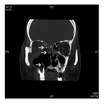
Figure 1: Computed tomography scan of the head in coronal cut.
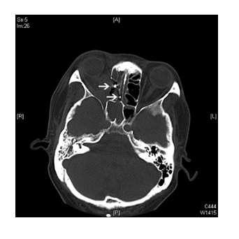
Figure 2: Computed tomography scan of the head in transverse cut
Two radiopaque radioactive seeds (arrowed) were seen at the level of ethmoid sinus. Right medial rectus muscle was displaced temporally by the bulky tumor mass.
Interviewed with his relatives, patient had left nasal cavity plasmacytoma history, with Cobalt-60 radiotherapy of 69.5Gy/65Fr in 1982; and right maxillary sinus sarcomatoid carcinoma with maxillectomy in 2015. Right facial recurrence in May 2017 was managed by surgical excision, yet lung metastasis was found in May 2018. Six cycles of docetaxel plus cyclophosphamide were given, followed by imatinib according to next-generation sequencing results.
In addition, they also volunteered the striking history of oncological implants two weeks ago in another city. Judging from their description, iodine-125 radioactive seeds implantation was suspected. Radioactivity measured around the face of the dead body at 50cm was 3·5uSv/hr. Luckily, none of the attending physicians were pregnant. However, all did not wear any protective shield upon handling of the patients. For radiation safety sake, cremation was not allowed, and burial for at least 2 years was required by local law. Disappointed by non-adherence to patient's cremation wish, deceased relatives expressed difficulties on finding a single burial place under the deadly coronavirus disease 2019 strike in the city.
Discussion
Radioactive implants were sometimes used as radiopharmaceuticals [4], such as gold-198 [5], iodine-125 [6,7,8], iodine-131 [9], radium-223 [10], and yttrium-90 [11] for prostate [10], lung [7], head and neck [8,9], thyroid [9], cervix cancer [12], choroidal melanoma [13], or hepatocellular carcinoma [11] etc. They come in different radiation dosage with variable half-lives [14], thus different implantation-to-cremation periods (Table 1).
|
Radiopharmaceutical |
Usual dosage range (GBq) |
Half life (days) |
|
Gold-198 |
12 |
2.7 |
|
Iodine-125 |
23 |
60 |
|
Iodine-131 |
35.5 |
8 |
|
Radium-223 |
3.5 |
11.4 |
|
Yttrium-90 |
1-2 |
2.7 |
Table 1: Radiation dosage and half-lives of different radiopharmaceuticals
Other than lifetime caring issues, doctors cares patients’ after-death wishes. Cremation, burial, donation, and mummification are some of the ways for body disposition. Although radioactive implant is usually given to those with reasonable life expectance, sudden death on these oncological patients is not predictable. Patient should seriously be briefed twice on their after-death arrangement before the implantation.
Conclusion
Patients with radiopharmaceuticals carries potential risk of radiation hazard towards their carers in life, or even towards cremation workers after their death. This article aims to update ophthalmologists on the properties of different radiopharmaceuticals.
Declaration of Interest
There is no conflict of interest to disclose
Funding:
There is no funding support for this manuscript
References
- Nitipir C, Niculae D, Orlov C, et al. Update on radionuclide therapy in oncology. Oncology Letters 14 (2017): 7011-7015.
- Shaikh MH, McMillan NA, Johnson NW. HPV-associated head and neck cancers in the Asia Pacific: a critical literature review & meta-analysis. Cancer Epidemiology 39 (2015): 923-938.
- Solares CA, Mason E, Panizza BJ. Surgical management of perineural spread of head and neck cancers. Journal of neurological surgery. Part B, Skull Base 77 (2016): 140.
- Bhattacharyya S, Dixit M. Metallic radionuclides in the development of diagnostic and therapeutic radiopharmaceuticals. Dalton Transactions 40 (2011): 6112-6128.
- Butler EB, Scardino PT, Teh BS, et al. The Baylor College of Medicine experience with gold seed implantation. InSeminars in Surgical Oncology 13 (1997): pp. 406-418.
- Li Q, Tian Y, Yang D, et al. Permanent iodine-125 seed implantation for the treatment of nonresectable retroperitoneal malignant tumors. Technology in Cancer Research & Treatment 18 (2019):1533033819825845.
- Huang Q, Chen J, Chen Q, et al. Computed tomographic-guided iodine-125 interstitial implants for malignant thoracic tumors. European Journal of Radiology 82 (2013): 2061-2066.
- Darakchiev BJ, Albright RE, Breneman JC, et al. Safety and efficacy of permanent iodine-125 seed implants and carmustine wafers in patients with recurrent glioblastoma multiforme. Journal of Neurosurgery 108 (2008): 236-242.
- Saini S, Tulla K, Maker AV, et al. Therapeutic advances in anaplastic thyroid cancer: a current perspective. Molecular Cancer 17 (2018): 154.
- Lien LM, Tvedt B, Heinrich D. Treatment of castration-resistant prostate cancer and bone metastases with radium-223 dichloride. International Journal of Urological Nursing 9 (2015): 3-13.
- Wu L, Shen F, Xia Y, et al. Evolving role of radiopharmaceuticals in hepatocellular carcinoma treatment. Anti-Cancer Agents in Medicinal Chemistry (Formerly Current Medicinal Chemistry-Anti-Cancer Agents) 16 (2016): 1155-1165.
- Kunos CA, Capala J, Kohn EC, et al. Radiopharmaceuticals for Persistent or Recurrent Uterine Cervix Cancer. Frontiers in Oncology 9 (2019).
- Peddada KV, Sangani R, Menon H, et al. Complications and adverse events of plaque brachytherapy for ocular melanoma. Journal of Contemporary Brachytherapy 11 (2019): 392.
- Gudkov SV, Shilyagina NY, Vodeneev VA, et al. Targeted radionuclide therapy of human tumors. International Journal of Molecular Sciences 17 (2016): 33.
Clinical Question Quiz:
Oncological patient with implantation of which of the following radioactive nuclides (with usual dosage range) five months before death is unsafe to undergo cremation with acceptable radioactivity level?
[A] Gold-198
[B] Iodine-125
[C] Radium-223
[D] Yttrium-90
Answer:
[B] Iodine-125
The 60-days half-life of Iodine-125 takes around 24 months to complete 12 half-lives for a usual dosage range of 2-3GBq to reach the safety level of 1 MBq for cremation.