Rovida S1*, Vetrugno L2, Naeem S3
1Department of Emergency Medicine, Linkoping University Hospital, Linkoping, Sweden
2Anesthesia and Intensive Care Unit, Department of Medicine, University of Udine, Udine, Italy
3Department of Emergency Medicine, Royal London Hospital, Barts Health NHS Trust, United Kingdom
*Corresponding Author: Dr. Serena Rovida, Department of Emergency Medicine, Linkoping University Hospital, Linkoping, Sweden
Received: 12 October 2020; Accepted: 09 November 2020; Published: 13 November 2020
Aneurysms are abnormal dilations in the vessel wall due breakdown in connective tissue. Aortic aneurysms are a common presentation in the emergency department (ED) and their incidence increases with age and associated risk factors. However, palmar artery aneurysms are a rare presentation. The increasing use of point of care ultrasound (PoCUS) has facilitated early diagnosis of aneurysms in the ED and has expedited their management. In this report we describe a case of palmar artery aneurysm in a 87 year old patient which was diagnosed with PoCUS in the ED.
Aneurysm; Ultrasound; Emergency department
1. Case Report
Peripheral artery aneurysms remain a rare presentation. Trauma to the vessel causes damage to the tunica media, resulting in a gradual dilatation of the vessel wall, hematoma formation and development of pseudoaneurysm [1, 2]. The majority of true aneurysms are due to repetitive trauma from occupational injuries or advanced atherosclerotic vessel disease, whereas the false (pseudo) aneurysms are mostly related to penetrating trauma with direct damage to the vessel wall [3-5]. Few cases of palmar arch aneurysms (PAA) have been reported in literature as shown in a recent systematic review and most of them were secondary to trauma [6]. Aneurysms can arise from any of the palmar arteries, however the ulnar artery and superficial arch are particularly prone to develop trauma due to their location and lack of palmar fascia in this area [3, 4]. The optimal management of these aneurisms is debated as several surgical or conservative approaches have been described [7].
We report a case of an 87-year-old right hand dominant lady who presented to the Emergency Medicine Department with one day history of progressive numbness and dyschromia of her right digits. No previous trauma, no occupational predisposition and no recent hand surgery were reported. The patient’s cardio pulmonary status was unremarkable. Physical examination of the right hand demonstrated marmorized phalanges of digits 2 to 5 and reduced sensitivity without any motor compromise (Figure 1). On the palmar side there was a round pulsatile compressible mass just distal to the hook of the hamate. Allen's test was negative bilaterally. A beside point of care ultrasound (PoCUS) bedside was performed by an Emergency Physician (EP) demonstrating the presence of a 24 x 17 mm pseudo-aneurysm of the palmar arch artery with mixed echogenicity and intramural hematoma. A visible floating flap was also noted (Figure 2a). The vascular surgeon was consulted, and the patient wasadmitted for further investigation and management. Angiography confirmed the presence of the superficial PAA. A coil embolization of both sides of the palmar arch artery was carried out with good outcome (Figure 3).
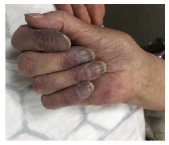
Figure 1: Marmorized digits of the right hand from 2nd to 5th with cyanotic distal phalanges.
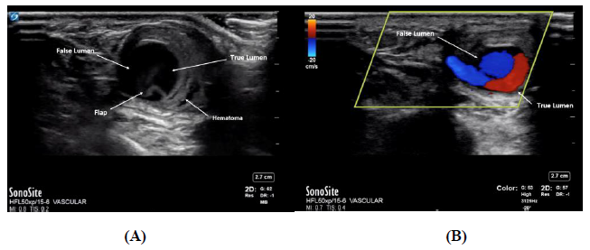
Figure 2: (A) B-mode transversal axis demonstrating the aneurysmal true and false lumen with intramural hematoma and flap. (B) Doppler Signal in transversal view showing the spontaneous echo contrast indicating turbulent blood flow within the pseudoaneurysm sac so called ‘Ying- Yang’ or ‘Pepsi’ sign.
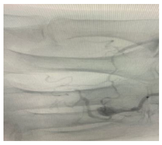
Figure 3: Arteriogram demonstrating abnormal flow in the superficial palmar arch artery.
In conclusion, PoCUS is a rapid imaging approach that can aid to formulate an accurate and real time diagnosis and it can be easily and safely performed by EPs. It can speed up the decision-making process when time really matters and an acute intervention such as early vascular reperfusion is needed. Nonetheless, its real time results can facilitate ruling out other major disorders that can present with similar clinical features. Ultrasound for PAA aneurysm is a relatively simple examination and it is often enough to confirm the diagnostic suspicion. However, the use of arteriography yields information regarding the collateral circulation and aids in preoperative planning.
Ethics approval
Not applicable.
Consent for Publication
Verbal consent was taken from the patient for the publication of this case.
Competing Interests
The authors declare that they have no competing interests.
Funding
No grants or fee was paid.
Authors' Contributions
SR, VL and SN interpreted the current literature and were involved in writing up the clinical letter. All authors read and approved the final manuscript.
References
- De Santis F, et al. Forearm and hand arteries’ aneurysms – a case report of bilateral true ulnar artery aneurysm in the hypothenar eminence and systematic review of the literature. Vascular 21 (2013): 169-176.
- Bouvet C, Bouddabous S, Beaulieu J. Aneurysms of the hand. Imaging and surgical technique. Hand Surgery and Rehabilitation 37 (2018).
- Kleinert HE, Burget GC, Morgan JA, et al. Aneurysms of the Hand. Arch Surg 106 (1973): 554-557.
- Shutze RA, Leichty J, Shutze WP. Palmar artery aneurysm. Proc (Bayl Univ Med Cent) 30 (2017): 50-51.
- L.AulicinoM.C., U.S.N.(Lt. Comdr.) P.M.J.HuttonM.C., U.S.N.R.(Lt. Comdr.) T.E.Du PuyM.C., U.S.N.(Capt.). True palmar aneurysms—A case report and literature review. The Journal of Hand Surgery 7 (1982): 613-616.
- Sheikh Z, Selvakumar S, Goon P. True aneurysm of the digital artery: a case report and systematic literature review. J Surg Case Rep (2020): rjz400.
- Igari K, Kudo T, Toyofuku T, et al. Surgical treatment of aneurysms in the upper limbs. Ann Vasc Dis 6 (2013): 637-641.