Zsuzsanna Arnold1,2*, Jennifer C Stabernak1,2, Verena Lenz1,2, Frieda-Maria Kainz3, Reinhard Moidl1,2, Thomas Aschacher1,2, Sebastian Tauber1,2, Georg Spinka4, Martin Grabenwöger1,2,5, Gabriel Weiss1,2,5
1Department of Cardiovascular Surgery, Clinic Floridsdorf, 1210 Vienna, Austria
2Institute of Cardiovascular Research, Karl Landsteiner Society, 1210 Vienna, Austria
3Cardiac Surgery, University Clinic St. Pölten, 3100 St. Pölten, Austria
4Department of Cardiology, Medical University Vienna, 1080 Vienna, Austria
5Medical Faculty, Sigmund Freud University, 1020 Vienna, Austria
*Corresponding Author: Zsuzsanna Arnold, Department of Cardiovascular Surgery, Clinic Floridsdorf, 1210 Vienna, Austria.
Received: 10 September 2026; Accepted: 17 September 2026; Published: 21 September 2026
Background: The use of biological prostheses for surgical aortic valve replacement (SAVR) in younger patients has increased despite guideline recommendations favoring mechanical valves. This study evaluated longterm outcomes, durability, and hemodynamic performance of biological aortic valve prostheses in patients younger than 60 years. Methods: Between January 2005 and December 2016, 256 consecutive patients younger than/ or 60 years old underwent biological SAVR. Early and long-term outcomes were analyzed retrospectively. Survival and freedom from reoperation were assessed using the Kaplan–Meier method as well as within a competing-risk framework. Multivariable analysis of reoperation was performed using the Fine-Gray sub distribution hazard model. Echocardiographic follow-up was evaluated to assess hemodynamic performance. Results: Mean age was 52.9 ± 7.2 years, and 24.6% of patients were female. Surgery was performed electively in 219 patients (85.5%), urgently in 28 (10.9%), and emergently in 9 (3.5%); infective endocarditis was present in 30 patients (11.7%). Thirty-day mortality was 4.7% overall and 1.9% for isolated procedures. Clinical follow-up was 99.2% complete, with a median duration of 9.6 years (2305 patient-years, maximum 20.6 years). Overall survival was 85.6% and 79.3% at 5 and 10 years, respectively. During follow-up, 49 patients (19.3%) underwent reoperation. Accounting for death as a competing event, the cumulative incidence of reoperation was 3.3%, 11.3%, and 35.7% at 5, 10, and 15 years, respectively. Reoperation occurred significantly earlier in younger patients (Gray’s test p < 0.001). In multivariable Fine–Gray analysis, younger age was the only independent predictor of reoperation (sub distribution hazard ratio SHR 0.94 per year, 95% CI 0.90–0.97; p = 0.004), whereas sex, infective endocarditis, prosthesis–patient mismatch, and prosthesis size were not significant predictors. Smaller prostheses showed higher transvalvular gradients, but prosthesis size was not associated with reoperation risk. Conclusions: Biological SAVR in patients younger than 60 years provided favorable long-term survival and acceptable durability. Younger age at the time of surgery was strongly associated with earlier need for reoperation. Although smaller prosthesis sizes were associated with less favorable hemodynamic performance, this did not translate into a higher rate of reoperation.
transcatheter aortic valve replacement
fortune journals articles
Introduction
In the era of transcatheter aortic valve replacement (TAVR) the number of young patients receiving biological surgical aortic valve replacement (SAVR) has increased substantially. Compared with mechanical valves, bioprostheses are associated with a lower risk of bleeding but a higher likelihood of reoperation [1-3]. Current guidelines generally recommend mechanical valves for younger patients; consequently, long-term outcome data on biological prostheses in consecutive, unselected younger cohorts remain limited [4,5]. However, increasing patient preference and emerging evidence supporting the safety of transcatheter valve-in-valve procedures have contributed to a shift toward the use of biological valves in progressively younger patients [6,7]. Notably, current American and European guidelines differ substantially in their recommendations for prosthesis selection in patients aged 50–70 years. The 2025 ESC/EACTS guidelines recommend mechanical prostheses in patients younger than 60 years and bioprostheses in those older than 65 years, with individualized decision-making in patients aged 60–65 years (Class IIa). In contrast, the 2020 ACC/AHA guidelines recommend mechanical valves in patients younger than 50 years and bioprostheses in those older than 65 years, with an individualized approach in patients aged 50–65 years (Class IIa). In both guidelines, shared and informed decision-making is a Class I recommendation [4,5]. These differences highlight the ongoing uncertainty in prosthesis selection in middle-aged patients and underscore the need for robust real-world data on the durability of bioprosthetic valves in younger populations, in order to better inform individualized decision-making in this age group. Structural valve deterioration (SVD) remains the major limitation to the durability of bioprosthetic valves, potentially leading to reoperation and reduced survival. While clinically relevant SVD is relatively uncommon in elderly patients, its impact is considerably greater in younger populations [8]. Beyond its effect on survival, SVD is associated with impaired quality of life, increased hospitalizations for heart failure, and a higher need for reinterventions, thereby imposing a substantial burden on healthcare systems [9-11]. In the contemporary TAVR era, with regulatory approval extending indications to lower-risk patients, long-term outcome data from real-world, unselected cohorts are of particular importance, especially in younger individuals. Ultimately, prosthesis selection should consider not only mortality but also quality of life, functional status (NYHA class), the risk of heart failure and rehospitalization, and the likelihood of future reinterventions. Therefore, this study aimed to evaluate long-term clinical outcomes and hemodynamic performance of biological aortic valve prostheses in young adults.
Methods
Study population
This retrospective study was approved by the local ethics committee (EK 20-105-VK). Between January 2005 and December 2016, all consecutive patients younger than / or 60 years old who underwent biological SAVR at our institution were included. Both isolated and combined procedures, as well as all types of biological prostheses, were considered. Patients with a history of prior cardiac surgery or infective endocarditis were not excluded. In-hospital data were collected retrospectively. Follow-up was conducted through active cross-sectional assessment via direct patient contact or structured telephone interviews, supplemented by review of electronic medical records. Autopsy reports or documented causes of death were available in 45% of deceased patients; for the remaining cases, causes of death were obtained through telephone interviews with the patients’ primary care physicians.
Echocardiographic assessment
Transthoracic echocardiography was performed at our institution prior to discharge. Follow-up echocardiographic examinations were obtained either in our outpatient clinic or by the patients’ referring cardiologist and collected via telephone or e-mail request. Data on aortic valve morphology, mean and peak transvalvular gradients, maximal transvalvular velocity, and effective orifice area (when available) were analyzed.
Study endpoints
Primary endpoints included 30-day mortality, long-term overall survival, freedom from valve-related death and reoperation-free survival. Secondary endpoints included the need for reoperation stratified by patient age and prosthesis size. Hemodynamic performance was analyzed according to valve size. Definitions of structural and non-structural valve deterioration, prosthesis–patient mismatch (PPM), prosthetic valve endocarditis, early mortality, and valve-related mortality were based on contemporary guidelines and related consensus statements [12,13].
Statistical Analysis
Categorical variables are presented as frequencies (percentages), continuous variables as mean ± standard deviation or median (interquartile range), as appropriate. Overall survival and freedom from valve-related death were estimated using the Kaplan–Meier method, with the date of the index procedure defined as time zero. Because death precludes reoperation, reoperation was analyzed within a competing-risk framework: cumulative incidence functions were estimated with the Aalen–Johansen estimator, treating death without preceding reoperation as a competing event, and groups were compared using Gray's test. Kaplan–Meier estimates of freedom from reoperation are additionally reported to allow comparison with earlier series. Multivariable analysis of reoperation was performed using the Fine-Gray sub distribution hazard model, with 95% confidence intervals obtained from 500 bootstrap replicates; the corresponding cause-specific Cox model is reported alongside. Numbers at risk are given for all time-to-event analyses. A p value < 0.05 was considered statistically significant. Analyses were performed in Python 3.10 using the lifelines package together with implementations of the Aalen–Johansen estimator, Gray's test and the Fine–Gray model that were validated against the corresponding standard estimators.
Results
Patient characteristics
Between January 2005 and December 2016, 256 consecutive patients younger than 60 years underwent biological SAVR and were included in the analysis. The mean age at surgery was 52.9 ± 7.2 years, and 24.6% (n = 63) were female. Baseline patient characteristics are summarized in Table 1. Aortic stenosis was the primary indication for surgery in 67.2% of patients. Renal impairment was present in 31.3%, and infective endocarditis in 11.7% of cases. Urgent or emergent procedures accounted for 14.5% of all operations.
|
Patient characteristics |
n (%) |
||
|
Mean age ± SD (range), years |
52.9 ± 7.2 (23-60) |
Coronary artery disease |
72 (28.1) |
|
Male |
193 (75.4) |
Previous cardiac surgery |
14 (5.5) |
|
Mean BMI ± SD, kg/m2 |
27.7 ± 4.86 |
Mean EuroSCORE II ± SD (range), % |
3.4 ± 6.9 (0.5-60.9) |
|
Hypertension |
146 (57.0) |
LVEF ± SD, % |
55.7 ± 10.1 |
|
Dyslipidemia |
138 (53.9) |
LVEF < 50% |
47 (18.4) |
|
COPD |
72 (28.1) |
Mean gradient ± SD, mmHg |
50.0 ± 18.6 |
|
Diabetes mellitus |
48 (18.8) |
Aortic valve area ± SD, cm2 |
0.82 ± 0.28 |
|
NYHA functional class |
Moderate/severe aortic regurgitation |
124 (48.4) |
|
|
I-II |
132 (51.6) |
Bicuspid/unicuspid aortic valve |
143 (55.9) |
|
III-IV |
124 (48.4) |
Infective endocarditis |
30 (11.7) |
|
Renal impairment* |
80 (31.3) |
Urgent or emergent indication |
37 (14.5) |
Table 1: Baseline characteristics of the study population n = 256 (100%).
Note: Preoperative demographic, clinical, and echocardiographic characteristics of patients undergoing biological surgical aortic valve replacement. Continuous variables are presented as mean ± standard deviation (SD) with range where indicated, and categorical variables as number (percentage). *Renal impairment was defined as creatinine clearance <85 mL/min according to the Cockcroft–Gault formula or the need for dialysis. BMI = body mass index; COPD = chronic obstructive pulmonary disease; LVEF = left ventricular ejection fraction; NYHA = New York Heart Association; SD = standard deviation.
Operative data
Operative characteristics are presented in Table 2. Bicuspid or unicuspid aortic valve morphology was present in 55.9% of cases. Isolated SAVR was performed in 41.4% (n = 106), while 58.6% (n = 150) underwent concomitant procedures, including coronary artery bypass grafting (CABG n = 49), ascending or aortic root repair (n = 87), mitral or tricuspid valve surgery (n = 24), and other (n = 27). Concomitant procedure categories were not mutually exclusive. Biological prostheses of different types and sizes were implanted. The most frequently used valve model was CE-Perimount / Magna or Magna Ease accounting for 80.9% of cases. The median prosthesis size was 23 mm (range 19–27 mm). PPM occurred in 34% of patients, including 31.3% moderate and 2.7% severe PPM.
|
Concomitant procedures |
n (%) |
||
|
AVR only |
106 (41.4) |
||
|
+ CABG |
49 (19.1) |
||
|
+ mitral or tricuspid repair/replacement |
24 (9.4) |
||
|
+ aortic root/ascending repair |
87 (34) |
||
|
+ other cardiac |
20 (7.8) |
||
|
+ other non-cardiac |
7 (2.7) |
||
|
Valve size |
n (%) |
Prosthesis type |
n (%) |
|
19 mm |
17 (6.6) |
CE-Perimount/ Magna/ Magna Ease |
207 (80.9) |
|
21 mm |
47 (18.4) |
SJM Trifecta |
18 (7.0) |
|
23 mm |
77 (30.1) |
Stentless |
15 (5.9) |
|
24 mm |
2 (0.8) |
Other stented bovine |
9 (3.5) |
|
25 mm |
61 (23.8) |
Other stented porcine |
5 (2.0) |
|
27 mm |
52 (20.3) |
Other |
2 (0.8) |
Table 2: Procedural aspects of biological surgical aortic valve replacement.
Note: Distribution of concomitant procedures, implanted prosthesis sizes, and valve types in the study cohort. Values are presented as number (percentage). AVR = aortic valve replacement; CABG = coronary artery bypass grafting.
Early outcomes
Thirty-day mortality was 4.7% overall. Mortality rates according to surgical urgency were 2.7% for elective procedures and 16.2% for urgent or emergent indications. Thirty-day mortality according to procedural type was 1.9% for isolated procedures and 6.7% for combined procedures. Postoperative complications comprised stroke (2.7%), prolonged ventilation (7.4%), reoperation for bleeding (6.6%), and permanent pacemaker implantation (4.7%). The median length of postoperative hospital stay was 10 days (IQR 9–13).
Follow-up and survival
Clinical follow-up for mortality and reoperation was 99.2% complete, with two patients lost to follow-up. The median duration of follow-up was 9.6 years (IQR 5.4-12.4), corresponding to 2305 patient-years of follow-up. Valve-related deaths occurred in 23.3% of patients with infective endocarditis. Kaplan–Meier estimated overall survival was 85.6% at 5 years and 79.3% at 10 years. A sensitivity analysis was performed to assess the impact of classifying deaths of unknown cause as either valve-related or non-valve-related (Table 3).
|
Analysis |
Events |
5 years, % (95% CI) |
10 years, % (95% CI) |
15 years, % (95% CI) |
|
Unknown cause classified as valve-related |
17 |
5.2 (2.5–8.0) |
6.9 (3.6–10.3) |
8.2 (4.1–12.3) |
|
Unknown cause classified as non-valve-related |
13 |
3.6 (1.3–6.0) |
5.4 (2.4–8.4) |
6.6 (2.8–10.5) |
Table 3: Valve-related death, main and sensitivity analysis.
Note: Cumulative incidence of valve-related death with death from other causes treated as a competing event CI = confidence interval.
Structural valve deterioration and reoperation
SVD occurred in 95 patients (37.4%) during follow-up, including 18.9% severe and 18.5% moderate SVD. During follow-up, 49 patients (19.3%) underwent reoperation and 52 patients died without preceding reoperation. Accounting for death as a competing event, the cumulative incidence of reoperation was 3.3% (95% CI 1.0–5.5) at 5 years, 11.3% (95% CI 6.9–15.7) at 10 years and 35.7% (95% CI 26.2–45.2) at 15 years, with 193, 103 and 19 patients remaining at risk. The cumulative incidence of death without reoperation was 12.4%, 18.2% and 27.6% at the same time points. Kaplan–Meier estimates of freedom from reoperation, which do not account for the competing risk of death, were 96.4% (95% CI 94.0–98.9), 86.9% (95% CI 81.7–92.0) and 54.3% (95% CI 42.1–66.5). The two approaches were nearly identical during the first decade but diverged thereafter, the Kaplan–Meier method overestimating the incidence of reoperation at 15 years by 10 percentage points in absolute terms (Figure 1, Table 4).
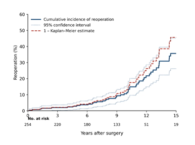
Figure 1A.
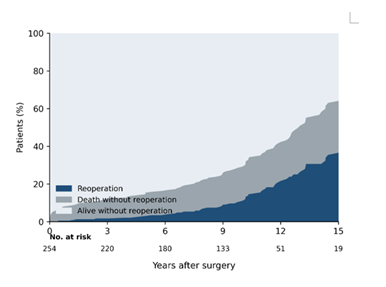
Figure 1B.
Figure 1: Cumulative incidence of reoperation after biological surgical aortic valve replacement in patients younger than 60 years, (A) estimated with the Aalen–Johansen estimator and treating death without preceding reoperation as a competing event. The solid line represents the cumulative incidence, the dotted lines the 95% confidence interval. The dashed line shows the complement of the Kaplan–Meier estimate, which does not account for the competing risk of death. Numbers at risk are displayed below the graph. (B) Stacked cumulative incidence of reoperation, death without preceding reoperation, and survival free from reoperation, in the overall cohort. Numbers at risk are displayed below the graph.
|
Years |
No. at risk |
Cumulative incidence of reoperation, % (95% CI) |
Cumulative incidence of death, % |
Freedom from reoperation (Kaplan–Meier), % (95% CI) |
|
5 |
193 |
3.3 (1.0–5.5) |
12.4 |
96.4 (94.0–98.9) |
|
10 |
103 |
11.3 (6.9–15.7) |
18.2 |
86.9 (81.7–92.0) |
|
12 |
51 |
21.7 (14.9–28.6) |
20.6 |
73.7 (65.5–82.0) |
|
15 |
19 |
35.7 (26.2–45.2) |
27.6 |
54.3 (42.1–66.5) |
Table 4: Reoperation in the overall cohort.
Note: Cumulative incidence estimated with the Aalen–Johansen estimator, death without preceding reoperation treated as a competing event. CI = confidence interval.
Age and size stratified analysis
The incidence of reoperation was analyzed according to patient age at the time of surgery (<40 years, 40–49 years, and 50–59 years). The cumulative incidence of reoperation differed markedly across age groups (Gray's test p < 0.001). At 10 years it was 29.1% in patients younger than 40 years, 26.4% in patients aged 40 to 49 years and 6.6% in patients aged 50 to 59 years. Estimates beyond 10 years should be interpreted with caution in the youngest group, in which only five patients remained at risk at 10 years (Figure 2A, Table 5). Reoperation was additionally evaluated according to prosthesis size (19–21 mm, 23–25 mm, and 27 mm). In contrast, no differences were observed between prosthesis size groups (Gray's test p = 0.43), with 10-year cumulative incidences of 10.8%, 12.8% and 7.6% for 19 to 21 mm, 23 to 25 mm and 27 mm prostheses (Figure 2B, Table 5).
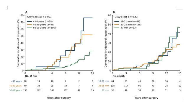
Figure 2: Cumulative incidence of reoperation stratified by patient age and prosthesis size, with death without preceding reoperation as a competing event. (A) Stratification by age at surgery (< 40, 40–49 and 50–59 years); groups were compared using Gray's test (p < 0.001). (B) Stratification by prosthesis size (19–21, 23–25 and 27 mm); no differences were observed (Gray's test p = 0.43). Numbers at risk are shown below each panel.
|
Group |
n |
Events |
5 years, % |
10 years, % (at risk) |
15 years, % (at risk) |
Gray's test |
|
< 40 years |
18 |
7 |
5.6 |
29.1 (5) |
76.3 (0) |
p < 0.001 |
|
40–49 years |
40 |
14 |
6.0 |
26.4 (16) |
51.8 (4) |
|
|
50–59 years |
196 |
28 |
2.6 |
6.6 (82) |
28.4 (15) |
|
|
19–21 mm |
64 |
14 |
3.3 |
10.8 (27) |
41.5 (4) |
p = 0.43 |
|
23–25 mm |
138 |
23 |
3.7 |
12.8 (53) |
28.2 (12) |
|
|
27 mm |
52 |
12 |
1.9 |
7.6 (23) |
46.6 (3) |
Table 5: Cumulative incidence of reoperation by age and prosthesis size. In patients younger than 40 years no patient remained at risk at 15 years; the corresponding estimate is not interpretable and should not be quoted.
In the multivariable Fine-Gray model, younger age at surgery was the only independent predictor of reoperation (sub distribution hazard ratio 0.94 per year of increasing age, 95% CI 0.90–0.97, p = 0.004). Female sex (SHR 1.89, 95% CI 0.75–4.35), infective endocarditis (SHR 0.94, 95% CI 0.27–2.32), PPM (moderate: SHR 1.06, 95% CI 0.51–2.14) and prosthesis size (SHR 1.06 per mm, 95% CI 0.92–1.24) were not significantly associated with the risk of reoperation. Of note, the hazard ratio for infective endocarditis decreased from 1.81 in the cause-specific Cox model to 0.94 in the sub distribution model, reflecting the high competing mortality in these patients (Table 6).
|
Covariate |
Cause-specific Cox, HR (95% CI) |
Fine–Gray, SHR (95% CI) |
p (Fine–Gray) |
|
Age at surgery, per year |
0.93 (0.90–0.97) |
0.94 (0.90–0.97) |
0.004 |
|
Female sex |
2.15 (0.93–4.98) |
1.89 (0.75–4.35) |
0.16 |
|
Infective endocarditis |
1.81 (0.79–4.15) |
0.94 (0.27–2.32) |
0.91 |
|
Prosthesis–patient mismatch, moderate |
1.25 (0.64–2.43) |
1.06 (0.51–2.14) |
0.83 |
|
Prosthesis–patient mismatch, severe |
0.44 (0.05–3.49) |
0.68 (0.00–9.41) |
0.76 |
|
Prosthesis size, per mm |
1.06 (0.91–1.24) |
1.06 (0.92–1.24) |
0.45 |
Table 6: Multivariable analysis of reoperation. 49 events. SHR = subdistribution hazard ratio; Fine–Gray 95% confidence intervals were obtained from 500 bootstrap replicates. CI = confidence interval.
Echocardiographic follow-up and hemodynamic performance
Transthoracic echocardiography at discharge demonstrated a mean transvalvular gradient of 17.4 ± 6.7 mmHg and a peak gradient of 29.2 ± 10.6 mmHg. A total of 181 follow-up echocardiographic examinations were available for analysis, corresponding to 74.8% of discharged patients. The mean interval between surgery and follow-up echocardiography was 8.1 ± 4.3 years. Hemodynamic parameters according to prosthesis size are summarized in Table 7. Mean and peak transvalvular gradients at discharge and follow-up as well as the incidence of severe PPM are reported for each valve size group. PPM was present in 34% of patients, including 2.7% severe and 31.3% moderate PPM.
|
Valve size (mm) |
Patients (n) |
Severe PPM, n (%) |
Mean gradient at discharge (mmHg) |
Mean gradient at follow-up (mmHg) |
Peak gradient at discharge (mmHg) |
Peak gradient at follow-up (mmHg) |
|
19 |
17 |
2 (11.8) |
24.9 |
34 |
41.1 |
52.2 |
|
21 |
47 |
4 (8.5) |
21.5 |
30 |
35.3 |
53.1 |
|
23-24 |
79 |
1 (1.3) |
17.2 |
23.9 |
28.9 |
40.6 |
|
25 |
61 |
0 |
15.5 |
21.3 |
26.1 |
35.6 |
|
27 |
52 |
0 |
12.6 |
22.0 |
22.0 |
35.0 |
Table 7: Hemodynamic performance according to prosthesis size.
Note: Postoperative transvalvular gradients measured at discharge and during follow-up stratified by prosthesis size. Values for gradients are presented as mean values. Severe PPM is reported as absolute numbers and percentage within each valve size group.
Discussion
The present study reports the long-term outcomes of biological SAVR in a consecutive cohort of patients younger than 60 years. With a median follow-up of nearly ten years and excellent follow-up completeness, our data provide real-world insights into survival, valve durability, and the need for reintervention in a population traditionally considered more suitable for mechanical prostheses. In this relatively young cohort, the choice of a biological prosthesis was predominantly driven by informed patient preference, reflecting an increasing desire to avoid lifelong anticoagulation and its associated impact on lifestyle and quality of life. Additional considerations included future pregnancy, concerns regarding adherence to anticoagulation particularly in patients with infective endocarditis or intravenous drug use—and relevant comorbidities associated with limited life expectancy. The present findings are in line with previous long-term studies investigating outcomes after SAVR in younger patients, while also adding important nuances. In the landmark study by Ruel et al., including patients younger than 60 years undergoing aortic valve replacement, no significant difference in long-term survival between mechanical and biological prostheses was observed, even at extended follow-up. Importantly, additional subgroup analyses in patients younger than 50 years yielded similar results. Furthermore, age was identified as an independent predictor of outcome, whereas prosthesis type itself was not consistently associated with long-term survival [14]. In contrast, long-term analyses of the Carpentier–Edwards Perimount valve by Bourguignon et al., including dedicated cohorts of patients aged ≤60 years, provide detailed age-stratified data on SVD and reoperation. These studies demonstrate a clear and progressive relationship between younger age at implantation and earlier onset of SVD, with median durability exceeding 20 years in older patients but substantially reduced in younger cohorts. Furthermore, competing risk analyses consistently show a steep increase in the cumulative incidence of reoperation with decreasing age at implantation [10,15-17]. Reoperation and death are competing events, and freedom from reoperation derived from the Kaplan–Meier method assumes that patients who die would otherwise have remained at risk for reoperation. In a cohort with a 15-year cumulative incidence of death without preceding reoperation of 27.6%, this assumption is not tenable and leads to systematic overestimation of the reoperation burden. The cumulative incidence function provides a more realistic estimate of the proportion of patients who will actually undergo a further operation, which is the quantity relevant to shared decision-making at the time of prosthesis selection, and it allows direct comparison with the competing-risk analyses reported by Bourguignon and colleagues. The divergence between the two approaches also explains why the apparent effect of infective endocarditis on reoperation disappears once the competing mortality of these patients is taken into account. The present study confirms these observations in a contemporary real-world cohort, demonstrating that younger age is the main determinant of earlier reoperation. Importantly, while smaller prosthesis sizes were associated with less favorable hemodynamic performance, this did not translate into a higher rate of reoperation, suggesting that patient-related factors, particularly age, may outweigh prosthesis-related factors in determining long-term durability. Overall survival in our cohort was favorable, with Kaplan–Meier estimates of 85.6% at five years and 79.3% at ten years. These findings are consistent with previous registry-based and institutional studies demonstrating acceptable long-term survival after biological SAVR in younger and middle-aged patients. Large observational analyses comparing biological and mechanical prostheses have reported no significant survival difference in selected patients aged 50–69 years, despite the traditionally stronger guideline preference for mechanical valves in younger individuals. Importantly, freedom from valve-related death remained high in our cohort, further suggesting that biological prostheses can provide satisfactory long-term clinical outcomes in appropriately selected younger patients [18,19]. Current international guidelines recommend mechanical valve prostheses for patients younger than 50 or 60 years because of their superior durability and the higher risk of SVD in biological valves [4,5]. Nevertheless, the use of bioprosthetic valves in younger patients has increased steadily over the last two decades [7]. Several factors contribute to this trend. First, patient preference plays an increasingly important role in valve selection, with many patients wishing to avoid lifelong anticoagulation and its associated lifestyle restrictions and bleeding risks. Second, specific patient populations may have relative contraindications to anticoagulation, including patients with poor treatment adherence or those with a history of infective endocarditis. In addition, women contemplating pregnancy frequently prefer biological prostheses to avoid the teratogenic risks and management challenges associated with vitamin K antagonist therapy during pregnancy. These considerations have contributed to a more individualized and patient-centered approach to prosthesis selection in contemporary clinical practice. Another factor influencing prosthesis choice is the rapid development of transcatheter valve technologies. The increasing availability and safety of transcatheter valve-in-valve procedures have altered the long-term management strategy for younger patients receiving biological prostheses [20,21]. In this context, implantation of a biological valve may be considered part of a staged lifetime management strategy, with the option of future transcatheter valve-in-valve therapy in case of SVD. However, this strategy must be approached cautiously in younger patients because valve-in-valve feasibility is influenced by the size and design of the original surgical prosthesis, the risk of residual gradients and PPM, coronary anatomy, and the uncertain consequences of multiple sequential valve procedures. SVD occurred in 37.4% of patients during follow-up, reflecting the well-known limited durability of biological prostheses in younger populations. Nevertheless, only 19.3% of patients underwent reoperation during follow-up. When accounting for death as a competing event, the cumulative incidence of reoperation was 3.3% at 5 years, 11.3% at 10 years, and 35.7% at 15 years. Kaplan–Meier estimates of freedom from reoperation were 96.4%, 86.9%, and 54.3% at the same time points, respectively. These findings are consistent with previously reported durability data for bioprosthetic aortic valves in younger patients, where structural degeneration typically becomes clinically relevant after the first decade following implantation. Our long-term reoperation profile aligns with established durability patterns reported in large surgical bioprosthesis series. In the very long-term experience with the Carpentier–Edwards Perimount valve, Bourguignon and colleagues reported freedom from reoperation due to SVD of approximately 88% at 10 years, ~71% at 15 years, and ~38% at 20 years, highlighting the expected acceleration of clinically relevant degeneration beyond the first decade [16]. In a similarly large-scale analysis, Johnston et al. emphasized that durability is strongly age-dependent, with younger recipients showing earlier degeneration and higher reintervention rates over time. Taken together, these data provide an important benchmark for interpreting real-world reoperation rates in younger patients receiving bioprostheses [22].
Age at the time of surgery had a significant influence on durability, with a markedly higher cumulative incidence of reoperation in younger patients. At 10 years, the cumulative incidence of reoperation was 29.1% in patients younger than 40 years, 26.4% in those aged 40–49 years, and 6.6% in patients aged 50–59 years. This observation is in line with the well-established relationship between younger age and accelerated SVD. The mechanisms underlying this relationship are likely multifactorial and may include greater calcium metabolism, a more active inflammatory and immune response, higher hemodynamic demands, and increased cumulative mechanical stress on the prosthetic leaflets. In multivariable analysis, younger patient age emerged as the only independent predictor of reoperation, whereas prosthesis size, PPM, sex, and infective endocarditis were not significantly associated with the risk of reintervention. Although previous large series and meta-analyses have identified PPM and elevated transvalvular gradients as additional potential risk factors for SVD, these associations were not observed in the present cohort [22-23]. An additional finding of the present study concerns the relationship between prosthesis size and hemodynamic performance. Smaller prosthesis sizes were associated with higher transvalvular gradients both at discharge and during long-term follow-up, whereas larger valve sizes demonstrated more favorable hemodynamic profiles. Although prosthesis size was not significantly associated with the risk of reoperation in our analysis, the observed hemodynamic differences may partly reflect a greater likelihood of PPM among recipients of smaller valves. Because PPM is determined by the effective orifice area of the implanted valve relative to the patient’s body size, prosthesis size alone should not be regarded as an equivalent measure. These findings emphasize the importance of achieving an adequate indexed effective orifice area during valve implantation. Surgical strategies aimed at minimizing PPM, including careful prosthesis selection and annular enlargement when necessary, may therefore remain particularly relevant in younger patients. In this population, prolonged exposure to elevated residual gradients may adversely affect ventricular remodeling and potentially influence long-term clinical outcomes and valve durability [24-26].
Limitations
Several limitations should be acknowledged. The retrospective single-center design introduces the possibility of selection bias in prosthesis choice and patient characteristics. Follow-up echocardiography was not available in all surviving patients, and the assessment of SVD therefore relied in part on clinical documentation and on information obtained from referring cardiologists. In four patients the cause of death could not be established and was conservatively classified as valve-related; a sensitivity analysis classifying these deaths as non-valve-related yielded a 10-year cumulative incidence of valve-related death of 5.4% instead of 6.9%, without affecting any conclusion. Data on anticoagulation, bleeding and thromboembolic events were not systematically collected, so the present analysis cannot address the trade-off between reoperation risk and bleeding risk that underlies prosthesis selection in this age group. The number of patients at risk beyond 15 years was small, particularly in the youngest age group, and late estimates are correspondingly imprecise. Finally, the absence of a concurrent mechanical valve control group precludes any comparative statement on the relative merits of the two prosthesis types.
Conclusions
Despite these limitations, the present analysis provides valuable long-term data on biological SAVR in a relatively young patient population. Biological SAVR was associated with favorable long-term survival. Valve durability was strongly influenced by patient age, with significantly earlier reoperation observed in younger individuals. Although smaller prosthesis sizes demonstrated less favorable hemodynamic performance, this did not translate into a higher rate of reoperation during follow-up. Careful patient selection and individualized decision-making therefore remain essential when considering biological prostheses in younger patients.
Funding:
This research received no external funding.
Conflict of interest:
none declared. Data available on request.
References
- Brennan JM, Edwards FH, Zhao Y, et al. Long-term safety and effectiveness of mechanical versus biologic aortic valve prostheses in older patients: results from the Society of Thoracic Surgeons Adult Cardiac Surgery National Database. Circulation 127 (2013): 1647-1655.
- Goldstone AB, Chiu P, Baiocchi M, et al. Mechanical or Biologic Prostheses for Aortic-Valve and Mitral-Valve Replacement. N Engl J Med 377 (2017): 1847-1857.
- Stassano P, Di Tommaso L, Monaco M, et al. Aortic valve replacement: a prospective randomized evaluation of mechanical versus biological valves in patients ages 55 to 70 years. J Am Coll Cardiol 54 (2009): 1862-1868.
- Praz F, Borger MA, Lanz J, et al. ESC/EACTS Scientific Document Group. 2025 ESC/EACTS Guidelines for the management of valvular heart disease. Eur Heart J 46 (2025): 4635-4736. Erratum in: Eur Heart J (2026).
- Otto CM, Nishimura RA, Bonow RO, et al. 2020 ACC/AHA guideline for the management of patients with valvular heart disease: executive summary: a report of the American College of Cardiology/American Heart Association Joint Committee on Clinical Practice Guidelines. Circulation 143 (2021): e35-e71.
- Dvir D, Webb JG, Bleiziffer S, et al. Transcatheter aortic valve implantation in failed bioprosthetic surgical valves. JAMA 312 (2014): 162-170.
- Isaacs AJ, Shuhaiber J, Salemi A, et al. National trends in utilization and in-hospital outcomes of mechanical versus bioprosthetic aortic valve replacements. J Thorac Cardiovasc Surg 149 (2015): 1262-1269.
- Rodriguez-Gabella T, Voisine P, Dagenais F, et al. Long-Term Outcomes Following Surgical Aortic Bioprosthesis Implantation. J Am Coll Cardiol 71 (2018): 1401-1412.
- Kilic A, Bianco V, Gleason TG, et al. Hospital readmission rates are similar between patients with mechanical versus bioprosthetic aortic valves. J Card Surg 33 (2018): 497-505.
- Bourguignon T, Bouquiaux-Stablo AL, Candolfi P, et al. Very long-term outcomes of the Carpentier-Edwards Perimount valve in aortic position. Ann Thorac Surg 99 (2015): 831-837.
- Senage T, Le Tourneau T, Foucher Y, et al. Early structural valve deterioration of Mitroflow aortic bioprosthesis: mode, incidence, and impact on outcome in a large cohort of patients. Circulation 130 (2014): 2012-2020.
- Capodanno D, Petronio AS, Prendergast B, et al. Standardized definitions of structural deterioration and valve failure in assessing long-term durability of transcatheter and surgical aortic bioprosthetic valves: a consensus statement from the European Association of Percutaneous Cardiovascular Interventions (EAPCI) endorsed by the European Society of Cardiology (ESC) and the European Association for Cardio-Thoracic Surgery (EACTS). Eur Heart J 38 (2017): 3382-3390.
- Varc-3 Writing C, Genereux P, Piazza N, et al. Valve Academic Research Consortium 3: updated endpoint definitions for aortic valve clinical research. Eur Heart J 42 (2021): 1825-1857.
- Ruel M, Chan V, Bédard P, et al. Very long-term survival implications of heart valve replacement with tissue versus mechanical prostheses in adults< 60 years of age. Circulation 116 (2007): I294-I300.
- Forcillo J, El Hamamsy I, Stevens LM, et al. The Perimount valve in the aortic position: twenty-year experience with patients under 60 years old. The Annals of Thoracic Surgery 97 (2014): 1526-1532.
- Bourguignon T, El Khoury R, Candolfi P, et al. Very long-term outcomes of the Carpentier-Edwards Perimount aortic valve in patients aged 60 or younger. The Annals of thoracic surgery 100 (2015): 853-859.
- Bourguignon T, Lhommet P, El Khoury R, et al. Very long-term outcomes of the Carpentier-Edwards Perimount aortic valve in patients aged 50–65 years. European Journal of Cardio-Thoracic Surgery 49 (2016): 1462-1468.
- Chiang YP, Chikwe J, Moskowitz AJ, et al. Survival and long-term outcomes following bioprosthetic vs mechanical aortic valve replacement in patients aged 50 to 69 years. Jama 312 (2014): 1323-1329.
- Schnittman SR, Adams DH, Itagaki S, et al. Bioprosthetic aortic valve replacement: revisiting prosthesis choice in patients younger than 50 years old. The Journal of Thoracic and Cardiovascular Surgery 155 (2018): 539-547.
- Webb JG, Mack MJ, White JM, et al. Transcatheter aortic valve implantation within degenerated aortic surgical bioprostheses: PARTNER 2 valve-in-valve registry. Journal of the American College of Cardiology 69 (2017): 2253-2262.
- Dvir D, Barbanti M, Tan J, et al. Transcatheter aortic valve-in-valve implantation for patients with degenerative surgical bioprosthetic valves. Current problems in cardiology 39 (2014): 7-27.
- Johnston DR, Soltesz EG, Vakil N, et al. Long-term durability of bioprosthetic aortic valves: implications from 12,569 implants. The Annals of thoracic surgery 99 (2015): 1239-1247.
- Ochi A, Cheng K, Zhao B, et al. Patient risk factors for bioprosthetic aortic valve degeneration: a systematic review and meta-analysis. Heart, Lung and Circulation 29 (2020): 668-678.
- Bilkhu R, Jahangiri M, Otto CM. Patient-prosthesis mismatch following aortic valve replacement. Heart 105 (2019): s28-s33.
- Yu W, Tam DY, Rocha RV, et al. Aortic root enlargement is safe and reduces the incidence of patient-prosthesis mismatch: a meta-analysis of early and late outcomes. Canadian Journal of Cardiology 35 (2019): 782-790.
- Sá MP, Jacquemyn X, Van den Eynde J, et al. Impact of prosthesis‐patient mismatch after surgical aortic valve replacement: systematic review and meta‐analysis of reconstructed time‐to‐event data of 122 989 patients with 592 952 patient‐years. Journal of the American Heart Association 13 (2024): e033176.