Matthew C. Martinez, Kevin Pierre, Charles Carpenter, Diego A L Garcia*
Department of Radiology, University of Florida, Gainesville, Florida, USA
*Corresponding Author: Diego A L Garcia, Department of Radiology, University of Florida Gainesville, FL, USA
Received: 01 August 2026; Accepted: 06 August 2026; Published: 21 August 2026
Background: Pseudomyogenic hemangioendothelioma (PHE) is a rare vascular neoplasm of intermediate malignancy that predominantly affects young males and frequently presents as multifocal disease involving the lower extremities. Osseous involvement is uncommon and may mimic metastatic disease or other multifocal skeletal disorders.
Case Presentation: A 17-year-old male presented with progressive pain involving the left ankle and foot. Initial radiographs demonstrated multiple poorly defined lytic lesions involving the distal tibia and several bones of the ipsilateral foot. Magnetic resonance imaging revealed extensive multifocal marrow-replacing lesions with avid enhancement and a small focus of extraosseous extension along the anterior aspect of the distal tibia. CT-guided biopsy confirmed pseudomyogenic hemangioendothelioma. Subsequent whole-body bone scintigraphy and FDG PET-CT demonstrated abnormal tracer uptake confined exclusively to the distal left lower extremity without evidence of disease elsewhere.
Conclusion: This case highlights a distinctive imaging phenotype characterized by multifocal lytic lesions involving contiguous bones of the distal lower extremity, limited extraosseous extension on MRI, and confinement to a single extremity on whole-body bone scintigraphy and FDG PET-CT. Recognition of this pattern may facilitate diagnosis and differentiation from systemic metastatic disease.
Pseudomyogenic hemangioendothelioma; Bone tumor; Ankle; Foot; MRI; PET-CT
Pseudomyogenic hemangioendothelioma articles; Bone tumor articles; Ankle articles; Foot articles; MRI articles; PET-CT articles.
1. Introduction
Pseudomyogenic hemangioendothelioma (PHE) is a rare vascular neoplasm classified by the World Health Organization as a tumor of intermediate malignancy [1]. The tumor predominantly affects young males during the second and third decades of life and demonstrates a marked predilection for the lower extremities [2]. Initially described as epithelioid sarcoma-like hemangioendothelioma [3]. PHE is now recognized as a distinct clinicopathologic entity characterized by recurrent FOSB-associated gene rearrangements and a tendency toward multifocal disease [2-4]. Unlike many musculoskeletal neoplasms, PHE frequently involves multiple lesions within a single anatomical region and may affect bone, skeletal muscle, subcutaneous tissue, and dermis [2]. Osseous involvement is relatively uncommon, and primary bone PHE is exceedingly rare, with fewer than 50 cases reported in English literature, most of which invade surrounding soft tissue [5,6]. Bony PHE may present a significant diagnostic challenge because the imaging appearance overlaps with metastatic disease, chronic osteomyelitis, Langerhans cell histiocytosis, epithelioid hemangioma, and other vascular neoplasms [7]. Classic imaging findings of bony PHE include well circumscribed, lytic lesions on plain radiographs and CT. They may cause cortical destruction, and pathological fractures. On MRI they are hypointense on T1-weighted images and hyperintense on T2 and STIR-weighted images. Bone Scintigraphy and FDG PET-CT generally show increased uptake [5,8-10]. The clustered distribution of lesions within a single extremity may create a particularly challenging diagnostic scenario because the appearance can mimic skeletal metastases despite the absence of systemic disease. The mainstay of treatment is surgical. Systemic chemotherapy, mTOR inhibitors, bisphosphonates and denosumab have also been used for treatment and symptom control in PHE with bony involvement [11-14]. We report a case of regional multifocal PHE involving the distal tibia, ankle, and multiple bones of the foot in a 17-year-old male. Whole-body bone scintigraphy and FDG PET-CT demonstrated disease confined to a single lower extremity despite extensive multifocal involvement.
2. Case Presentation
A 17-year-old previously healthy male presented with a six-month history of progressive pain involving the left ankle and foot. Symptoms were associated with intermittent limping and reduced participation in athletic activities. There was no history of trauma, fever, weight loss, night sweats, or other constitutional symptoms. Physical examination demonstrated focal tenderness over the distal tibia and hindfoot. No palpable soft tissue masses or cutaneous lesions were identified. Laboratory evaluation, including complete blood count, erythrocyte sedimentation rate, C-reactive protein, and serum biochemical profile, was unremarkable.
3. Imaging Findings
3.1. Radiography
Initial radiographs of the left ankle and foot demonstrated multiple poorly defined lytic lesions involving the distal tibial metaphysis, talus, calcaneus, navicular, cuboid, cuneiforms, and multiple metatarsals (Figure 1). Mild endosteal scalloping and cortical thinning were present without cortical destruction, aggressive periosteal reaction, matrix mineralization, or pathologic fracture. The clustered distribution of lesions across contiguous bones of the distal lower extremity suggested a regional process rather than a random skeletal distribution.
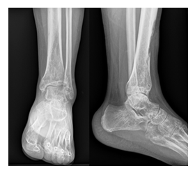
Figure 1: Initial radiographs of the left distal leg, ankle, and foot. Anteroposterior and lateral radiographs demonstrate multiple poorly defined lytic lesions involving the distal tibia and several bones of the left foot, including the talus, calcaneus, navicular, cuboid, cuneiforms, and metatarsals. No aggressive periosteal reaction, cortical destruction, matrix mineralization, or pathologic fracture is identified.
3.2. Magnetic Resonance Imaging
MRI was performed for local staging and lesion characterization. Sagittal and axial T1-weighted images demonstrated multiple marrow-replacing lesions involving the distal tibia, hindfoot, midfoot, and forefoot. Corresponding sagittal, axial, and coronal fat-suppressed T2-weighted images demonstrated markedly increased signal intensity throughout the lesions. Post-contrast axial fat-suppressed T1-weighted images demonstrated avid enhancement of the osseous lesions. A small focus of extraosseous extension was identified along the anterior cortex of the distal tibia (Figure 2). No large soft tissue mass, neurovascular encasement, or articular collapse was present. Overall, MRI demonstrated substantially greater disease burden than appreciated on radiography and confirmed extensive involvement of contiguous bones throughout the distal lower extremity.
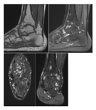
Figure 2: Magnetic resonance imaging of the left distal lower extremity. Sagittal T1-weighted (A), sagittal fat-suppressed T2-weighted (B), axial fat-suppressed T2-weighted (C), coronal fat-suppressed T2-weighted (D) images demonstrate multifocal marrow-replacing lesions involving the distal tibia, ankle, and multiple bones of the foot. The lesions are hypointense relative to normal marrow on T1-weighted images, hyperintense on fluid-sensitive sequences, and demonstrate avid enhancement following gadolinium administration. A small focus of extraosseous extension is present along the anterior aspect of the distal tibia.
3.3. Histopathologic Confirmation
CT-guided biopsy of a representative distal tibial lesion was performed. Microscopic examination demonstrated epithelioid and spindle cells with endothelial differentiation. Immunohistochemical analysis demonstrated diffuse positivity for ERG, CD31, and FOSB with retained INI1 expression, confirming the diagnosis of pseudomyogenic hemangioendothelioma [4,15].
3.4. Bone Scintigraphy
Following histopathologic diagnosis, whole-body bone scintigraphy using technetium-99m methylene diphosphonate (99mTc-MDP) was performed to assess the extent of skeletal disease. Bone scintigraphy demonstrated multiple foci of increased radiotracer uptake involving the distal left tibia, ankle, and multiple bones of the foot (Figure 3). No abnormal uptake was identified elsewhere in the appendicular or axial skeleton.
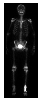
Figure 3: Whole-body 99mTc-MDP bone scintigraphy. Anterior and posterior projections demonstrate multifocal increased radiotracer uptake involving the distal left tibia, ankle, and multiple bones of the foot. No abnormal radiotracer uptake is identified elsewhere in the appendicular or axial skeleton.
3.5. FDG PET-CT
FDG PET-CT was subsequently performed for systemic staging. PET-CT demonstrated increased metabolic activity corresponding to all known lesions identified on radiographs, MRI, and bone scintigraphy (Figure 4). No additional FDG-avid lesions were identified elsewhere in the skeleton, soft tissues, thorax, abdomen, pelvis, or contralateral extremities. The combined scintigraphic and PET findings confirmed multifocal disease confined to a single lower extremity.
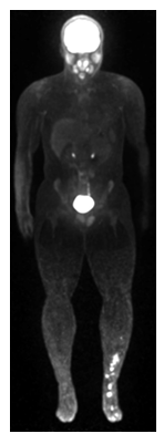
Figure 4: Whole-body FDG PET maximum-intensity projection (MIP) and fused PET-CT images demonstrate hypermetabolic lesions corresponding to the known sites of disease within the distal left lower extremity. No additional FDG-avid lesions are identified elsewhere in the body, confirming confinement of disease to a single extremity.
4. Discussion
Pseudomyogenic hemangioendothelioma is an uncommon vascular neoplasm that most frequently affects young males during the second and third decades of life [2]. Recognition of its characteristic imaging appearance is important because diagnosis is often delayed and lesions may be mistaken for infection, metastatic disease, or other primary bone tumors.
The most distinctive feature of the present case is the remarkable discordance between disease burden and disease distribution. Radiographs and MRI demonstrated extensive multifocal osseous involvement of the distal tibia, ankle, and foot, while both bone scintigraphy and FDG PET-CT confirmed confinement of disease to a single lower extremity. This imaging phenotype may represent an underrecognized manifestation of regional multifocal dissemination in pseudomyogenic hemangioendothelioma and may help distinguish this entity from metastatic disease.
Previous studies have emphasized the tendency of PHE to present as multifocal disease involving multiple lesions within the same anatomical region [2,7,16]. In our patient, more than a dozen lesions involved contiguous bones extending from the distal tibia through the ankle and foot, creating an appearance that initially suggested a disseminated skeletal process despite the absence of disease elsewhere. Radiographs demonstrated multiple lytic lesions with relatively nonaggressive features, consistent with previous descriptions of osseous PHE [7]. MRI proved particularly valuable because it demonstrated substantially greater disease burden than radiography alone and identified a small focus of extraosseous extension. Similar observations have been reported in prior imaging studies of PHE involving bone [16,17]. Whole-body bone scintigraphy and FDG PET-CT provided complementary staging information. Both modalities demonstrated disease limited to the distal left lower extremity despite extensive multifocal involvement. The absence of additional lesions elsewhere supports the concept of regional multifocal dissemination rather than conventional hematogenous metastatic spread. This was similarly described by Liu in a 20-year-old man whom FDG PET-CT showed numerous multilayer, multifocal lesions in a single distal lower extremity, which may be a distinctive imaging finding of PHE with bony involvement [18]. Histopathologic confirmation remains essential because imaging findings are not pathognomonic. The differential diagnosis includes multifocal osteomyelitis, Langerhans cell histiocytosis, epithelioid hemangioma, epithelioid hemangioendothelioma, epithelioid sarcoma, leiomyosarcoma, non-ossifying fibroma, osteoblastoma, giant cell tumor and metastatic disease [5,7,8,19,20]. Demonstration of endothelial differentiation together with strong nuclear FOSB expression is highly supportive of PHE and reflects the recurrent molecular alterations characteristic of this entity [4,15]. Additional immunohistochemical markers that support the diagnosis of PHE include CD31, ERG, FLI1 positivity and CD34 negativity [8,10,20]. This case expands the spectrum of reported imaging presentations of PHE and highlights a distinctive pattern of extensive regional multifocal osseous disease involving contiguous bones of the distal lower extremity while remaining confined to a single anatomical territory on whole-body imaging.
5. Conclusion
Pseudomyogenic hemangioendothelioma should be considered in the differential diagnosis of multifocal lytic lesions involving the distal lower extremity in adolescents and young adults. The combination of lesions affecting contiguous bones of the distal tibia, ankle, and foot, together with confinement to a single extremity on whole-body bone scintigraphy and FDG PET-CT, may represent a distinctive imaging phenotype of regional multifocal dissemination. MRI, bone scintigraphy, FDG PET-CT, and histopathologic correlation play complementary roles in establishing the diagnosis.
Learning Points
- Pseudomyogenic hemangioendothelioma may present as extensive multifocal osseous disease involving contiguous bones of a single extremity.
- MRI frequently demonstrates substantially greater disease burden than conventional radiography.
- Whole-body bone scintigraphy and FDG PET-CT can demonstrate regional confinement despite extensive multifocal involvement.
- Recognition of a clustered locoregional distribution pattern may facilitate differentiation from metastatic disease and guide appropriate biopsy planning.
References
- WHO Classification of Tumours Editorial Board. Soft Tissue and Bone Tumours. Lyon: International Agency for Research on Cancer (5th edtn), (2020).
- Hornick JL, Fletcher CDM. Pseudomyogenic hemangioendothelioma: a distinctive, often multicentric tumor with indolent behavior. Am J Surg Pathol 35 (2011): 190-201.
- Billings SD, Folpe AL, Weiss SW. Epithelioid sarcoma-like hemangioendothelioma. Am J Surg Pathol 27 (2003): 48-57.
- Walther C, Tayebwa J, Lilljebjörn H, et al. A novel SERPINE1-FOSB fusion gene results in transcriptional upregulation of FOSB in pseudomyogenic haemangioendothelioma. J Pathol 232 (2014): 534-540.
- Sun Y, Zhao M, Lao IW, et al. The clinicopathological spectrum of pseudomyogenic hemangioendothelioma: report of an additional series with review of the literature. Virchows Arch 477 (2020): 231-240.
- Takada N, Oebisu N, Yao H, et al. Pseudomyogenic hemangioendothelioma of the tibia: a case report. Int J Surg Case Rep 138 (2026): 802-807.
- Inyang A, Mertens F, Puls F, et al. Primary pseudomyogenic hemangioendothelioma of bone. Am J Surg Pathol 40 (2016): 587-598.
- Caballero GA, Roitman PD. Pseudomyogenic hemangioendothelioma (epithelioid sarcoma-like hemangioendothelioma). Arch Pathol Lab Med 144 (2020): 529-533.
- Pradhan D, Schoedel K, McGough RL, et al. Pseudomyogenic hemangioendothelioma of skin, bone and soft tissue: a clinicopathological, immunohistochemical, and fluorescence in situ hybridization study. Hum Pathol 71 (2018): 126-134.
- Plouznikoff N, Mourad SA, Turpin S, et al. Case report: 18F-FDG PET/CT of pseudomyogenic hemangioendothelioma. Clin Nucl Med 45 (2020): 368-369.
- Brance ML, Cóccaro NM, Roitman P, et al. Pseudomyogenic hemangioendothelioma with bone and soft tissue involvement with favorable response to pamidronate: a case report and systematic review of the literature. Arch Osteoporos 17 (2022): 28.
- Pranteda G, Magri F, Muscianese M, et al. The management of pseudomyogenic hemangioendothelioma of the foot: a case report and review of the literature. Dermatol Ther 31 (2018): e12725.
- Pasricha S, Sharma A, Pruthi M, et al. Multifocal primary pseudomyogenic hemangioendothelioma of bone managed with denosumab: a rare case with diagnostic and therapeutic challenge. J Cancer Res Ther 18 (2022): 817-819.
- Kikili Cİ, Köylü B, Kemik F, et al. The mTOR pathway inhibition with everolimus in pseudomyogenic hemangioendothelioma harboring SERPINE1-FOSB gene fusion: a case report and review of the literature. Anticancer Drugs 36 (2025): 770-774.
- Hung YP, Fletcher CDM, Hornick JL. FOSB is a useful diagnostic marker for pseudomyogenic hemangioendothelioma. Am J Surg Pathol 41 (2017): 596-606.
- Dianat S, Eslami V, Mirzaei A, et al. Pseudomyogenic hemangioendothelioma: a case report and review of the literature. Radiol Case Rep 14 (2019): 997-1001.
- Otani S, Yamamoto N, Hayashi K, et al. Pseudomyogenic hemangioendothelioma of bone treated with denosumab: a case report. BMC Cancer 19 (2019): 975.
- Liu Y. Pseudomyogenic hemangioendothelioma: distinctive FDG PET/CT findings with numerous multilayer lesions in a single distal extremity. Clin Nucl Med 45 (2020): 248-249.
- Al-Qaderi A, Mansour AT. Pseudomyogenic hemangioendothelioma. Arch Pathol Lab Med 143 (2019): 763-767.
- Dianat S, Yousaf H, Murugan P, et al. Pseudomyogenic hemangioendothelioma: a case report and review of the literature. Radiol Case Rep 14 (2019): 1228-1232.