Jun Wang1, Feng Dai1,2, Xiaoming Liu3, Wen Ju1, Huageng Liang1*, Xiaoping Zhang1*
1Department of Urology, Union Hospital, Tongji Medical College, Huazhong University of Science and Technology, Wuhan, China
2Jianli county people’s hospital, Jianli, China
3Department of Radiology, Union Hospital, Tongji Medical College, Huazhong University of Science and Technology, Wuhan, China
*Corresponding Author: Xiaoping Zhang, Department of Urology, Union Hospital, Tongji Medical College, Huazhong University of Science and Technology, 1277 Jiefang Avenue, Wuhan, Hubei Province, 430022, People’s Republic of China
Huageng Liang, Department of Urology, Union Hospital, Tongji Medical College, Huazhong University of Science and Technology, 1277 Jiefang Avenue, Wuhan, Hubei Province, 430022, People’s Republic of China
Received: 22 December 2021; Accepted: 13 January 2022; Published: 18 January 2022
Some appendicitis with the appendix in rare localization shows atypical presentations, leading to diagnostic difficulties. We firstly report one case of retroperitoneal appendicitis encroaching on the ureter. The diagnosis was interpreted as ureter cancer by the MRU (Magnetic Resonance Urography) and PET (Positron Emission Computed Tomography), whereas the intraoperative finding and pathological examination finally confirmed that the hydronephrosis was caused by the retroperitoneal appendicitis. Herein, the case report we presented will provide a demonstration for the subsequent differential diagnosis of ectopic appendicitis.
Appendicitis; Appendix; Ureter cancer
Abbreviations:
MRU: Magnetic Resonance Urography; PET: Positron Emission Computed Tomography
1. Introduction
The common anatomical positions of the appendix are in the anterior ileum, the posterior ileum, the pelvic cavity, the posterior cecum, the inferior cecum and the lateral cecum [1, 2]. However, some appendixes present rare localization, and the corresponding appendicitis show atypical symptoms or infrequent complications, resulting in diagnostic difficulties. We firstly report one case of retroperitoneal appendicitis, where the inflammatory mass of the appendicitis encroached on the ureter and caused a hydronephrosis. It was difficult to distinguish these medical findings of the appendicitis from ureter cancer.
2. Case Report
A 54-year-old male patient was admitted to the urology department with paroxysmal pain in the right flank. The patient had no history of medication and surgery. No abdominal pain or rebound pain was found by physical examinations. The MRU demonstrated a 2.0 cm × 2.8cm × 3.5cm mass with unclear boundary around the right ureter at the L4 level, and the ipsilateral hydronephrosis with pelvic dilation about 3cm, the mass was considered as ureteral carcinoma (Figure 1). The PET revealed an irregular soft tissue with clearly visible increased metabolism at the L3-L4 level, and the ipsilateral hydronephrosis with pelvic dilation about 3.2 cm, the mass was also considered as ureteral cancer (Figure 2). The ureteroscopy examination showed that the ureteral lumen was smooth and there was a stenosis about 3cm in length in the ureter. The ureteroscope could pass through the stenosis and enter the pelvic. The ureteroscopy examination suggested that the stenosis was caused by oppression outside of the ureter. Intraoperative findings displayed that there was a hard, non-enveloped 4 cm × 3 cm mass down to the appendix which located in the retroperitoneum, the mass is closed to the peritoneum and around the upper ureter. The pathological examination revealed that the mass consisted of hyperplastic fibrous tissue, infiltrated lymphocytes and plasma cells (Figure 3). Thus, the mass was confirmed as inflammatory mass of appendicitis.
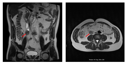
Figure 1: The mass around the right ureter with ipsilateral hydronephrosis in MRU. The red arrow revealed the localization of the mass.
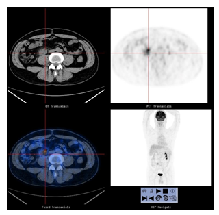
Figure 2: The mass around the right ureter with increased metabolism in PET.
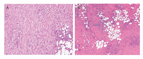
Figure 3: The pathological examination of the excised appendix. (A) Plasma cell infiltration in appendiceal mucosa and fat metaplasia under mucosa were revealed. (B) Hyperplastic fibrous tissue, lymphocytes and plasma cell infiltration were showed in tissue around appendix.
3. Discussion
Retroperitoneal appendicitis encroaching the ureter, as far as we know, is the first time reported. Symptoms of peritoneal irritation in case of retroperitoneal appendicitis are not obvious. This patient only presented the waist pain. Due to the followed PET and MRU examination, the patient was considered to suffer from ureteral cancer. There are two types of ureter cancer, exogenous and endogenous ureter cancer [3]. Endogenous ureteral cancer can be easily diagnosed through ureteroscopy [4]. Exogenous ureteral cancer is, in most cases, caused by different metastatic cancers, such as metastatic breast cancer and gastric cancer causing bilateral hydronephrosis [5, 6]. Since neither endogenous ureteral cancer was found in the ureteroscopy examination, nor other cancers were found in this patient, the diagnosis of ureteral cancer was suspicious even before surgery.
During the exploratory laparotomy, the mass was found around the ureter and continuous to the retroperitoneal appendix, so the mass was reasoned from the appendix. After separating the mass and protecting the ureter carefully, pathological examination confirmed the mass as inflammatory tissue originated from the infected appendix. The therapy of ureteral cancer usually requires the resection of the ureter or the resection of the ipsilateral kidney, the whole ureter and a part of bladder. Precise diagnosis is needed to differentiate a retroperitoneal appendicitis around the ureter, since the symptoms seem to be the same, but the therapy is totally different. Patients with appendicitis and ureter cancer suffer from differently therapeutic procedure and clinical result. The retroperitoneal appendicitis around the ureter and ureter cancer should be carefully distinguished due to similar result of medical imaging and in absence of specific symptoms. The pre-operatively pathological exam is useful for the differential diagnosis.
Acknowledgement
Not applicable
Statement of Ethics
This study protocol was reviewed and approved by Wuhan Union hospital, written informed consent for publication of their clinical details and clinical images was obtained from the patient.
Conflict of Interest Statement
The author reports no conflicts of interest in this work.
Funding Sources
There is no funding source.
Author Contributions
J.W, F.D, contributed to acquisition and analysis of data, and preparation of manuscript. XM. L analyzed the PET and MRI data. XP.Z, HG.L and W.J diagnosed the surgical sample, wrote the manuscript and gave final approval of the version to be published. All authors read and approved the final manuscript.
Data Availability Statement
The datasets used during the current study are available from the corresponding author on reasonable request.
References
- Evrimler S, Okumuser I, Unal N. Computed Tomography (CT) Findings of a Diagnostic Dilemma: Atypically Located Acute Appendicitis. Pol J Radiol 81 (2016): 583-588.
- Takabatake K, Ikeda J, Furuke H, et al. A case of a horseshoe appendix. Surg Case Rep 2 (2016): 140.
- Kondo T, Hara I, Takagi T, et al. Template-based lymphadenectomy reduces the risk of regional lymph node recurrence among patients with upper/middle ureteral cancer. Int J Clin Oncol 22 (2017): 145-152.
- Joo M, Chang SH, Kim H, et al. Idiopathic segmental ureteritis, misdiagnosed as ureteral cancer preoperatively: a case report with literature review. Pathol Int 60 (2010): 779-783.
- Kane AJ, Wang ZJ, Qayyum A, et al. Frequency and etiology of unexplained bilateral hydronephrosis in patients with breast cancer: results of a longitudinal CT study. Clin Imaging 36 (2012): 263-266.
- Thomas MH, Chisholm GD. Retroperitoneal fibrosis associated with malignant disease. Br J Cancer 28 (1973): 453-458.