Interactions between HLA immunogenetic profile, undiagnosed mild immunodeficiencies and inflammatory comorbidities in the amplification of post-vaccinal responses
TCHAME Claudric Roosvelt*,1, Adrien Fapeingou TOUNKARA2, MENGUE ESSINDI Annie3, ATANGANA YENE Roxanne Lucienne1, EWANE Olivier4, TCHOKFE NDOULA Shalom3
1Ministry of public health Cameroon / DLMEP
2UNICEF Cameroon
3Ministry of public health Cameroon / PEV
4OMS Cameroon
*Corresponding author: TCHAME Claudric Roosvelt, Ministry of public health Cameroon / DLMEP.
Received: 28 August 2026; Accepted: 31 August 2026; Published: 07 September 2026
Background. From smallpox variolation practiced in imperial China in the 10th century to the messenger RNA vaccines deployed in 2020, vaccination ranks among the most effective public health interventions in the history of medicine. Nevertheless, the rare but clinically significant occurrence of serious adverse events following immunization (AEFI) remains a major challenge for contemporary pharmacovigilance. Identifying the individual determinants of susceptibility represents a leading scientific and ethical priority.
Objective. To critically analyze the available scientific literature in order to determine the extent to which three families of host factors the HLA immunogenetic profile, undiagnosed mild immunodeficiencies, and chronic inflammatory comorbidities contribute to the occurrence of serious AEFI, and to identify biomarkers and prevention strategies likely to improve vaccine safety.
Methods. Narrative and critical literature review. The PubMed/MEDLINE, Scopus, Web of Science, and Google Scholar databases were searched for the period 1990–2025. Original articles, systematic reviews, meta-analyses, and case series describing a link between vaccination and HLA profiles, mild immunodeficiencies, or inflammatory comorbidities were included. Editorials without data and exclusively animal studies were excluded.
Results. The data converge toward an integrative model in which serious AEFI result from the interaction between a platform-specific vaccine stimulus and a permissive host background. Certain HLA alleles (HLA-A*03:01, HLA-DRB1*11:04, HLA-DQB1*06) modulate antigen presentation and predispose to inadequate or autoreactive immune responses. Mild immunodeficiencies, particularly type I interferon deficits and dysregulation of regulatory T lymphocytes, compromise immune-braking mechanisms. Chronic inflammatory comorbidities, diabetes, hypertension, pre-existing autoimmune diseases raise the baseline level of inflammation and lower the threshold for triggering a pathological response. Four etiological categories of serious AEFI are identified: (A) with a dominant cause linked to the vaccine platform; (B) multifactorial metabolic; (C) mixed, resulting from the interaction between a vaccine mechanism and a latent host susceptibility; (D) with a mechanism not yet elucidated.
Conclusion. The systematic integration of HLA typing, type I interferon assays, and cytokine profiling into pharmacovigilance algorithms would refine individual causal assessment and pave the way for personalized vaccination. Multicenter prospective studies, including African populations underrepresented in the literature, are needed to validate these biomarkers in clinical practice.
serious AEFI; HLA; mild immunodeficiency; inflammatory comorbidities; pharmacovigilance; vaccination; COVID-19; biomarkers; vaccine safety
serious AEFI articles; HLA articles; mild immunodeficiency articles; inflammatory comorbidities articles; pharmacovigilance articles; vaccination articles; COVID-19 articles; biomarkers articles; vaccine safety articles
Introduction
The history of vaccination is inseparable from that of humanity's struggle against infectious scourges. Long before the term "vaccine" entered the medical lexicon, ancient civilizations had intuitively understood that controlled exposure to a pathogen could confer lasting protection. As early as the 10th century, Chinese physicians practiced variolation by insufflating dried scabs from smallpox pustules into children's nostrils a technique that crossed the Silk Road to reach Constantinople and, from there, Enlightenment Europe [1]. Lady Mary Wortley Montagu, wife of the British ambassador to the Sublime Porte, observed this practice in 1718 and had it applied to her own children, thereby introducing variolation into the English aristocracy [2]. Nearly a century later, on May 14, 1796, Edward Jenner inoculated an eight-year-old boy, James Phipps, with the contents of a cowpox pustule taken from the hand of a milkmaid, Sarah Nelmes, demonstrating empirically for the first time that a benign infection could protect against a lethal disease [3]. This founding gesture inaugurated the modern vaccine era.
The centuries that followed were punctuated by unprecedented public health triumphs. The Black Death, which had decimated nearly one-third of the European population in the 14th century and caused lasting economic collapse [4]; the 1918 Spanish influenza, responsible for 50 to 100 million deaths [5]; and smallpox, eradicated in 1980 after a global campaign coordinated by the World Health Organization (WHO) and thereby preventing about two million deaths per year [6], all helped forge the conviction that vaccination is one of the most effective public health interventions ever devised. Global infant mortality, which stood at 93 deaths per 1,000 live births in 1990, fell to 37 in 2020 a dramatic decline largely attributable to the expansion of vaccination coverage [7].
Yet the COVID-19 pandemic was an unexpectedly brutal reminder of the fragility of this collective protection. Although the messenger RNA (mRNA) and adenoviral-vector vaccines were developed, evaluated, and deployed at an unprecedented speed in the history of pharmacology the BNT162b2 vaccine received emergency use authorization on December 11, 2020, less than a year after the publication of the SARS-CoV-2 genomic sequence [8] their administration on a scale of billions of doses inevitably brought to light rare but clinically significant adverse events. Post-mRNA myocarditis, occurring at an estimated frequency of 1 to 5 cases per 100,000 doses in young men [9], and vaccine-induced immune thrombotic thrombocytopenia (VITT), associated with adenoviral-vector vaccines [10], are the most thoroughly documented examples.
According to the World Health Organization (WHO) classification, adverse events following immunization (AEFI) are defined, based on their cause, in five distinct etiological categories [11]: vaccine product-related reactions, the occurrence of which is caused or precipitated by one or more properties inherent to the vaccine product itself; vaccine quality defect-related reactions, caused or precipitated by one or more quality defects of the vaccine product, including the administration device provided by the manufacturer; immunization error-related reactions, resulting from improper handling, prescription, or administration of the vaccine, and inherently preventable in nature; immunization anxiety-related reactions (ISRR, Immunization Stress-Related Response), resulting from anxiety or stress associated with the act of vaccination; and finally, coincidental events, caused by a factor independent of the vaccine itself, an immunization error, or vaccination-related anxiety. Among these categories, the reactions intrinsically linked to the vaccine attract the greatest scientific and public attention, as they directly call into question the very nature of the product and shape vaccine confidence.
The central question that emerges from this observation is the following: what differentiates individuals who develop a serious AEFI from the vast majority of vaccinated subjects who do not? If the entire population is exposed to the same vaccine antigen but only a few individuals develop a pathological response, it is logical to consider that the difference lies less in the vaccine itself than in the way the body regulates its immune response. Contemporary scientific research converges on three particularly relevant families of host factors.
(1) The HLA immunogenetic profile, which modulates the presentation of vaccine antigens to T lymphocytes and influences the quality, intensity, and specificity of the adaptive immune response. Some HLA alleles are associated with a better vaccine response, while others predispose to inadequate, exaggerated, or autoreactive responses. For instance, certain HLA-DRB1 and HLA-DQB1 alleles have been associated with non-response to the hepatitis B vaccine [12], and others with susceptibility to VITT [13].
(2) Undiagnosed mild immunodeficiencies, which weaken the natural immune-braking mechanisms and favor disproportionate inflammatory responses. CTLA-4 or A20 (TNFAIP3) haploinsufficiency, partial type I interferon deficits, and dysregulation of regulatory T lymphocytes are paradigmatic examples [14,15].
(3) Chronic inflammatory comorbidities diabetes, arterial hypertension, obesity, pre-existing autoimmune diseases which raise the basal level of systemic inflammation and may increase vaccine reactogenicity by lowering the threshold for triggering a pathological response [16].
The aim of the present article is to critically analyze the available scientific literature in order to understand the extent to which these three families of host factors interact to contribute to the occurrence of serious AEFI, and to identify biomarkers and prevention strategies likely to improve vaccine safety and sustain public confidence.
Methods
Type of review
The present study constitutes a narrative and critical literature review. Unlike a systematic review or a meta-analysis, the objective is not to quantify associations or statistically combine results, but rather to select, analyze, and critique the existing scientific work in order to identify the strengths, limits, and zones of uncertainty regarding the role of host factors in the occurrence of serious AEFI. This approach, although it does not meet the criteria for the full reproducibility of systematic reviews, offers the flexibility necessary to integrate data from heterogeneous disciplinary fields immunogenetics, fundamental immunology, pharmacovigilance, clinical epidemiology and to formulate integrative hypotheses [17].
Literature search strategy
The PubMed/MEDLINE, Scopus, Web of Science, and Google Scholar databases were systematically searched for the period from 1990 to 2025. This time window was chosen so as to include foundational works on HLA genetics and vaccine variability, while also covering the abundant literature generated by the COVID-19 pandemic. Only articles published in English or French were retained. The search terms, used alone or in combination using Boolean operators, included: "adverse events following immunization," "AEFI," "vaccine safety," "HLA," "human leukocyte antigen," "genetic susceptibility," "immune deficiency," "immune dysregulation," "primary immunodeficiency," "autoimmune disease," "inflammatory comorbidity," "myocarditis," "anaphylaxis," "thrombosis," and "VITT."
Selection criteria
Original articles, literature reviews, case studies, and case series describing a link between vaccination and (a) particular HLA profiles; (b) mild immunodeficiencies or immune regulation abnormalities; (c) chronic inflammatory comorbidities were included. Editorials without data, exclusively animal studies without direct clinical relevance, and works not describing serious AEFI were excluded. Titles and abstracts were first examined to discard manifestly off-topic articles. The full texts of potentially relevant articles were then analyzed to confirm their inclusion. Selection was carried out by at least two independent readers in order to limit subjectivity bias.
Method of analysis and critique
For each retained article, the following elements were systematically recorded: objectives, type of study and methodology, size and characteristics of the study population, main results, and limits and biases identified by the authors or by the authors of the present review. A narrative critique was then conducted to compare results across studies, identify zones of consensus, controversy, or scientific gap, and highlight normal and pathological situations. The articles were grouped and discussed according to the three families of host factors studied. The synthesis is presented thematically: each section first sets out the results of the available studies, then a critical discussion that emphasizes the scientific robustness of the data, methodological biases, clinical and public health implications, and avenues for future research.
Results and Discussion
The underlying causes of serious AEFI remain partly unexplained. Establishing a direct causal link between a hospitalization and the administration of a vaccine often proves complex because of the presence of multiple confounding factors: a patient may have a latent infection, an underlying metabolic condition, or a pre-existing immune susceptibility whose expression temporally associated with vaccination may create the illusion of an adverse effect when it is in fact only a coincidence. In this context, understanding the biological, immunological, and genetic factors likely to influence both vaccine effectiveness and tolerance represents a major scientific objective, essential for distinguishing the expected physiological responses from the dysregulated responses that may induce serious AEFI [18].
Influence of the HLA system on vaccine response
The HLA (Human Leukocyte Antigen) system, also known as the major histocompatibility complex (MHC), is a set of highly polymorphic genes located on the short arm of chromosome 6 (region 6p21.3). It constitutes the cornerstone of adaptive immune recognition, enabling the immune system to distinguish "self" from "non-self." The discovery of this system goes back to the pioneering work of Jean Dausset in 1958, who identified the first human leukocyte antigen work that earned him the Nobel Prize in Physiology or Medicine in 1980 [19]. The HLA system is subdivided into three functional classes: class I molecules (HLA-A, HLA-B, HLA-C), expressed on all nucleated cells, present endogenous peptides particularly of viral or tumoral origin to CD8+ cytotoxic T lymphocytes; class II molecules (HLA-DR, HLA-DQ, HLA-DP), expressed on antigen-presenting cells (macrophages, dendritic cells, B lymphocytes), present exogenous peptides to CD4+ helper T lymphocytes, thereby triggering cytokine production and activation of the humoral response; class III genes encode complement proteins (C2, C4, factor B) and cytokines (TNF-α, TNF-β) involved in inflammatory regulation [20]. The polymorphism of the HLA system with more than 30,000 alleles catalogued to date constitutes one of the most remarkable genetic features of the human genome [21]. This diversity, shaped by millions of years of parasitic selection pressure, gives each individual a unique combination of HLA molecules inherited from their parents. In the context of vaccination, this polymorphism directly influences the ability to present vaccine antigens to T lymphocytes, thus modulating the intensity, quality, and duration of the immune response. Some alleles favor a robust and protective response, while others lead to an attenuated, inadequate, or, in some cases, autoreactive and potentially pathological response.
HLA and variability of the anti-SARS-CoV-2 vaccine response
Bolze et al. (2022), in a cohort study of more than 100,000 participants vaccinated against COVID-19, demonstrated that carriers of the HLA-A*03:01 allele exhibited significantly increased reactogenicity after administration of the BNT162b2 (Pfizer-BioNTech) vaccine, manifesting as increased local cytotoxicity, release of pro-inflammatory cytokines, and a higher incidence of fever and chills. This increased reactogenicity was not observed or was significantly less pronounced with the mRNA-1273 (Moderna) vaccine, suggesting a specific interaction between certain vaccine epitopes and HLA allelic variants [22]. Mentzer et al. (2023), in a study published in Nature Medicine, showed that carriers of the HLA-DQB1*06 allele developed significantly higher anti-Spike/RBD antibody titers after vaccination against SARS-CoV-2, conferring increased protection against infection [23]. These results were corroborated by the work of Esposito et al. (Communications Medicine), confirming the association between certain HLA alleles and the variability of neutralizing antibody titers [24].
HLA and vaccine non-response
The question of vaccine non-response has been studied in particular in the context of the hepatitis B vaccine. Wang et al. (2021), in a study published in Frontiers in Immunology, explored the association between certain class II HLA alleles and a prolonged or absent response to the hepatitis B vaccine. The HLA-DPB1*05 and HLA-DQB1*02 alleles were identified as factors associated with non-response to the booster vaccine, suggesting that the HLA-DP and HLA-DQ molecules affect the presentation of peptides derived from the HBs antigen as well as the persistence of B and T immune memory [12]. These observations are of particular importance in public health, since vaccine non-response, although less spectacular than serious AEFI, constitutes a major determinant of individual and collective vaccine failure.
HLA and vaccine-induced immune thrombotic thrombocytopenia (VITT)
Petito et al. (2025), in a case-control study, analyzed the association between HLA alleles and vaccine-induced immune thrombotic thrombocytopenia (VITT) caused by adenoviral-vector vaccines. Carriers of the HLA-DPB1*17:01, HLA-DQA1*05:01, and HLA-DRB1*11:04 alleles had a significantly increased risk of developing this rare but potentially fatal autoimmune syndrome [13]. The proposed pathophysiological mechanism rests on the formation of complexes between free adenoviral DNA and platelet factor 4 (PF4), followed by phagocytosis of these complexes by macrophages, presentation of PF4-derived peptides by class II HLA molecules to CD4+ T lymphocytes, and subsequent activation of autoreactive B lymphocytes producing pathogenic anti-PF4 antibodies [10,13]. This immune-pathological cascade illustrates in a paradigmatic way how the convergence of a vaccine-related factor (free adenoviral DNA interacting with PF4) and a host-related factor (permissive HLA alleles) can give rise to a serious AEFI.
Innate inflammation and immune regulation
Innate immunity, the body's first line of defense, relies on the recognition of conserved molecular structures specific to pathogens (PAMPs, pathogen-associated molecular patterns) and to endogenous danger signals (DAMPs, damage-associated molecular patterns) by pattern recognition receptors (PRRs), expressed in particular on dendritic cells, macrophages, and epithelial cells. Their activation triggers a rapid and nonspecific inflammatory response that constitutes the immune context within which the subsequent activation of the adaptive response via the HLA system takes place [25]. Wang et al. (2023), in a mechanistic systematic review, analyzed the role of innate immunity in the severity of SARS-CoV-2 infection. Their main hypothesis posits that the virus blocks PRRs via the ORF6 and NSP1 proteins, preventing early detection and delaying the production of type I interferons (IFN-I). This delayed innate response favors uncontrolled viral replication, followed by an excessive compensatory response manifesting as a cytokine storm characterized by massive secretion of IL-6, TNF-α, and IL-1β, neutrophilia, and diffuse tissue damage [26]. Silva et al. (2022) confirmed these observations by showing that the early absence of IFN-I constitutes a major factor determining the severity of COVID-19, a deficit that may be exacerbated by the presence of neutralizing autoantibodies directed against IFN-α and IFN-ω, identified in 10 to 15% of patients with critical forms [27]. These anti-IFN-I autoantibodies constitute a paradigm of acquired mild immunodeficiency, clinically silent before exposure to the virus but decisive for the clinical course.
The regulation of the balance between innate and adaptive immunity is ensured by a complex network of cellular and molecular mediators. Cronkite and Strutt (2018) showed that Th1 and Th17 lymphocytes, by producing pro-inflammatory cytokines (IFN-γ, IL-17), can amplify the innate response by activating macrophages, neutrophils, and dendritic cells, creating a vicious circle of inflammation. Regulatory T lymphocytes (Tregs), through the secretion of IL-10 and TGF-β, normally constitute the physiological brake on this cascade. During severe infections or intense antigenic stimulation, this regulation may become insufficient, leading to a Th1/Th17-Treg imbalance that turns a controlled immune response into a pathological one [28]. Elliott et al. (2014) further showed that the inappropriate activation of PRRs (TLR, NLR, RLR) constitutes a major driver of chronic inflammatory diseases and severe infections, underscoring the importance of fine regulation of innate immunity in preventing disproportionate responses [29].
Mild immunodeficiencies and vaccine response
Immunodeficiency, long conceptualized as a factor reducing the immune response, in fact plays a paradoxical role in the context of AEFI. Chun et al. (2022), in a systematic review on the antibody response of HIV-positive patients to anti-COVID-19 vaccines, observed that patients with a CD4 count <200-350/mm³ had significantly reduced rates of seroconversion and lower neutralizing antibody titers [30]. Tan et al. (2023) confirmed that immunocompromised populations show prolonged infections, more vaccine failures, and may become reservoirs for the emergence of variants [31]. These observations suggest that severe immunodeficiency does not, strictly speaking, constitute an amplifying factor for AEFI, but rather a factor for vaccine failure. By contrast, mild immunodeficiencies CTLA-4 haploinsufficiency, partial IFN-I deficits, Treg dysregulation can, by selectively weakening the immune-braking mechanisms without abolishing the capacity to respond, create a permissive ground favorable to autoreactive responses and serious AEFI [14,15].
Inflammatory comorbidities and AEFI
Prevalence of comorbidities in vaccinated subjects
Pre-existing comorbidities represent an unavoidable variable in the assessment of vaccine safety. In the phase III clinical trials of the BNT162b2 (Pfizer-BioNTech) vaccine, 20.3% of participants had at least one comorbidity, dominated by arterial hypertension, type 2 diabetes, and chronic pulmonary diseases [8]. In Cameroon, among the 25,417 participants enrolled in the anti-COVID-19 vaccination campaign, 1.8% reported at least one comorbidity at enrolment hypertension accounting for 53% of cases, followed by diabetes (15%) and chronic respiratory diseases (13%); 1.7% were on concomitant medication and 0.9% of women were pregnant at the time of vaccination [32]. A global pharmacovigilance analysis of several hundred million doses of mRNA vaccines revealed that a pre-existing comorbidity was documented in a substantial proportion of reported AEFI cases [33].
Post-vaccinal autoimmune AEFI
Post-vaccinal myocarditis, the most heavily publicized complication of mRNA vaccines, has been the subject of considerable scientific attention. Florek and Sokolski (2024), in a narrative review, observed that these myocarditides are rare, predominate in young men, and occur principally after the second dose an observation immunologically consistent with the phenomenon of anamnestic recall [34]. Stowe et al. (2023) confirmed a significantly increased risk of myocarditis in patients aged 16 to 39, with a modulation of risk according to vaccine type [35]. Choi et al. and Zheng et al. respectively documented post-vaccinal immune thrombocytopenia and post-vaccinal autoimmune hepatitis, two conditions sharing a common mechanism of molecular mimicry between viral (vaccinal) proteins and platelet or hepatic autoantigens, with a genetic susceptibility associated with HLA-DRB1 [36, 37].
Post-vaccinal metabolic decompensation
Sá-Ferreira et al. (2022) and Ganakumar et al. (2022) described the occurrence of diabetic ketoacidosis during COVID-19 as a multifactorial phenomenon involving systemic inflammation that induces insulin resistance via IL-6 and TNF-α, activation of lipolysis, and possible dysfunction of pancreatic β cells linked to ACE2 expression [38,39]. Watanabe et al. (2024) and Yakou et al. (2022) reported episodes of post-vaccinal ketoacidosis in diabetic patients treated with sodium-glucose cotransporter type 2 inhibitors (SGLT-2 inhibitors), underscoring the need for enhanced metabolic surveillance in this at-risk population [40,41]. TNF-α, produced in excess during the cytokine storm linked to the IFN-I deficit [26,27], constitutes the molecular mediator through which the dysregulation of innate immunity can induce insulin resistance and precipitate ketoacidosis in the diabetic patient [38,39].
The following figure provides an overview of the mechanisms underlying severe AEFI
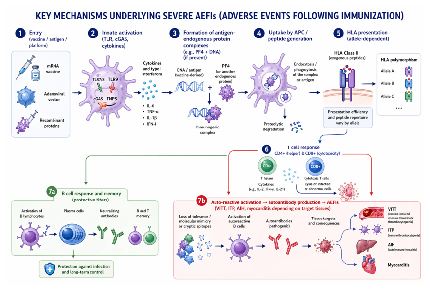
Figure 1: Key mechanisms underlying severe AEFIs.
Etiological models and classification of serious AEFI
Etiological models
A central question emerges when one examines serious AEFI as a whole: do they fall under a uniform causal model, or under fundamentally distinct etiological architectures? Two major causal models can be invoked to account for them.
The first is an additive multicausal model, in which several risk factors, genetic, metabolic, immunological, environmental add up to progressively increase the probability of an adverse event, without any one of them being sufficient on its own. This model is classically that of metabolic diseases and certain autoimmune diseases, in which pathology emerges from the convergence of multiple determinants.
The second is a threshold model with a dominant cause, in which a principal immunological or infectious mechanism must be activated beyond a certain threshold to trigger a pathological state. In this model, a single or predominant causal factor explains most of the pathological variance, even if modulating cofactors (genetic background, hormonal status) modify its clinical expression.
Scientific data show that serious AEFI do not form a homogeneous set and cannot be reduced to either of these models. Some rest on an identifiable principal mechanism: VITT associated with adenoviral-vector vaccines results from the formation of PF4-polyanion complexes and the production of anti-PF4 autoantibodies; post-mRNA myocarditis is attributed to excessive IFN-I-type immuno-inflammatory activation in a particular genetic or hormonal context; the infectious complications of live attenuated vaccines result from uncontrolled viral replication. Other serious AEFI fall within a multicausal model: glycemic decompensation or ketoacidosis in a diabetic patient may be precipitated by the conjunction of vaccine-induced inflammation, fever, and metabolic stress.
Furthermore, the vaccine platform profoundly modulates the nature of the underlying mechanism. Live attenuated vaccines (MMR, varicella, yellow fever) carry rare immune-infectious risks dominated by viral replication; mRNA vaccines (Pfizer-BioNTech, Moderna) induce intense, brief innate activation via the TLR7 and MDA5 receptors; adenoviral-vector vaccines (AstraZeneca, Janssen) combine innate activation with specific interaction with platelet factor 4. This diversity of mechanisms across vaccine platforms confirms that a unified approach to serious AEFI would be reductive, and that any classification must integrate vaccine type as a structuring variable.
In the following sections, we propose criteria allowing serious AEFI to be classified into several operational categories.
Criteria for AEFI classification
To ground an operational classification of serious AEFI, five complementary criteria can be retained:
Causal weight of the vaccine. Does the vaccine mechanism on its own explain the bulk of the pathophysiology (e.g., anti-PF4 autoantibodies in VITT), or does it represent only one contribution among others? In the first case, the AEFI is termed "dominant cause"; in the second, "multifactorial."
Number of necessary cofactors. An AEFI with a dominant cause may occur with a single modulating cofactor (e.g., a permissive HLA background), whereas a multifactorial AEFI requires the convergence of several determinants (inflammation + metabolic stress + hormonal imbalance).
Specificity of the vaccine-AEFI association. Is the AEFI linked to a single vaccine platform (VITT to adenoviral vectors), or observed with vaccines of different nature (subacute thyroiditis after mRNA, inactivated, or live attenuated vaccines)?
Experimental reproducibility. A mechanism is all the more considered dominant if it can be reproduced in vitro or in animal models, independently of the host background.
Reversibility through action on a single factor. Is modifying a single component of the vaccine (removal of the adenoviral vector, optimization of the mRNA sequence) sufficient to significantly reduce the incidence of the AEFI? If so, a principal mechanism is at work.
Classification of AEFI
On the basis of these criteria, serious AEFI can be divided into four categories.
Category A — Dominant cause
The principal causal mechanism is linked to the vaccine platform and explains the bulk of the pathophysiology. Host cofactors modulate the clinical expression without being sufficient on their own.
VITT. Formation of PF4-polyanion complexes and anti-PF4 autoantibodies, specific to adenoviral vectors. The permissive HLA background is a modulating cofactor, but the vaccine mechanism remains the principal determinant.
Post-mRNA myocarditis. Excessive innate activation (TLR7, MDA5) and disproportionate IFN-I response. Male sex, young age, and certain HLA variants amplify the risk without being its principal cause.
Characteristics: experimentally reproducible mechanism, strong specificity for a platform, potential reversibility through modification of the vaccine formulation.
Category B — Multifactorial AEFI
The vaccine acts as a nonspecific trigger (inflammation, fever, metabolic stress) on a fragile pre-existing background. No single factor is sufficient on its own.
Ketoacidosis in the diabetic patient. Vaccine-induced inflammatory stress destabilizes a precarious glycemic balance. The vaccine does not cause the condition: it precipitates a decompensation in a vulnerable host.
Post-vaccinal subacute thyroiditis. The transient immune activation reveals a pre-existing subclinical thyroid susceptibility. This mechanism is not platform-specific: it has been described after mRNA, inactivated, and live attenuated vaccines.
Characteristics: low specificity for a platform, convergence of several determinants, low reversibility through modification of the vaccine alone.
Category C — Mixed AEFI (vaccine × host susceptibility interaction)
The AEFI results from the interaction between an immunological mechanism linked to the vaccine and a pre-existing but latent host susceptibility. Neither the vaccine alone nor the host background alone is sufficient to produce the disease.
Infectious complications of live attenuated vaccines (VAPP, measles inclusion-body encephalitis, YEL-AVD). The effector mechanism is uncontrolled viral replication, but these complications occur almost exclusively in immunodeficient individuals (T-cell deficit, HIV, iatrogenic immunosuppression). Immunodeficiency does not produce the disease, viral replication does but it constitutes a necessary prerequisite. The AEFI emerges only from the encounter between the live vaccine and the immunodeficient host.
Post-vaccinal ITP. Molecular mimicry or polyclonal B-cell activation triggering autoimmune destruction of platelets in predisposed individuals (HLA variants, family history of autoimmunity).
Post-vaccinal autoimmune hepatitis. Loss of hepatic tolerance in carriers of susceptibility markers (anti-smooth muscle, low-titer anti-LKM1).
Post-vaccinal pericarditis. Profile distinct from myocarditis, with a more pronounced autoimmune component and a stronger dependence on the host background than on the platform.
Post-Pandemrix narcolepsy. Interaction between the AS03 adjuvant, H1N1 epitopes, and the HLA-DQB1*06:02 background, leading to autoimmune destruction of hypocretin neurons.
Characteristics: joint requirement of a vaccine signal and a host susceptibility, variable specificity for the platform, partial reproducibility in transgenic models.
Category D — Unresolved mechanism
Transitional category for AEFI whose mechanism remains incompletely understood. As data accumulate, these AEFI migrate toward categories A, B, or C.
Post-vaccinal Guillain-Barré syndrome. Molecular mimicry is suspected but not formally demonstrated. The diversity of associated vaccines (influenza, COVID-19, meningococcal) suggests potentially distinct mechanisms.
Synthesis: Serious AEFI by vaccine platform
Table 1 presents a synthetic view of the main serious AEFI according to vaccine platform, etiological category, estimated frequency, principal mechanism, associated conditions, and host cofactors. This synthesis incorporates the classification proposed in section 3.4.3 and allows an immediate comparative reading, useful for individual causal assessment and for the personalization of vaccine strategy.
Table 1: Classification of serious AEFI by vaccine platform and etiological category.
|
Vaccine platform |
Cat. |
Estimated frequency |
Main mechanism |
Associated conditions |
Host cofactors |
|
Adenoviral vectors (AstraZeneca, Janssen) |
A Dominant cause |
~1/50,000 to 1/100,000 doses |
Formation of PF4-polyanion complexes and anti-PF4 autoantibodies Specific to adenoviral vectors |
VITT Cerebral and splanchnic venous thromboses |
Permissive HLA background (HLA-DPB1*17:01, HLA-DQA1*05:01, HLA-DRB1*11:04) |
|
mRNA (BNT162b2, mRNA-1273) |
A Dominant cause |
~1/10,000 to 1/20,000 (men 16-29 years, 2nd dose) |
Excessive innate activation (TLR7, MDA5) Disproportionate IFN-I response |
Myocarditis Pericarditis (autoimmune component → cat. C) |
Male sex, young age HLA variants Recent SARS-CoV-2 infection |
|
All types (platform-nonspecific) |
B Multifactorial |
Variable, depends on metabolic background |
Nonspecific inflammatory stress (fever, IL-6, TNF-α) Destabilization of a precarious metabolic balance |
Diabetic ketoacidosis Glycemic decompensation |
Pre-existing diabetes (type 1, type 2 on SGLT-2 inhibitors) Elevated HbA1c Intercurrent infection |
|
All types (mRNA, inactivated, live attenuated) |
B Multifactorial |
Variable, latent thyroid background |
Transient immune activation revealing pre-existing subclinical thyroid susceptibility |
Post-vaccinal subacute thyroiditis |
Subclinical anti-thyroid autoantibodies (anti-TPO, anti-Tg) Genetic predisposition Family history of thyroid disease |
|
Live attenuated (MMR, OPV, yellow fever) |
C Mixed |
VAPP: ~1/750,000 YEL-AVD: ~1/250,000 |
Uncontrolled viral replication in immunodeficient hosts Neither vaccine alone nor immunodeficiency alone is sufficient |
VAPP Measles inclusion-body encephalitis Yellow fever vaccine-associated viscerotropic disease (YEL-AVD) |
Primary or acquired immunodeficiency (T-cell deficit, HIV, iatrogenic immunosuppression) Genetic deficits of innate immunity |
|
Variable (mRNA, inactivated, live attenuated) |
C Mixed |
Variable |
Molecular mimicry or polyclonal B-cell activation Loss of tolerance in predisposed hosts |
Post-vaccinal ITP Autoimmune hepatitis Autoimmune pericarditis |
HLA variants Pre-existing autoantibodies (anti-smooth muscle, anti-LKM1, anti-platelet) Family history of autoimmunity |
|
Adjuvanted (AS03) (Pandemrix, H1N1) |
C Mixed |
~1/18,400 (children/adolescents, Scandinavia) |
Interaction of AS03 adjuvant × H1N1 epitopes × HLA-DQB1*06:02 Autoimmune destruction of hypocretin neurons |
Type 1 narcolepsy |
HLA-DQB1*06:02 (near-mandatory) Genetic predisposition to narcolepsy |
|
Variable (influenza, COVID-19, meningococcal) |
D Unresolved |
Variable ~1-2/1,000,000 doses |
Molecular mimicry suspected but not formally demonstrated Potentially distinct mechanisms across platforms |
Guillain-Barré syndrome ADEM |
Undetermined History of post-vaccinal GBS (relative contraindication) |
Note: Frequencies are orders of magnitude derived from the main pharmacovigilance studies.
VITT = vaccine-induced immune thrombotic thrombocytopenia; VAPP = vaccine-associated paralytic poliomyelitis; YEL-AVD = yellow fever vaccine-associated viscerotropic disease; ITP = immune thrombocytopenic purpura; GBS = Guillain-Barré syndrome; ADEM = acute disseminated encephalomyelitis; SGLT-2 = sodium-glucose cotransporter type 2 inhibitors; ATCD = history.
Biological diagnosis and decision-making algorithm
In line with the recommendations of the WHO causality assessment framework for adverse events following immunization, the Expanded Programme on Immunization (EPI) of Cameroon defines an AEFI as any unfavorable medical event, unforeseen sign, abnormal laboratory result, symptom, or disease occurring after vaccination, regardless of any established causal link with the vaccine product [11,42]. The surveillance system rests on a three-tier architecture: passive surveillance via spontaneous reporting, weekly active surveillance by AEFI Surveillance Focal Points, and community-based surveillance involving Community Health Workers [42]. Any serious AEFI must be reported within 24 hours and undergo an initial investigation within 24 to 48 hours by a multidisciplinary team. The data are centralized in the national database before transmission to VigiBase®, the WHO international pharmacovigilance database [42].
The assessment of the causal link is conducted at two complementary levels. At the population level, the assessment seeks to answer the following question: "Can this vaccine actually cause this manifestation?" It draws on six criteria derived from the Bradford Hill method: the temporal relationship between vaccination and the event (the only absolutely necessary criterion), the strength of the observed statistical association, the dose-effect relationship reflecting the increase in risk with the dose administered, the consistency and replication of evidence across different studies, the specificity of the association reflecting the characteristic nature of the vaccine event link, and biological plausibility, which assesses whether a known pathophysiological mechanism can explain the occurrence of the manifestation [11,43]. At the individual level, the assessment follows a four-step structured process: verification of case eligibility, completion of a checklist exploring the arguments for and against a causal link (search for alternative causes, compatibility of the time window, existence of similar documented reactions, availability of direct biological evidence, individual modifying factors), application of a hierarchical decision-making algorithm, and final classification (consistent, inconsistent, indeterminate, or unclassifiable) [11,42]. In Cameroon, this classification is carried out by the National AEFI Expert Committee (CNEM), an independent and multidisciplinary body strengthened in 2021 in the context of the COVID-19 pandemic, with strong involvement from clinicians [42].
However, the quality of this classification depends closely on the availability of direct biological evidence, the nature of which varies according to the vaccine platform. For adenoviral-vector vaccines, the diagnosis of VITT relies on the detection of anti-PF4 antibodies combined with elevated D-dimers. For mRNA vaccines, myocarditis and pericarditis are confirmed by the assay of cardiac troponins and CRP, supplemented by cardiac imaging (MRI). For live attenuated vaccines, abnormal viral replication requires confirmation by PCR or viral culture. In the case of an excessive inflammatory response, the cytokine profile (IL-6, TNF-α, IFN-γ) makes it possible to assess its intensity, and HLA typing offers the possibility of identifying subjects with individual genetic susceptibility. Thus, the diagnosis of serious AEFI cannot be reduced to the mere application of the WHO algorithm: it requires the systematic integration of biological markers specific to each vaccine platform into the individual causal assessment process.
Conclusion and Perspectives
The present review highlights an emerging paradigm: serious AEFI do not result from an intrinsic defect of the vaccine, but from the complex interaction between a platform-specific vaccine stimulus and a permissive host background, defined by the convergence of immunogenetic factors (HLA alleles), immunoregulatory factors (IFN-I deficits, Treg dysregulation, CTLA-4 haploinsufficiency), and inflammatory factors (pre-existing metabolic and autoimmune comorbidities). This triad constitutes what we propose to call "silent immune vulnerability" a state that is clinically inapparent in the absence of antigenic stimulation, but capable of unmasking pathological susceptibility upon vaccination.
Several practical implications follow from this analysis. First, the integration of HLA typing, anti-IFN-I autoantibody assays, and cytokine profiling into pharmacovigilance algorithms would refine individual causal assessment and move beyond the current WHO classification, which rests primarily on epidemiological and temporal criteria. Second, the identification of predictive biomarkers would pave the way for personalized vaccination or, failing that, individualized post-vaccination surveillance in subjects identified as bearers of at-risk profiles. Third, strengthening pharmacovigilance surveillance in low- and middle-income countries, particularly in sub-Saharan Africa where populations are underrepresented in clinical trials and pharmacovigilance studies, is both an ethical and a scientific necessity.
The limitations of this review must be acknowledged. As a narrative review, it does not claim to be exhaustive nor to achieve the full reproducibility of a systematic review. The majority of available data come from Caucasian and Asian populations, limiting the generalizability of identified HLA associations to African populations, whose HLA polymorphism is the most diverse in the world. Multicenter prospective studies, integrating large-scale HLA genotyping, systematic anti-IFN-I autoantibody assays, and longitudinal follow-up of inflammatory biomarkers, are needed to validate these associations in clinical practice and to transform the concept of silent immune vulnerability into an operational tool for prevention.
References
- Needham J. Science and Civilisation in China, Volume 6, Part VI: Medicine; Cambridge University Press: Cambridge UK (2000).
- Grundy I. Lady Mary Wortley Montagu: Comet of the Enlightenment; Oxford University Press: Oxford UK (1999).
- Jenner E. An Inquiry into the Causes and Effects of the Variolae Vaccinae; Sampson Low: London UK (1798).
- Benedictow OJ. The Black Death 1346-1353: The Complete History; Boydell Press: Woodbridge UK (2004).
- Taubenberger JK, Morens DM. 1918 Influenza: The Mother of All Pandemics. Infect. Dis 12 (2006).
- Fenner F, Henderson DA, Arita I, et al. Smallpox and Its Eradication; World Health Organization: Geneva, Switzerland (1988).
- United Nations Inter-Agency Group for Child Mortality Estimation. Levels and Trends in Child Mortality: Report 2021; UNICEF: New York NY USA (2021).
- Polack FP, Thomas SJ, Kitchin N, et al. Safety and Efficacy of the BNT162b2 mRNA Covid-19 Vaccine. Engl. J Med 383 (2020): 2603-2615.
- Mevorach D, Anis E, Cedar N, et al. Myocarditis after BNT162b2 mRNA Vaccine against Covid-19 in Israel. Engl. J Med 385 (2021): 2140-2149.
- Greinacher A, Thiele T, Warkentin TE, et al. Thrombotic Thrombocytopenia after ChAdOx1 nCov-19 Vaccination. Engl. J Med 384 (2021): 2092-2101.
- World Health Organization. Causality Assessment of an Adverse Event Following Immunization (AEFI): User Manual for the Revised WHO Classification, 2nd ed.; World Health Organization: Geneva, Switzerland, (2020).
- Wang WC, Lin YS, Chang YF, et al. Association of HLA-DPA1, HLA-DPB1, and HLA-DQB1 Alleles with the Long-Term and Booster Immune Responses of Young Adults Vaccinated against the Hepatitis B Virus as Neonates. Immunol 12 (2021): 710414.
- Petito E, Bury L, Antunes Heck L, et al. Association of Human Leucocyte Antigen Loci with Vaccine-Induced Immune Thrombotic Thrombocytopenia: Potential Role of the Interaction between Platelet Factor 4-Derived Peptides and MHC-II. J. Haematol 206 (2025): 290-295.
- Schubert D, Bode C, Kenefeck R, et al. Autosomal Dominant Immune Dysregulation Syndrome in Humans with CTLA4 Mutations. Med 20 (2014): 1410-1416.
- Zhou Q, Wang H, Schwartz DM, et al. Loss-of-Function Mutations in TNFAIP3 Leading to A20 Haploinsufficiency Cause an Early-Onset Autoinflammatory Disease. Genet 48 (2016): 67-73.
- Pellini R, Venuti A, Pimpinelli F, et al. Initial Observations on Age, Gender, BMI and Hypertension in Antibody Responses to SARS-CoV-2 BNT162b2 Vaccine eClinicalMedicine 36 (2021): 100928.
- Green BN, Johnson CD, Adams A. Writing Narrative Literature Reviews for Peer-Reviewed Journals: Secrets of the Trade. Chiropr. Med 5 (2006): 101-117.
- Heinz FX, Stiasny K. Distinguishing Features of Current COVID-19 Vaccines: Knowns and Unknowns of Antigen Presentation and Modes of Action. npj Vaccines 6 (2021): 104.
- Dausset J. Iso-Leuco-Anticorps. Acta Haematol 20 (1958): 156-166.
- Klein J, Sato A. The HLA System. First of Two Parts. Engl J Med 343 (2000): 702-709.
- Robinson J, Barker DJ, Georgiou X, et al. IPD-IMGT/HLA Database. Nucleic Acids Res 48 (2020): D948-D955.
- Bolze A, Neveux I, Schiabor Barrett KM, et al. HLA-A*03:01 Is Associated with Increased Risk of Fever, Chills, and Stronger Side Effects from Pfizer-BioNTech COVID-19 Vaccination. HGG Adv 3 (2022): 100084.
- Mentzer AJ, O'Connor D, Bibi S, et al. Human Leukocyte Antigen Alleles Associate with COVID-19 Vaccine Immunogenicity and Risk of Breakthrough Infection. Med 29 (2023): 147-157.
- Esposito M, Minnai F, Copetti M, et al. Human Leukocyte Antigen Variants Associate with BNT162b2 mRNA Vaccine Response. Med 4 (2024): 63.
- Iwasaki A, Medzhitov R. Control of Adaptive Immunity by the Innate Immune System. Nat Immunol 16 (2015): 343-353.
- Wang Z, Cheng F, Xu Y, et al. Role of innate immunity in SARS-CoV-2 infection. Biosaf Health 5 (2023): 280-288.
- Silva MJA, Rodrigues YC, Lima KVB, et al. Innate Immunity to SARS-CoV-2 Infection: A Review. Epidemiol Infect 150 (2022): e142.
- Cronkite DA, Strutt TM. The Regulation of Inflammation by Innate and Adaptive Lymphocytes. Immunol Res (2018): 1467538.
- Elliott DE, Siddique SS, Weinstock JV. Innate Immunity in Disease. Gastroenterol Hepatol 12 (2014): 749-755.
- Chun HM, Milligan K, Agyemang E, et al. A Systematic Review of COVID-19 Vaccine Antibody Responses in People with HIV. Open Forum Infect. Dis 9 (2022): ofac579.
- Tan TT, Ng HJ, Young B, et al. Effectiveness of Vaccination against SARS-CoV-2 and the Need for Alternative Preventative Approaches in Immunocompromised Individuals: A Narrative Review of Systematic Reviews. Expert Rev Vaccines 22 (2023): 341-365.
- Expanded Programme on Immunization of Cameroon. Annual AEFI Surveillance Report; Ministry of Public Health: Yaoundé, Cameroon (2023).
- Rosenblum HG, Gee J, Liu R, et alet al. Safety of mRNA Vaccines Administered during the Initial 6 Months of the US COVID-19 Vaccination Programme: An Observational Study of Reports to the Vaccine Adverse Event Reporting System and v-safe. Lancet Infect. Dis 22 (2022): 802-812.
- Florek K, Sokolski M. Myocarditis Associated with COVID-19 Vaccination. Vaccines 12 (2024): 1193.
- Stowe J, Miller E, Andrews N, et al. Risk of Myocarditis and Pericarditis after a COVID-19 mRNA Vaccine Booster and after COVID-19 in Those with and without Prior SARS-CoV-2 Infection: A Self-Controlled Case Series Analysis in England. PLoS Med 20 (2023): e1004245.
- Choi PY-I, Hsu D, Tran HA, et al. Immune Thrombocytopenia Following Vaccination during the COVID-19 Pandemic. Haematologica 107 (2022): 1193-1196.
- Zheng H, Zhang T, Xu Y, et al. Autoimmune Hepatitis after COVID-19 Vaccination. Immunol 13 (2022): 1035073.
- de Sá-Ferreira CO, da Costa CHM, Guimarães JCW, et al. Diabetic Ketoacidosis and COVID-19: What Have We Learned So Far? J. Physiol. Endocrinol. Metab 322 (2022): E44-E53.
- Ganakumar V, Jethwani P, Roy A et al. Diabetic Ketoacidosis (DKA) in Type 1 Diabetes Mellitus (T1DM) Temporally Related to COVID-19 Vaccination. Diabetes Metab. Syndr 16 (2022): 102371.
- Watanabe Y, Akaike E, Tokunaga Y, et al. Acute Pancreatitis Complicated with Diabetic Ketoacidosis Following COVID-19 mRNA Vaccination: A Case Report. Rural Med 19 (2024): 199-203.
- Yakou F, Saburi M, Hirose A, et al. A Case Series of Ketoacidosis after Coronavirus Disease 2019 Vaccination in Patients with Type 1 Diabetes. Endocrinol 13 (2022): 840580.
- Expanded Programme on Immunization of Cameroon. AEFI Surveillance and Causality Assessment Guide; Ministry of Public Health/WHO: Yaoundé, Cameroon (2023).
- Hill AB. The Environment and Disease: Association or Causation? Proc R Soc Med 58 (1965): 295-300.