Vikas Bhatia MD*, DNB, DM, Lokesh Singh MD*, Jyoti Gupta MD, Uma Debi MD
Department of Radio diagnosis, PGIMER Chandigarh, India
*Corresponding Author: Dr. Vikas Bhatia, Department of Radio diagnosis, PGIMER Chandigarh, India
Received: 14 March 2020; Accepted: 02 April 2020; Published: 09 April 2020
Small bowel obstruction; Infraumbilical; Computed tomography
1. Clinical Image
A 68 year old obese female patient presented to emergency department with a hard palapable lump in periumbilical region for last 7 days. She complains of recurrent vomiting with constipation and obstipation for last three days. Clinical examination revealed an approximately 5 × 8 cms sized, firm, smooth marginated, nontender, non yielding mass in infraumbilical region (arrow; Figure 1A) which does not reduce on pressing. No gurgling sound heard on auscultation. Contrast enhanced CT scan revealed presence of an approximately 2.6 cms sized ventral defect in abdominal wall in left para median infraumbilical location through which small bowel (distal jejunal loop), omental fat and properitoneal fat herniated out into a sac measuring 5 × 7 × 9 cms in AP × TR × CC dimensions (arrow; Figure 1B). There was twisting of bowel loop at neck of sac and proximal dilatation of duodenojejunal loops. Mild mural thickening was seen herniated bowel loop with adjacent fat stranding. No fluid collection within the sac. Distal bowel loops were predominantly collapsed. Radiological impression of a ventral incarcerated hernia with proximal small bowel obstruction was made. Abdominal wall hernias are commonly complicated by strangulation or incarceration. Early diagnosis of these complications is important to ensure early management. Multi-detector row computed tomography (CT) is particularly useful for the evaluation of these hernias and associated complications due to its multiplanar capabilities [1, 2].
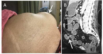
Figure 1A: Hard palpable lump in infraumbilical region.
Figure 1B: Ventral defect in abdominal wall in left para median infraumbilical location through which small bowel (distal jejunal loop), omental fat and properitoneal fat herniated out into a sac.
Patient Consent
Informed consent was taken from the patient
Conflicts of Interest and Funding
Nil
References
- Aguirre DA, Casola G, Sirlin C. Abdominal wall hernias: MDCT findings. AJR Am J Roentgenol 183 (2004): 681-690.
- Zalcman M, Sy M, Donckier V, et al. Helical CT signs in the diagnosis of intestinal ischemia in small-bowel obstruction. AJR Am J Roentgenol 175 (2000): 1601-1607.