Gautam A Taralekar1*, Girish Shinde1, Sasha Martyres1, Sachin G Shetti2, Lokesh Singh3
1Department of Orthopaedics, Bharati Vidyapeeth (Deemed to be University) Medical College and Hospital, Sangli, Maharashtra, India
2Department of Physiotherapy, Bharati Vidyapeeth (Deemed to be University) Medical College and Hospital, Sangli, Maharashtra, India
3Department of Radiodiagnosis, ESIC PGIMSR, New Delhi, India
*Corresponding Author: Gautam Taralekar, Department of Orthopaedics, Bharati Vidyapeeth (Deemed to be University) Medical College and Hospital, Sangli, Maharashtra, India
Received: 13 December 2020; Accepted: 23 December 2020; Published: 11 January 2021
Introduction: The surgical treatment of Rotator cuff repair ranges from open to arthroscopic assisted mini open repair to full arthroscopic repair. The anterior approach or lateral deltoid approach has its advantages and disadvantages depending upon the site and the extent of the rotator cuff tear.
Aim and objective: To assess the functional outcome of mini-open anterolateral approach for degenerative rotator cuff tear by trans-osseous sutures in elderly in terms of pain and range of movements.
Methodology: The present study was prospective observational study conducted on Patients with degenerative rotator cuff tears presenting at Department of Orthopaedics. Patients underwent mini-open anterolateral approach for degenerative rotator cuff tear by trans-osseous sutures. Follow-up of patients was done immediate after suture removal, at six weeks, at twelve weeks, twenty-four weeks following the surgery. Pre-op and Final follow up evaluation was done for each patient using VAS scale and range of movement. Shoulder index using ASES scores were noted.
Results and discussion: Majority of the patients (52%) were males and (48%) were females. 44% cases were between 61 to 70 years of age. In our study in follow up at 6,12, 24 weeks, range of motion was found to be increasing during subsequent follow ups. (p<0.001) The average ASES scores improved from 20.83 to 81.15 (Range 58-67) at final follow up (p<0.0001).
Degenerative rotator cuff tear repair; Transosseous suture; Mini-open antero-lateral approach
1. Introduction
Four muscles namely, Supraspinatus, Teres minor, Infraspinatus, and Subscapularis together in conjunction form the Rotator cuff. Rotator cuff tears are most commonly seen in the Supraspinatus. Rotator cuff tears can be because of trauma (traumatic) or because of some degenerative pathology (degenerative) mostly seen in elderly. This study deals with treatment of degenerative rotator cuff tears. Patients have symptoms ranging from pain to decrease in range of movements affecting the activities of daily living. The surgical treatment of Rotator cuff repair ranges from open to arthroscopic assisted mini open repair to full arthroscopic repair [1-3]. Recently, arthroscopic rotator cuff repair with advantages of less post-operative pain, low deltoid morbidity and faster rehabilitation is popular and satisfactory outcomes are reported by many. Although arthroscopy has its advantages, it has a steep learning curve. Also, the cost of the armamentarium required makes it a costly affair. As a result, Rotator cuff tears are treated commonly using a mini open technique using different approaches [4-6]. Many such studies have been documented comparing the results of open and arthroscopic techniques for repair. The anterior approach or lateral deltoid approach has its advantages and disadvantages depending upon the site and the extent of the rotator cuff tear. We planned this study taking into consideration elderly population (50 years and above) using a modified Antero-lateral approach which would also allow an acromioplasty whenever required [7]. Also a small deltoid split was used to access larger tears extending more anteriorly or posteriorly. Degenerative Rotator cuff tears are commonly encountered in the study region. The outcome is observed to be exclusively dependent upon the modality of treatment as well as the follow up. Thus, the study of these fractures has got a clinical outcome importance.
1.1 Aim and objective
To assess the functional outcome of mini-open anterolateral approach for degenerative rotator cuff tear by trans-osseous sutures in elderly in terms of pain and range of movements.
2. Material and Methods
The present study was prospective observational study conducted at Dept. of Orthopaedics at a Tertiary Care Centre during January 2018 to June 2019. Study population was Patients with degenerative rotator cuff tears presenting at Department of Orthopaedics.
2.1 Inclusion Criteria
- All patients of age above 50 years with rotator cuff injury and operated with the technique
- Patients who were willing to participate in the study.
2.2 Exclusion Criteria
Patients who were not willing to participate in study.
Approval for the study was obtained from the Institutional Ethics Committee of medical college. A valid written consent was taken from the patients after explaining study to them. Sample size was calculated by taking all the patients with proximal humerus fractures admitted over a period of last 1 ½ year. Total 25 patients were studied. At the arrival of the patient with these degenerative tears, a careful history was elicited from the patients and/or attendants about age, sex, pain, details of problems with activities of daily living, duration, were obtained through an interview. Patients were evaluated for associated medical problems and associated injuries and were addressed. Patients were subjected to clinical and local examination. These findings were recorded on predesigned and pretested proforma. Patients underwent all necessary investigations for anaesthetic assessment. MRI scan was carried out to study the tear size location and extent along with the other relevant findings. Goniometer was used to document the range of movements of the affected and unaffected shoulder. CT scan studies were not carried out in any of our patients. The local examination of injured shoulder was done to look for the attitude, swelling, restricted range and loss of function. Any nerve injury was also carefully looked for and noted.
All patients were operated within seven days of the injury. Patients placed in supine position on operating table with sandbag under the same side of side scapula. Operating limb was draped free, because it is moved during the procedure. Regional block (interscalene block) or general anaesthesia as per the anaesthesiologists preference. After palpating antero-lateral edge of acromion and 4-5 cm incision was taken over it. Superficial dissection was carried out between the anterior and middle part of deltoid (raphe). The deltoid attached to the anterior edge was elevated or detached using the cautery to expose the rotator cuff. Anterior inferior acromion was osteotomised to carry out acromioplasty either by using an osteotome or a high speed burr. This was followed by removal of the hypertrophied pathological bursal tissue. Traction stay sutures were used to tag the torn tendon. By rotating the arms the tear geometry and the reduction on the footprint of greater tuberosity were confirmed. Using a currette and a rasp a trough was made in the footprint of the greater tuberosity. Depending upon the size of the tear, 2-4 tunnel were drilled in the greater tuberosity which exited laterally. 1 to 3 horizontal mattress sutures using ethylon no.2 non absorbable suture were tied on the lateral surface again depending upon the size of the tear. The remaining lateral surface was approximated with 1 or 2 de-loading sutures taken through the tendon. For massive tears, along with the technique used above, the anterior and posterior edges were also sutured to each other. Trans-osseous osteo-periosteal sutures using no.1 vicryl (absorbable suture) were taken to prevent avulsion of deltoid from the acromion. Skin was sutured using ethilon 3.0. Compression dressing was done using dynaplast. Immobilization of the operated shoulder was done using an arm sling pouch before shifting the patient from the OT.
Patients began pendulum exercises and passive range of motion exercises one day after surgery. Appropriate antibiotics as well as analgesics were used. Sutures were removed by 10th post-operative day. Depending upon the pain, pendulum exercises were begun as soon as possible. At first week passive range of motion were started. Active range of motion was started at 3 weeks postoperatively 3 times a day for 20 repetitions. Active assisted movements started up to 900 abduction with no forced external rotation. At sixth to eighth week, full range of movements with active exercises including rotator cuff muscle strengthening exercises were started. At the end of six months the patients were examined clinically assessed for range of motion. The patients with shoulder stiffness were given physiotherapy for 1 to 2 weeks on outpatient basis.
Follow-up of patients was done immediate after suture removal, at six weeks, at twelve weeks, twenty-four weeks following the surgery. Pre-op and Final follow up evaluation was done for each patient using VAS scale and range of movement. Reduction in pain and clinical outcome were the parameters taken into consideration respectively. Completion of the ASES questionnaire, both pre-operatively and after follow up was a pre-requisite. The overall results were calculated as Excellent >/= 90; Good 80-89; fair 70-79; Poor < 70 which were obtained after calculating shoulder index using ASES system [8]. Data obtained was coded and entered into Microsoft Excel spreadsheet. ‘p’ value of less than or equal to 0.05 was considered as statistically significant.
3. Results
In this study most of the patients presented with age between 61 to 70 years (44%) followed by >70 years (36%). The mean age was 56.7 years. In the present study, 52% of the patients were males and 48% were females. In our study, 68% of the patients were dominant side and 32% non-dominant side. Out of total 25 patients, 36% of the patients was presented with crescent tear, reverse L 4%, L shaped 16%, Trapezoidal 24% and Massive 20%. Figure 1 shows comparison of flexion and extension score at follow up period. Flexion at presentation was 45.40 ± 32.50. Flexion score improved at follow up of 6 weeks (90 ± 27.39) and 12 weeks (114.40 ± 21.03). A=it again decreased at 24 weeks (46.40 ± 3.96). Post operatively, flexion score significantly improved (p<0.001). Extension score of patients at presentation was 19.60 ± 7.63. This score improved significantly at 6 weeks (28.40 ± 5.90), 12 weeks (38 ± 4.56) and 24 weeks (47.20 ± 5.02). (p<0.001). Figure 2 shows comparison of external rotation and internal rotation at different follow up periods. Mean External rotation at presentation was 22.80 ± 17.80, at 6 weeks follow up it was 31.60 ± 15.32. External rotation score were 40.72 ± 12.42 and 50.40 ± 9.99 at 12 weeks and 24 weeks respectively. Thus we can say that external rotation was improved over follow up periods after the surgery (p<0.001). Mean Internal rotation score of patients at presentation was 18.80 ± 7.26. it improved to upto 46.40 ± 3.96 at 24 weeks follow up. This difference was statistically significant (p<0.001). Mean adduction at presentation was 21.20 ± 20.88. Adduction movement increased over time upto 24 weeks (49.60 ± 12.90). Surgery significantly improved adduction (p<0.001) (Table 2). Table 3 shows comparison of abduction at different follow up periods. Mean abduction significantly improved from 46.80 ± 36.14 at presentation to 146.80 ± 18.76 at 24 weeks follow up (Table 3). In our study, the average ASES scores improved from 20.23 (Range 58-67) to 81.15 (range 0-40) at final follow up (p<0.0001). Majority of the patients in our study were having excellent outcome (40%) followed by good (24%), out of 25 patients, fair outcome was observed in 20% patients and poor outcome was observed in 16% patients (Table 4).
|
Sr no |
Age group (years) |
Frequency |
Percentage |
|
1 |
50-60 |
05 |
20 |
|
2 |
61-70 |
11 |
44 |
|
3 |
>70 |
09 |
36 |
|
4 |
Total |
25 |
100 |
Table 1: Distribution of patients according to age group.
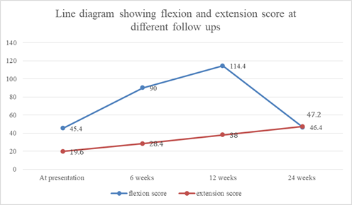
Figure 1: Line diagram showing Flexion and extension score at follow up.
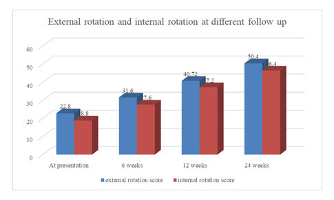
Figure 2: Line diagram showing external rotation and internal rotation at follow up.
|
Sr no |
Adduction |
Mean ± SD |
P value |
|
1 |
Range of movement at presentation |
21.20 ± 20.88 |
(Base line) |
|
2 |
6th week |
31 ± 18.31 |
<0.001 |
|
3 |
12th weeks |
40.40 ± 15.54 |
<0.001 |
|
4 |
24th weeks |
49.60 ± 12.90 |
<0.001 |
Table 2: Comparison of adduction at different follow up.
|
Sr no |
Abduction |
Mean ± SD |
P value |
|
1 |
Range of movement at presentation |
46.80 ± 36.14 |
(Base line) |
|
2 |
6th week |
82.80 ± 28.65 |
<0.001 |
|
3 |
12th weeks |
117.20 ± 21.32 |
<0.001 |
|
4 |
24th weeks |
146.80 ± 18.76 |
<0.001 |
Table 3: Comparison of abduction at different follow up.
|
Sr no |
Final ASES score |
Frequency |
Percentage |
|
1 |
Excellent |
10 |
40% |
|
2 |
Fair |
5 |
20% |
|
3 |
Good |
6 |
24% |
|
4 |
Fair |
4 |
16% |
Table 4: Distribution of patients according to Final ASES score.
4. Discussion
In this study most of the patients presented with age between 61 to 70 years (44%) followed by > 70 years (36%). The mean age was 56.7 years. Similar to our study Burks et al. [9] and Grasso et al. [10] found mean age of 56 and 57 years respectively. In a study of Charourset et al. [11] mean age of the patients was 59 years. In our study out of 25 patients, 13 were male 12 were female men had significantly higher or better ASES score than female. In our study 7 out of 11 patients with large sized tears (7-large 5 massive) had satisfactory outcome. Our study results were comparable to previous studies showing more than 80% satisfactory results. Romeo et al. reported that tears more than equal to 5cm were associated with poorer outcome [12]. However, Baysel et al. [13] included 17 patients with massive tears in their prospective study of 84 patients and found statistically significant improvement in shoulder scores in these patients as well. Our study results of 25 patients with tear of all sizes prospectively, who underwent a mini-open technique with significant improvement in range of motion and ASES scores pre-op (20.23 to 81.15). Our results were comparable to Baysal et al. who had reviewed 84 patients with tears of all sizes prospectively, who underwent a mini open repair with significant improvement in shoulder scores and range of motion. Our results are also comparable with the studies of Hanusch et al. [14] Chun et al. [15], Duraide et al. [16] and who concluded that there was significant decrease in pain and shoulder score after a mini open repair & demonstrated that mini open techniques can be effectively used for even large and massive tears.
After meticulous post-operative care patients were followed ups at 6, 12, 24 weeks and range of motion was found to be increasing during subsequent follow ups. The average ASES scores improved from 20.23 (Range 58-67) to 81.15 (range 0-40) at final follow up (p<0.0001). Most of the patients has excellent outcome (40%), fair (20%), good (24%), poor (16%) outcome. Similar results were observed in Burks et al. and Sugaya et al. [17]. Park et al. observed ASES score of 41.3 and it improved to 92.9 at the end of follow up [18]. Thus, anterolateral approach for mini open repair using trans-osseus sutures provides satisfactory results. This approach not only allows visualization of all types of tears but it also allows their repair and thus can help avoid major surgeries.
However, this study was a single centric study carried out only for a year which may be a short time to draw any inference as cases was not a large enough number to strengthen the significance of the differences in the functional outcome. Also, the participating surgeons were having relatively less experience with an all arthroscopic technique which also has to be taken into consideration. In view of ASES scores this method can be definitely employed whenever needed rationally. However, longer and larger studies will be needed including a proper RCT with prespecified hypothesis and confirmatory post-operative scans for ongoing studies, before any definitive guidelines are suggested for the treatment of these tears.
Conclusion
Anterolateral approach for mini open repair using trans-osseus sutures provides satisfactory results in terms of pain reduction and improved range of movement in elderly for degenerative rotator cuff tear.
Conflicts of Interests
None.
Acknowledgements
None.
References
- Nho S. Systematic Review of Arthroscopic Rotator Cuff Repair and Mini- Open Rotator Cuff Repair. The Journal of Bone and Joint Surgery (American) 89 (2007): 127.
- Osti L, Papalia R, Paganelli M, et al. Arthroscopic vs mini- open rotator cuff repair. A quality of life impairment study. Int Orthop 34 (2010): 389-394.
- Razmjou H. Evaluating Equivalency of Treatment Effectiveness: The Example of Arthroscopic and Mini-Open Rotator Cuff Repairs. Hand Clinics 25 (2009): 67-70.
- Hata Y, Saitoh S, Murakami N, et al. A less invasive surgery for rotator cuff tear: Mini-open repair. Journal of Shoulder and Elbow Surgery 10 (2001): 11-16.
- Pearsall A, Ibrahim K, Madanagopal S. The results of arthroscopic versus mini-open repair for rotator cuff tears at mid-term follow-up. Journal of Orthopaedic Surgery and Research 2 (2007).
- Cho C, Song K, Jung G, et al. Early Postoperative Outcomes Between Arthroscopic and Mini-open Repair for Rotator Cuff Tears. Orthopedics 35 (2012): 1347-1352.
- Mohtadi N, Hollinshead R, Sasyniuk T, et al. A Randomized Clinical Trial Comparing Open to Arthroscopic Acromioplasty with Mini-Open Rotator Cuff Repair for Full-Thickness Rotator Cuff Tears. The American Journal of Sports Medicine 36 (2008): 1043-1051.
- Richards RR, An KN, Bigliani LU, et al. A standardized method for the assessment of shoulder function. Journal of Shoulder and Elbow Surgery 3 (1994): 347-352.
- Burks RT, Crim J, Brown N, et al. A prospective randomized clinical trial comparing arthroscopic single- and double-row rotator cuff repair: magnetic resonance imaging and early clinical evaluation. Am J Sports Med 37 (2009): 674-682.
- Grasso A, Milano G, Salvatore M, et al. Single-row versus double-row arthroscopic rotator cuff repair: a prospective randomized clinical study. Arthroscopy 25 (2009): 4-12.
- Charousset C, Grimberg J, Duranthon LD, et al. Can a double-row anchorage technique improve tendon healing in arthroscopic rotator cuff repair? A prospective, nonrandomized, comparative study of double-row and single-row anchorage techniques with computed tomographic arthrography tendon healing assessment. Am J Sports Med 35 (2007): 1247-1253.
- Romeo AA, Hang DW, Bach BR Jr, et al. Repair of full thickness rotator cuff tears. Gender, age, and other factors affecting outcome. Clin Orthop Relat Res 367 (1999): 243-255.
- Baysal D, Balyk R, Otto D, et al. Functional outcome and health-related quality of life after surgical repair of full-thickness rotator cuff tear using a miniopen technique. Am J Sports Med 33 (2005): 1346-1355.
- Hanusch BC, Goodchild L, Finn P, et al. Large and massive tears of the rotator cuff: functional outcome and integrity of the repair after a mini-open procedure. J Bone Joint Surg Br 91 (2009): 201-205.
- Chun J, Kim S, Kim J. Arthroscopically Assisted Mini-Deltopectoral Rotator Cuff Repair. Orthopedics 31 (2008): 1-6.
- Duralde X, Greene R. Mini-open rotator cuff repair via an anterosuperior approach. Journal of Shoulder and Elbow Surgery 17 (2008): 715-721.
- Sugaya H, Maeda K, Matsuki K, et al. Functional and structural outcome after arthroscopic full-thickness rotator cuff repair: single-row versus dual-row fixation. Arthroscopy 21 (2005): 1307-1316.
- Park MC, Tibone JE, ElAttrache NS, et al. Part II: biomechanical assessment for a footprint-restoring transosseous-equivalent rotator cuff repair technique compared with double-row repair technique. J Shoulder Elbow Surg 16 (2007): 469-476.