Maria Gorete de Andrade and Paolo Ruggero Errante*
Veterinary Medicine Course. Faculdades Integradas Campos Salles, São Paulo, Brazil
* Corresponding Author: Paolo Ruggero Errante, Veterinary Medicine Course Faculdades Integradas Campos Salles, São Paulo, Brazil
Received: August 10, 2026; Accepted: August 17, 2026; Published: August 21, 2026
Subvalvular aortic stenosis is considered a common congenital heart disease in dogs, characterized by the presence of fibrous tissue below the aortic valve, which causes stenosis of the left ventricular outflow tract. Clinical findings include arrhythmias, left-sided congestive heart failure, endocarditis, exercise intolerance, syncope, and sudden cardiac death. The echocardiographic examination is considered the gold standard, and the pharmacological treatment used in veterinary medicine includes the use of β-adrenergic blockers such as propranolol or atenolol. This study aimed to report a case of aortic stenosis in a female Rottweiler adopted at four months of age with a history of a heart murmur; at eight months of age was attended by a cardiologist veterinary specialist, an echocardiogram was performed, resulting in a diagnosis of subvalvular aortic stenosis, and the dog has been treated with atenolol to date.
Congenital Heart Disease; Subaortic Stenosis; Aortic Valve; Dogs; Echocardiography.
Congenital Heart Disease articles; Subaortic Stenosis articles; Aortic Valve articles; Dogs articles; Echocardiography articles.
Introduction
Subvalvular aortic stenosis, or subaortic stenosis, is the most common congenital heart disease in dogs, accounting for 24% of cases of canine congenital heart disease [1]. Subvalvular aortic stenosis is characterized by a localized narrowing or reduction in the dimensions of the outflow tract, causing pressure overload. It is a congenital defect that affects large, purebred dogs such as Boxers, Golden Retrievers, Rottweilers, German Shepherds, Newfoundlands, and Dogue de Bordeaux, although it can also affect mixed breeds [2-4]. Aortic stenosis occurs due to the presence of a fibrous ring located just below the aortic semilunar valve that impedes left ventricular emptying [5]. Dogs with aortic stenosis may be clinically asymptomatic or may exhibit exercise intolerance, collapse, or syncope. The predominant physical finding in animals affected by this condition is the presence of a systolic murmur, best heard at the base of the heart; in moderate to severe cases, femoral pulses may be weakly palpable [3-6]. Diagnostic tests include the electrocardiogram, which demonstrates a left-axis shift or ventricular ectopy, and chest radiography, which may reveal a normal cardiac silhouette and left atrial enlargement, with dilation of the ascending aorta [5-7]. However, echocardiography is considered the gold-standard examination, demonstrating in addition to subvalvular aortic stenosis, left ventricular wall thickening and mitral regurgitation [8]. Pharmacological treatment includes the use of beta-adrenergic blockers such as propranolol or atenolol, which reduce the risk of sudden death, decrease myocardial oxygen demand, and suppress the onset of ventricular arrhythmias during physical exercise [9]. Treatment with furosemide and enalapril, on the other hand, is indicated in asymptomatic cases [10].
Case Report
In June 2022, a four-month-old female Rottweiler with a history of a heart murmur was adopted by her current owner; at eight months of age, she underwent a veterinary cardiological clinical evaluation. The tutor reported that the dog in any moment present signals of dispneia, fatigue or cough (Figure 1). During the physical examination, the dog appeared active and well-hydrated, with a normal body condition score (3/5) and pink mucous membranes; vital signs included a respiratory rate of 35 movements per minute, a heart rate of 120 beats per minute, and a body temperature of 37.5°C. Capillary refill time was under 2 seconds, and the heart rhythm was regular, though a mild systolic murmur was noted. A veterinary cardiologist confirmed a grade II systolic murmur at the aortic valve area, raising suspicion of left ventricular outflow tract obstruction.
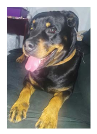
Figure 1: Physical appearance of a female Rottweiler with eight-months-old age presenting subvalvular aortic stenosis.
A complete blood count, measurement of alanine aminotransferase (ALT), alkaline phosphatase (AF), glucose, urea, creatinine, type I urine, chest X-ray, electrocardiogram and doppler echocardiography were performed. In the blood count, a normal number of erythrocytes and leukocytes was observed. The dosages of ALT, FA, glucose, urea and creatinine were considered normal for canine species. A Type I urinalysis was performed on a sample obtained via cystocentesis; the presence of bilirubin, glucose, ketone bodies, and occult blood was not observed. The chest X-ray revelead enlargement of the cardiac silhouette along the apicobasal axis, with chamber remodeling and bulging in the cranial region due to prominence of aortic arch (Figure 2).
Enlargement of the cardiac silhouette, most evident along the apicobasal axis, indicating chamber remodeling. Bulging in the cranial region due to prominence of the aortic arch
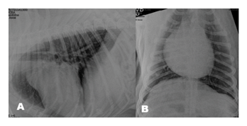
Figure 2: Chest X-ray. A) Right lateral and B) ventrodorsal projections.
Enlargement of the cardiac silhouette, most evident along the apicobasal axis, indicating chamber remodeling. Bulging in the cranial region due to prominence of the aortic arch
The electrocardiogram showed a slight prolongation of QRS complex duration, suggestive of nonspecific intraventricular conduction delay. A negative T wave with increased amplitude relative to the R wave was observed, characterizing a nonspecific ventricular repolarization abnormality (Figure 3).
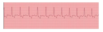
Figure 3: Slight prolongation of QRS complex duration, and a negative T wave with increased amplitude relative to the R wave.
Echocardiography was performed using M-mode, spectral Doppler (pulsed-wave, continuous-wave, and color), and tissue Doppler, employing an Ultramedic® Infinit 7V system and a 5-7 MHz sector transducer. Doppler echocardiography showed left atrial remodeling, moderate concentric left
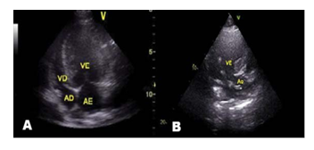
Figure 4: Echocardiographic images of a Rottweiler with subvalvular aortic stenosis.
- Evidence of left atrial remodeling and moderate concentric left ventricular hypertrophy.
- Moderate aortic valve insufficiency with a fibrous ring in the left ventricular outflow tract.
ventricular hypertrophy with the presence of a fibrous ring in the left ventricular outflow tract (Pyle-Patterson type II classification) (Figure 4) with hemodynamic repercussions in the left atrium. Color flow mapping demonstrated the presence of turbulent blood flow and high aortic flow velocity, consistent with subvalvular aortic stenosis (Figure 5).
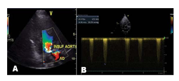
Figure 5: 2D Doppler echocardiographic images of a Rottweiler with subvalvular aortic stenosis.
- Echocardiographic image from the right parasternal window, five-chamber longitudinal view of an 8-month-old female Rottweiler, demonstrating blood turbulence in the left ventricular outflow
- Echocardiographic image (left parasternal window, apical five-chamber view) of an 8-month-old female Rottweiler; the arrow indicates high velocity and a pressure gradient in the left ventricular outflow tract, characterizing mild subaortic stenosis.
Since the animal showed no clinical signs of subvalvular aortic stenosis, pharmacological therapy with atenolol (1 mg/kg, orally, twice daily) was initiated to avoid the risk of cardiac arrhythmias and sudden death. Until the date, the dog has not exhibited symptoms of coughing, dyspnea, or mucosal cyanosis. Because the risk of future complications, echocardiography, chest X-rays, and electrocardiography are performed semiannually to monitor the subvalvular aortic stenosis.
Discussion
Subvalvular aortic stenosis is considered a common congenital heart disease in dogs, caused by the presence of a fibrous ring located below the aortic valve, which creates stenosis and impedes complete blood outflow from the left ventricle [3]. In subvalvular aortic stenosis, the presence of poorly organized fibrous tissue is observed forming near or below the aortic valve, located 2 to 5 mm caudoventral to the valve, projecting 1 to 2 mm into the lumen from the endocardial surface [5,6]. It has been postulated that the presence of a pronounced aortoseptal angle, or a misalignment between the aortic root and the interventricular septum, they are responsible for generating shear stress that leads to the proliferation of fibrous tissue [11,12], and stenosis [13]. Subvalvular aortic stenosis can be classified into different degrees, proposed by Pyle & Patterson [14]. Grade 1 involves thickening or slight elevation of septal endocardium, forming small nodules 1 to 2 mm in size, sometimes present on the ventricular surface of aortic cusps; in grade 2, the endocardium appears more thickened and elevated, forming a discrete fibrous ridge that extends partially along the left ventricular outflow tract. In more severe cases, such as grade 3, a prominent, thick fibrous ring, band, or ridge is observed completely encircling the left ventricular outflow tract (tunnel stenosis) [14]. In our study, it was observed that Rottweiler presented with grade II disease, based on the anatomical classification derived from necropsy and catheterization studies proposed by Pyle & Patterson. Although the semilunar valves are formed during embryonic development from the subendocardial cushions of the truncus arteriosus, the heart continues to undergo developmental changes during the perinatal and neonatal periods [15,16]. Thus, from a clinical standpoint, the disease cannot be diagnosed at birth, as patients cannot be reliably classified into a severity category until 6 to 12 months of age. In our study, however, since the dog showed no clinical signs of the disease, the diagnosis was established solely through Doppler echocardiography. Subaortic valve stenosis compromises blood flow, causing an increase in aortic blood flow velocity and pressure overload on the left ventricle, leading to left ventricular concentric hypertrophy, diastolic dysfunction, and mitral regurgitation [3,4]. Subendocardial ischemia/necrosis and fibrosis may also occur due to thickening of intramural coronary arteries, left ventricular myocardial insufficiency, and/or left heart failure. Other observed cardiac abnormalities include post-stenotic aortic dilation and aortic insufficiency. Left ventricular pressure overload [17] can cause exercise intolerance, syncope, and sudden death due to ventricular arrhythmias resulting from subendocardial ischemic/fibrotic changes [18]. During physical examination and cardiac auscultation, animals affected by subvalvular aortic stenosis may exhibit a systolic murmur, best heard at the base of the left ventricle. Other findings include a palpable carotid or left parasternal thrill and a high-frequency early diastolic murmur if aortic insufficiency is present [1-4]. In our study, the presence of a cardiac murmur was observed, consistent with descriptions in the literature [5,6]. In moderate to severe cases, femoral pulses may be weak. Due to these clinical manifestations, the average life expectancy of untreated dogs with severe subvalvular aortic stenosis is 19 months [19]. Diagnosis is established through a comprehensive medical history, physical examination, and echocardiogram [7-13]. In some cases, blood tests, chest X-rays, an electrocardiogram (ECG), and/or a 24-hour ambulatory electrocardiogram may be indicated. On chest radiographs, findings indicative of left ventricular enlargement may include elevation of the carina, loss of the caudal waist, and a more perpendicular caudal cardiac border. Pulmonary vascular markings are generally normal in subvalvular aortic stenosis [20]. In our study, enlargement of the cardiac silhouette was observed, most notably along the apicobasal axis, indicating cardiac chamber remodeling and cranial bulging due to prominence of the aortic arch. According to the literature, in cases of aortic stenosis, the electrocardiogram may appear normal, but it may also offer information consistent with left ventricular hypertrophy, with R waves of greater amplitude, and with supraventricular or ventricular arrhythmia [21]. However, in our study, electrocardiographic findings indicated the presence of slight prolongation of the QRS complex duration and a negative T wave with increased amplitude relative to the R wave. Transthoracic echocardiography is the primary diagnostic tool for congenital heart disorders, used in conjunction with complementary tests such as chest radiography and electrocardiography. Doppler echocardiography is highly capable of classifying the severity of subvalvular aortic stenosis [22]. The fundamental aspect of echocardiographic assessment of dogs with subclinical aortic stenosis is the evaluation of the left ventricular outflow tract and the stenotic region below the aortic valve using two-dimensional (2D) imaging, M-mode, and Doppler [7,8]. Based on two-dimensional assessment, it is possible to recognize anatomical abnormalities and classify subaortic stenosis according to autopsy and catheter studies conducted by Pyle and Patterson [14]. In this case, ultrasound allowed the report to be classified as type II subvalvular aortic stenosis due to the absence/slight thickening of the endocardium. In Doppler evaluation, turbulent blood flow can be observed, with high velocity in the lumen of the aortic artery and a pressure gradient above normal in the left ventricular outflow tract, in addition to the presence of aortic valve insufficiency and moderate diastolic impairment [6]. The severity of a case is defined by the measured aortic velocity and the calculated pressure gradient, since as stenosis progresses, the blood flow velocity increases [23]. It is accepted that a dog is considered to have aortic stenosis when it presents a flow velocity through the aorta greater than 2.5 m/s [11,12]. In our study, the echocardiogram revealed normal movement of the aortic valve cusps, with the presence of a subaortic fibromuscular ring with turbulent flow causing concentric hypertrophy of the left ventricle with hemodynamic repercussions in the left atrium. The differential diagnosis for subvalvular aortic stenosis includes pulmonary stenosis, Tetralogy of Fallot, and ventricular septal defect [24-26]. The treatment used for dogs with stenosis is pharmacological, employing β-adrenergic blockers (propranolol or atenolol) to reduce the risk of sudden death, myocardial oxygen demand, and suppress ventricular arrhythmias during exercise [27]. Beta-adrenergic blockers decrease the patient's heart rate and increase myocardial perfusion. Atenolol, which is cardioselective, tends to be the beta-blocker of choice [3,28]. In our study, the dog is treated twice daily with atenolol to control arrhythmias and prevent sudden death. Due to the risk of future complications in animals with subvalvular aortic stenosis, echocardiography, thoracic radiography, and electrocardiography were recommended every six months.
Conclusion
Among congenital heart diseases in dogs, subvalvular aortic stenosis stands out due to its high prevalence; it is often diagnosed at a later stage, particularly in medium to large-size purebred dogs. It is crucial for clinical veterinarians to perform a thorough physical examination and refer young animals to a veterinary cardiologist for further diagnostic testing. Among these tests, Doppler echocardiography is considered the gold standard for diagnosing subvalvular aortic stenosis. Early identification of subvalvular aortic stenosis allows for periodic echocardiographic monitoring of disease progression and the implementation of pharmacological treatment, when necessary, thereby improving the quality and expectancy of life of affected dogs.
References
- Ontiveros ES, et al. Congenital cardiac outflow tract abnormalities in dogs: prevalence and pattern of inheritance from 2008 to 2017. Front Vet Sci 6 (2019):1-10.
- Buchanan The history of veterinary cardiology. J Vet Cardiol 15(2013):65-85.
- Ontiveros ES, Stern JA Genetics of canine subvalvular aortic stenosis (SAS). Canine Med Genet 8 (2021): 1-9.
- Chetboul V, et Congenital pulmonic and aortic stenosis in Newfoundland dogs: results of a 14-years French cardiovascular screening program (921 dogs). PLoS One 20 (2025): 1-13.
- Crofton AE et al. Subvalvular aortic stenosis: learning from human and canine clinical research. Cardiol Res 14 (2023) :319-333.
- O'Grady MR et al. Canine congenital aortic stenosis: a review of the literature and commentary. Can Vet J 30 (1989): 811-815.
- Thomas WP, et al. Recommendations for standards in transthoracic two-dimensional echocardiography in the dog and cat. Echocardiography Committee of the Specialty of Cardiology, American College of Veterinary Internal Medicine. J Vet Intern Med 7 (1993): 247-252.
- Bussadori C, et al. Guidelines for the echocardiographic studies of suspected subaortic and pulmonic stenosis. J Vet Cardiol 2 (2000): 15-22.
- Eason BD, et Influence of beta blockers on survival in dogs with severe subaortic stenosis. J Vet Intern Med 28 (2014): 857-862.
- Magalhães GH, et al. Aortic stenosis in a Shih Tzu dog. A case report. Enciclopédia biosfera, Centro Científico Conhecer 10 (2041): 1020-1026.
- Belanger MC, et al. Association between aortoseptal angle in Golden Retriever puppies and subaortic stenosis in adulthood. J Vet Intern Med 28 (2014): 1498-1503.
- Quintavalla C, et al. Aorto-septal angle in Boxer dogs with subaortic stenosis: an echocardiographic Vet J 185 (2010): 332-337.
- Oyama MA, Thomas Two-dimensional and M-mode echocardiographic predictors of disease severity in dogs with congenital subaortic stenosis. J Am Anim Hosp Assoc 38(2002):209-215.
- Pyle RL, Patterson DF, Chacko The genetics and pathology of discrete subaortic stenosis in the Newfoundland dog. Am Heart J 92 (1976): 324-334.
- Höllmer M, et al. Aortic stenosis in the Dogue de Bordeaux. J Small Anim Pract 49 (2008): 432-437.
- Stern JA, et al. Familial subvalvular aortic stenosis in Golden Retrievers: inheritance and echocardiographic findings. J Small Anim Pract. 201253 (2012): 213-216.
- Kienle RD, Thomas WP, Pion PD. The natural clinical history of canine congenital subaortic stenosis. J Vet Intern Med 8 (1994): 423-431.
- Oliveira P, et Retrospective review of congenital heart disease in 976 dogs. J Vet Intern Med 25 (2011): 477-483.
- Kienle RD, Thomas WP, Pion PD. The natural clinical history of canine congenital subaortic stenosis. J Vet Intern Med 8 (1994): 423-431.
- Aronson E, McCaw D. Congenital cardiac disease in dogs. Mod Vet Pract 65 (1984): 287-291.
- Owens EJ, et al. Echocardiographic and electrocardiographic effects of atenolol versus sotalol in dogs with severe subaortic stenosis. J Vet Cardiol 41(2022):199-208.
- Tidholm A, et al. Current use of real-time three-dimensional transthoracic echocardiography in J Vet Cardiol 51 (2024): 97-104.
- Castro MG, et Echodopplercardiographic retrospective study of the main cardiopathies diagnosed in dogs. Arq Bras Med Vet Zootec 61 (2009):1238-1241.
- Scansen BA. Advances in the treatment of pulmonary valve stenosis. Vet Clin North Am Small Anim Pract 53 (2023): 1393-1414.
- Valdeomillos E, et al. Animals models of repaired Tetralogy of Fallot: current applications and future perspectives. Can J Cardiol 35 (2019): 1762-1771.
- Graczyk S, et al. A comprehensive review of canine and feline ventricular septal defects- from pathogenesis to long-term follow-up. Animals (Basel) 15 (2025): 1-32.
- Eason BD, et Influence of beta blockers on survival in dogs with severe subaortic stenosis. J Vet Intern Med 28 (2014): 857-862.
- Galderisi M, D'Errico Beta-blockers and coronary flow reserve: the importance of a vasodilatory action. Drugs 68 (2008): 579-590.