Yayun Liang and Salman M. Hyder*
Department of Pathobiology and Integrative Biomedical Sciences, 1520 E Rollins Road, Columbia, MO 65211, USA
*Corresponding Author: Dr. Salman M. Hyder, Department of Pathobiology and Integrative Biomedical Sciences, 1520 E Rollins Road, Columbia, MO 65211, USA.
Received: 16 September 2026; Accepted: 23 September 2026; Published: 30 September 2026
Triple-negative breast cancers (TNBC) lack ER, PR and Her2neu, proteins that are commonly targeted in breast cancer therapies. As a consequence, women who suffer from TNBC have poor prognosis and limited treatment options, and are usually administered toxic, non-specific chemotherapeutic agents. Drug-resistance almost always occurs, leading to tumor metastasis, the main cause of patient death. New non-toxic therapeutic strategies to control metastasis of TNBC are urgently needed. Cholesterol is an essential structural and functional component of cell membranes that is necessary for the growth of both primary tumors and metastatic colonies that reside in distant organs. Therefore, inhibiting cholesterol production is an attractive therapeutic strategy. We targeted oxidosqualene cyclase (OSC), an enzyme that occurs downstream of HMGCoA-reductase in the cholesterol biosynthetic pathway, to disrupt tumor metastasis. Potent small molecule inhibitors of OSC have been identified. RO 48-8071 (4′-[6-(allylmethylamino)hexyloxy]-4-bromo-2′-fluorobenzophenone fumarate) (RO), has emerged as a useful chemotherapeutic agent for treating several forms of primary tumors, including those of TNBC origin. We developed a mouse model for studying lung metastasis using an aggressive MDA-MB-231 variant (LM2) TNBC cell line that produces detectable metastatic colonies in the lungs in approximately 4–6 weeks. Female nu/nu mice, 5–6 weeks old, were inoculated with tumor cells via tail vein. Five days after inoculation animals received RO or vehicle alone every other day for two weeks. Since RO induces estrogen receptor β, we also tested an ERβ interacting natural compound, liquiritigenin, as a therapeutic agent. Animals were then sacrificed, and both lungs harvested and immediately fixed in Bouin’s fixative. Metastatic colonies on the lung surface were identified and counted under a dissecting microscope. We observed that RO effectively reduced lung metastasis without affecting animal weight. Liquiritigenin was ineffective either alone or in combination with RO to suppress metastasis. In our study, which is the first to demonstrate that disruption of cholesterol biosynthesis in a model of human TNBC leads to reduced metastasis in lungs, we also found that RO treatment downregulated MetAp2, a proangiogenic protein that promotes the survival of tumor cells. Further studies will determine mechanisms through which RO suppresses TNBC metastasis to the lungs.
RO 48-8071; Triple-negative breast cancer; lung metastasis; Liquiritigenin
RO 48-8071 articles; Triple-negative breast cancer articles; lung metastasis articles; Liquiritigenin articles
1. Introduction
Triple-negative breast cancers (TNBC) comprise 15–20% of all tumors of the breast and lack estrogen receptor (ER), progesterone receptor (PR), and human epidermal growth factor receptor (HER2/neu) [1,2]. Current treatments for non-triple negative breast cancers target these three proteins to control tumor growth, since they are generally hormone-responsive and stimulated by the protein-ligand interactions of ER, PR, and/or HER2. TNBC is subclassified into various phenotypes, though all TNBC phenotypes are associated with a significantly poorer clinical outcome because they are difficult to treat [1,3,4], and cancers of this type that escape adjuvant treatment and surgery generally emerge as particularly aggressive tumors. In addition, Perou et al. reported that women with basal-like (specifically, triple-negative) breast cancers have an increased risk of tumor recurrence compared with women with other cancer subtypes [5] TNBC frequently metastasizes to the viscera (ie, lung, brain, and liver) and, to a lesser extent, bone [6-9]; in addition, the rate of cancer recurrence 3–5 years post-therapy is greater in women with TNBC than in their non-triple-negative counterparts [8, 9]. For these reasons, new therapeutic options for TNBC that are less toxic and more effective are needed.
In the present report, we describe the effects of a cholesterol biosynthesis inhibitor (RO) on lung metastasis of TNBC. We previously showed that RO inhibits progression of TNBC [10] and herein, we demonstrate that RO reduces TNBC metastasis to the lungs. Furthermore, we correlate the inhibitory effect of RO with reduction of MetAP2, a protein that promotes angiogenesis, an essential component of growth and metastasis of tumor cells [11]. We previously reported on the ability of RO to induce ERβ [10] and we showed that an ERβ interacting natural compound, liquiritigenin (LQ), a non-toxic naturally occurring plant flavonoid, enhanced RO effects in suppressing TNBC progression [12]. Because there are few clinically accepted standardized chemotherapeutic options to combat metastatic TNBC, we conducted studies to ascertain whether LQ might enhance the anti-metastatic properties of RO. Our findings show that while RO alone suppressed the ability of TNBC cells to metastasize to the lungs, LQ, either with or without RO, does not prevent metastasis. We therefore propose that RO should be further studied as a non-toxic compound to suppress metastasis of TNBC until better combination therapy options are available.
2. Materials and Methods
2.1 Reagents
RO 48-8071 (RO) was provided by Roche Pharmaceuticals (Basel, Switzerland) for in vivo studies, and was synthesized as previously described [13,14]. RO for in vitro studies was purchased from Sigma-Aldrich (St Louis, MO) and dissolved in phosphate-buffered saline (PBS). LQ was dissolved in DMSO for in vitro studies and in 50% DMSO/15% ethanol/35% PBS for in vivo studies. Pierce bicinchoninic acid (BCA) protein reagents were obtained from Fisher Scientific (Pittsburgh, PA, USA). MetAP2 (Abcam, ab124953, 1:1000 dilution), GAPDH (Cell Signal, product number, 1:1000 dilution) were used in the Western blots described below.
2.2 Cell lines and culture
The MDA-MB-231 (4175) LM2 breast cancer cell line is a metastatic subpopulation isolated from TNBC MDA-MB-231 cells in Dr. Joan Massague’s lab [15] and maintained in DMEM supplemented with 10% fetal bovine serum (FBS; Sigma-Aldrich) at 37°C/5% CO2. Cells were harvested with 0.05% trypsin-EDTA (Invitrogen). For in vitro experiments, cells were allowed to reach 50–60% confluence, washed in phosphate-buffered saline (PBS), and maintained in DMEM/F12 or DMEM supplemented with 10% FBS for 24 hours prior to treatment.
2.3 Microarray
Both the parental MDA-MB-231 cells and their derivative MDA-MB-231 LM2 cells were cultured in DMEM/F12 +10% FBS. Prior to treatment with RO cells were washed and lifted in DMEM/F12 + 5% FBS. Cells were treated for 24 h with either PBS or 10 µM RO and RNA prepared using the RNAzol (Molecular Research Center, Inc) reagent protocol. Briefly, samples were homogenized in RNAzol and RNAse free water was added to sediment DNA and proteins. RNA was precipitated with isopropanol, and the resulting RNA pellet washed with 75% ethanol. RNA integrity was determined by evaluation of the 260/280 and 260/230 ratio using Nanodrop. RNA isolated from the three dishes was combined prior to cDNA synthesis using the RT2 First Strand Kit (Qiagen, Cat# 33001). The Human Metastasis – RT2 Profiler PCR array (Qiagen, Cat# PAHS-028ZA) was used to survey 84 genes related to metastasis. Using the RT2 SYBR Green Mastermix, 500ng cDNA was amplified. Analysis was performed with an Eppendorf Mastercycler ep Realplex 2. Relative gene expression was determined using the following formula: Fold-change in gene expression, 2(-ΔΔCt), where ΔΔCt = ΔCt (RO10 treated sample) – ΔCt (control samples)}, where ΔCt = Ct (gene of interest) – Ct (HKG), where Ct represents the cycle threshold.
2.4 BCA protein assay in preparation for Western Blotting
MDA-MB-231 and MDA-MB-231 (4175) LM2 cells were washed twice with PBS, then harvested, pelleted, and resuspended in 300 µL lysis buffer (50 mM Tris/HCl, pH 8, 150 mM NaCl, and 1% Nonidet P-40). Protein concentration was determined using a microplate reader to measure absorbance at 562 nm with a bovine serum albumin standard (Cat # 23209; Thermo Fisher Scientific; Waltham, MA, USA). Experiments were performed in triplicate, and samples were analyzed in duplicate.
2.5 Western blotting
Cells were grown in a humidified atmosphere of 5% CO2 at 37oC in 10 mm cell culture plates using phenol red-free Dulbecco’s modified Eagle’s medium (DMEM/F12) supplemented with 10% fetal bovine serum. When they had reached 50-60% confluence, cells were washed with PBS and treated for 24 h with RO 48-8071 (10- and 25 µM). Whole cell protein was extracted using 40 µL complete lysis buffer per dish following the manufacturer’s guidelines (Active Motif, USA). Protein aliquots (25 µg) were separated by SDS-PAGE, transferred to polyvinylidene difluoride membrane and blotted with the following human specific antibodies: MetAP2 (Abcam, ab124953, 1:1000 dilution), GAPDH (Cell Signal, 1:1000 dilution). Protein bands were detected and quantified by blotting with anti-rabbit-HRP secondary antibody, or anti-mouse-HRP secondary antibody (1:1000 dilution). Chemi-luminescent signals were detected using the Bio Rad Chemi Doc XRS system.
2.6 TNBC lung metastasis xenograft model
All animal studies were approved by the Animal Care and Use Committee at the University of Missouri, Columbia. Female athymic nu/nu nude mice, 6 weeks old (18–22 g), were purchased from Harlan Sprague-Dawley, Inc (Indianapolis, IN, USA). Mice were housed in a laminar air-flow cabinet under specific pathogen-free conditions. All facilities were approved by the American Association for Accreditation of Laboratory Animal Care in accordance with current U.S. federal regulations and standards. LM2 cells were harvested by trypsinization and washed twice with DMEM/F12 or DMEM medium. Cell pellets were then suspended in medium and injected intravenously (i.v.) into the tail vein. MDA-MB-231 (4175) LM2 cell line was injected at 2.0 x 10⁵ cells per 150 µL medium without FBS (Figure 1). RO (20 or 40 mg/kg) was initially given iv for three days, then by ip for a further 4 days, and finally every other day by ip for a further 15 treatments as described in Figure 1. For studies involving LQ, mice were administered RO at 20 mg/kg and LQ at 20 mg/kg. RO, LQ and vehicle control were given intraperitoneally (i.p.) on the fifth day post-inoculation of cells and continued every day for five days. This was followed by ip injections every other day for 14 more treatments, followed by three more treatments every day. Control animals were injected with the same volume of vehicle (70% DMSO/30% PBS). Animals were weighed every 2–4 days throughout the study. Lungs were harvested following sacrifice of animals on Day 41 and fixed in Bouin’s Fixative Solution (Cat # 1120-32; Ricca Chemical Company; Arlington, TX, USA), per the manufacturer’s recommendations. Superficial lung colonies were counted using a stereoscopic dissection microscope and the results from two independent researchers were averaged, then statistically analyzed using SigmaPlot software, version 12.5 (San Jose, CA, USA).
2.7 Statistical analysis
Statistical significance was tested using t-test or one-way analysis of variance (ANOVA) followed by a Newman-Keuls multiple comparison test to determine the difference in mean between groups. If normality failed, the data were tested using a nonparametric one-way ANOVA on ranks (Kruskal-Wallis), followed by the Newman-Keuls nonparametric multiple comparisons test or Dunn’s Method in the presence of an uneven n. The Student’s t-test was used to report the difference between two group means; when normality failed, the Mann-Whitney rank sum test was used. Animal weight comparisons were analyzed using a Two-way repeated measures ANOVA. Data were reported as means ± standard error of the mean (SEM). For all comparisons, P ≤ 0.05 was regarded as statistically significant, and analyses were performed using SigmaPlot 12.5 software.
3. Results
3.1 RO 48-8071 (RO) inhibits metastasis of human TNBC cells in a mouse model
To determine the effectiveness of RO as an anti-metastatic compound that might be used to combat TNBC metastasis, we utilized a xenograft metastasis model that mimics secondary-site colonization (Figure 1). We inoculated female nu/nu mice, 5–6 weeks old, via tail vein with LM2 cells, a variant with a molecular signature specific to lung metastasis. Five days after tumor cell inoculation animals received 20 mg/kg (n=6), 40 mg/kg (n=7) RO (ip) or vehicle alone (n=7) as described in Figure 1. Following sacrifice, both lungs were harvested and immediately fixed in Bouin’s fixative. Metastatic colonies on the lung surface were identified and counted under a dissecting microscope. Number of metastatic colonies was significantly reduced from 56 + 7 (Mean + SEM) in control mice injected with vehicle alone, to 28 + 5 and 6 + 1 in animals given 20 mg/kg RO and 40 mg/kg RO, respectively (Figure 2A and 2B; p<0.05, ANOVA). Our study also showed that RO effectively reduced lung metastasis without affecting animal weight (Figure 2C).
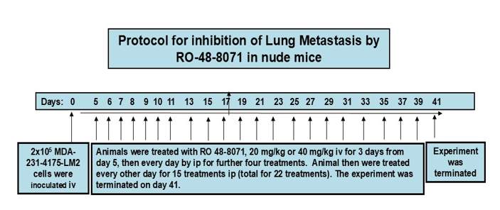
Figure 1: Experimental design and treatment protocol for examining the effects of different doses of RO 48-8071 (RO) on MDA-MD-231-4175 induced lung metastasis in nude mice.
MDA-231-4175-LM2 cells, (2x10⁵) in 0.1ml culture medium (without FBS) were injected into mice through the tail vein (iv) at day zero. Animals were given iv injections of RO (20 mg/kg or 40 mg/kg) on days 5-7, then daily intraperitoneal (ip) injections on days 8-11, and finally ip injections every other day for 30 days (15 treatments), for a total of 22 RO treatments. The experiment was terminated on day forty-one.
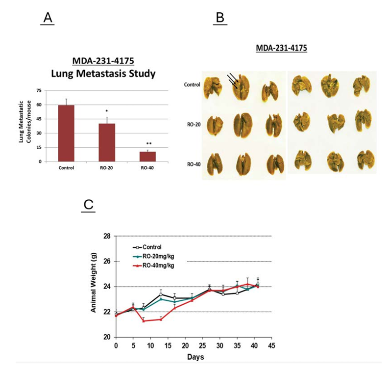
Figure 2: Dose-dependent inhibition by RO of metastatic colony formation in lungs. (A) At the end of the experiment, animals were sacrificed and lungs with metastatic colonies from each mouse harvested, washed with PBS, and fixed in Bouin’s Fixative Solution for 5-7 days. Metastatic colonies were counted using a stereoscopic dissection microscope, results from two independent researchers tallied and results averaged and analyzed statistically using Sigma Plot software, version 12.5. Figure 2A shows that both doses of RO significantly inhibited lung metastasis. *: significantly different to control group; **: significantly different to both control and RO 20 mg/kg groups, P< 0.05, by ANOVA. (B) Representative images of metastatic colonies on lungs from each group. Arrows indicate metastatic colonies. (C) Animal weights were monitored throughout the experiment. No significant weight differences were observed between groups, though animal weights in the 40 mg/kg RO group were slightly, though not significantly lower during the first week of treatment and were back to normal by the second week.
3.2 RO inhibits MetAp2 protein in vitro in TNBC cells
In order to address possible molecular mechanisms involved in RO-mediated suppression of lung metastasis, we treated cells with RO and used a metastasis array as described in materials and methods to determine changes in gene expression. While several changes in gene expression were observed, MetAp2 mRNA expression was reduced most significantly (not shown). MetAp2 is a protein involved in angiogenesis and cell survival [11], and since its expression was inhibited by RO, we used Western blot analysis to ascertain whether MetAp2 protein was also reduced. As shown in Figure 3A and B, MetAp2 protein was reduced in both parental MDA-MB-231 cells and in MDA-MB-231 LM2 cells that metastasize to the lungs.
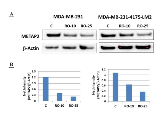
Figure 3: A-B: RO inhibited MetAp2 expression in both MDA-MB-231 and MDA-MB-231-4175 cell lines in a dose-dependent manner. (A) MDA-MB-231 and MDA-MB-231-4175 cells were treated with 10µM or 25µM RO for 24hrs. Cells were washed with PBS and whole cell lysates prepared. Western blot analysis was performed as described in Materials and Methods. Western blots show that RO inhibited MetAp2 expression in a dose-dependent manner. (B) Scan and quantitation of 3A, showing dose-dependent inhibition of MetAp2 expression by RO.
3.3 LQ does not enhance anti-metastatic activity of RO in TNBC cells
Previously we showed that a natural compound, LQ, arrested TNBC growth, most likely by interacting with ERβ, which is induced by RO [12]. In this study we determined whether LQ has the capacity to potentiate the effects of RO and suppress metastasis additively or synergistically. Figure 4 shows the protocol used to administer LQ and RO in nude mice that were injected with MDA-MB-231 LM2 cells.
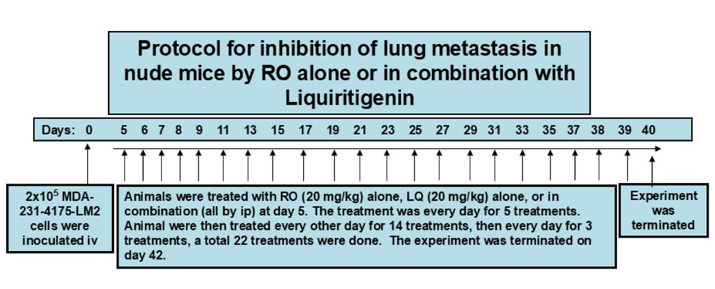
Figure 4: Experimental design and protocol for RO and LQ combination treatment against MDA-MB-231-4175 induced lung metastasis in nude mice.
MDA-231-4175-LM2 cells (2x10⁵) in 0.1ml culture medium (without FBS) were injected into mice via tail vein (iv) at day zero. Animals were treated ip with RO (20 mg/kg) alone, LQ (20 mg/kg) alone, or a combination of both on days 5-9, after which, injections continued every other day from day 11 to 37, followed by three more daily treatments. The experiment was terminated on day 40.
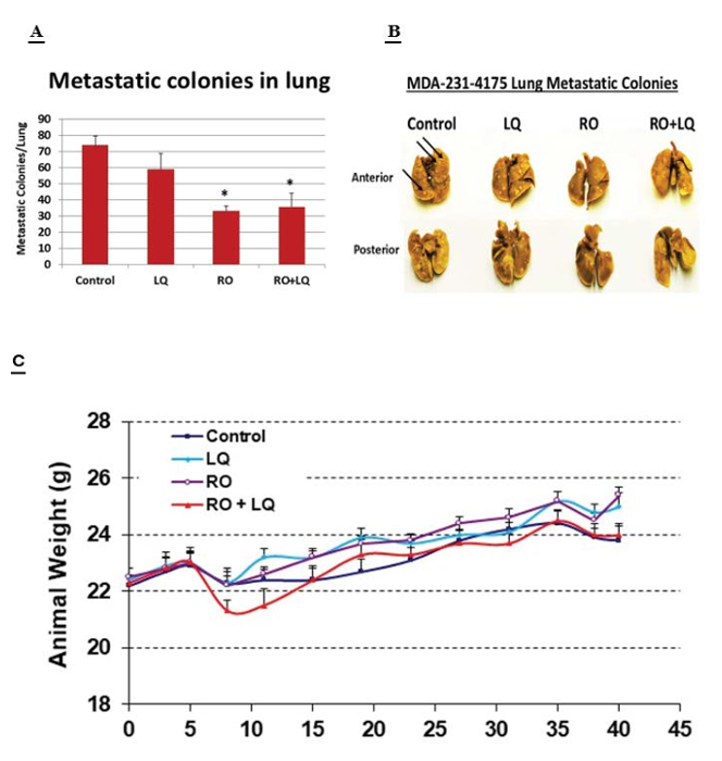
Figure 5: A-C: RO alone and a combination of RO + LQ inhibited lung metastatic colony formation in nude mice. (A) At the end of the experiment, animals were sacrificed and metastatic colonies from the lungs of each mouse harvested, washed with PBS, and fixed in Bouin’s Fixative Solution for 5-7 days. Metastatic colonies were counted using a stereoscopic dissection microscope. Results from two independent researchers were averaged and analyzed statistically by Sigma Plot software, version 12.5. *: significant differences compared with control group, P< 0.05, by ANOVA. (B) Representative images of metastatic colonies from both front and back of lungs from each group. (C) Animal weights were monitored throughout the experiment. No significant weight differences were observed between groups, though weights of animals in the RO + LQ group were slightly lower during the first week of treatment. However, this did not reach significance and weights recovered in the second week of treatment.
Figure 5A shows that when administered alone, RO suppressed metastasis. However, LQ alone did not affect metastasis, and a combination of RO + LQ had no enhanced effect over and above the antimetastatic activity of RO alone. Figure 5B shows the distribution of lung colonies following RO or LQ treatment, while Figure 5C illustrates the effect of treatment on animal weight. No significant adverse effects were observed in the time frame tested.
4. Discussion
Breast cancer is a deadly disease that afflicts millions of women worldwide and almost 20% of detected tumors are of the TNBC type. Because no recognized targeted chemotherapeutic approaches exist for TNBC, patients diagnosed with this type of tumor have a poor prognosis [16,17]. While significant progress has been made in understanding TNBC, considerable challenges remain in its diagnosis and treatment. Continued research, coupled with a multidisciplinary approach, will be vital in unraveling the complexities of TNBC and improving outcomes for affected patients. The integration of novel therapeutic strategies and a commitment to personalized medicine may pave the way for more effective interventions in this challenging area of breast cancer. Current treatments for TNBC are highly toxic, and resistant tumors often arise and metastasize despite the use of aggressive chemotherapy regimens [18]. Given the urgent need for safer and more effective treatments to combat metastatic TNBC, we embarked on a study of the cholesterol biosynthesis inhibitor RO, which we have previously shown to be non-toxic and effective against a variety of different types of cancer cell, including prostate and colorectal cancer cells, both in vitro and in vivo. To our knowledge, the capacity of RO to inhibit metastasis of TNBC cells to the lungs has not been previously reported, though in earlier studies we showed that RO inhibits expression of VEGF, which is associated with promoting cellular migration and neovascularization [19], both indispensable steps in tumor metastasis. Herein, we examined whether RO might block metastasis of tumors originating from TNBC cells and found that it significantly inhibited metastasis of MDA-231 (4175) LM2 TNBC cells with no observed toxicity. Compounds such as RO have the benefit of being either non-toxic, or of low toxicity, and therefore confer significant potential advantages over currently used drugs.
In addition to inhibiting metastasis, we showed that RO suppresses expression of MetAp2, a protein that is associated with angiogenesis and, furthermore, that levels of MetAp2 protein are reduced by RO in TNBC cells. Angiogenesis is a vital component of tumor cell growth and metastasis, since new blood vessels facilitate the delivery of nutrients to tumors as well as the dissemination of cancer cells to distant sites. As previously mentioned, in an earlier report we showed that RO inhibits expression of VEGF [20], which is also fundamental to the formation of new blood vessels; thus, by quashing both VEGF and MetAp2, RO appears to possess considerable anti-angiogenic activity and should therefore be considered as a potentially important tool for suppressing the growth and expansion of metastatic colonies. The possibility of interaction between VEGF and MetAp2 warrants further investigation.
We previously showed that RO induced ERβ in TNBC breast cancer cells and that LQ, a naturally occurring ERβ ligand, enhanced the ani-cancer properties of RO [21]. Consequently, we conducted studies to determine whether LQ also enhances the anti-metastatic capacity of RO. Disappointingly, LQ did not inhibit metastasis of TNBC cells, nor did it potentiate the anti-metastatic activity of RO, suggesting that metastatic colonies are either resistant to LQ mediation, possibly due to a dose effect, or that levels of ERβ available for interaction with LQ within metastatic colonies are insufficient, precluding potentiation of RO effects. Such scenarios remain to be investigated and may lead to hitherto unexplored areas of study in which combination therapy is used to combat TNBC metastasis.
While its precise mechanisms of action remain to be determined, we contend that these studies support further investigation of RO as an anti-metastatic agent that could be used to combat early, as well as late-stage TNBC, including its metastasis.
Acknowledgements
We would like to thank Dr. Joan Massague from Memorial Sloan-Kettering Cancer Center for providing the MDA-MB-231 LM2 cell line. The reported studies were made possible through generous support from donors at the Ellis Fischel Cancer Center, University of Missouri-Columbia. We would also like to thank Dr. Carolyn Henry for her invaluable support during the completion of this project. SMH is the Zalk Missouri Professor of Tumor Angiogenesis.
Disclosure
The authors report no conflicts of interest in this work.
References
- Arvold ND, Taghian AG, Niemierko A, et al. Breast cancer subtype approximation, and local recurrence after breast-conserving therapy. J. Clinical Oncology 29 (2011): 3885-3891.
- Lehmann BD, Bauer JA, Chen X, et al. Identification of human triple-negative breast cancer subtypes and preclinical models for selection of targeted therapies. J. Clinical Investigation 121 (2011): 2750-2767.
- Lund MJ, Butler EN, Hair BY, et al. Age/race differences in HER2 testing and in incidence rates for breast cancer triple subtypes. Cancer 116 (2010): 2549-2559.
- Metzger-Filho O, Sun Z, Viale G, et al. Patterns of recurrence and outcome according to breast cancer subtypes in lymph node–negative disease: Results From International Breast Cancer Study Group Trials VIII and IX. J. Clinical Oncology 31 (2013): 3083-3090.
- Perou CM, Sørlie T, Eisen MB, et al. Molecular portraits of human breast tumours. Nature 406 (2000): 747-752.
- Minn AJ, Gupta GP, Siegel PM, et al. Genes that mediate breast cancer metastasis to lung. Nature 436 (2005): 518-524.
- Rodríguez-Pinilla SM, Sarrió D, Honrado E, et al. Prognostic significance of basal-like phenotype and fascin expression in node-negative invasive breast carcinomas. Clinical Cancer Res 12 (2006): 1533-1539.
- Liedtke C, Mazouni C, Hess KR, et al. Response to neoadjuvant therapy and long-term survival in patients with triple-negative breast cancer. J Clinical Oncology 26 (2008): 1275-1281.
- Dent R, Hanna WM, Trudeau M, et al. Pattern of metastatic spread in triple-negative breast cancer. Breast Cancer Res. Treat 15 (2009: 423-428.
- Lian Y and Hyder SM. Oxidosqualene cyclase Inhibitor RO 48-8071 increases functional ERβ levels in triple-negative breast cancer cells. Journal of Cancer Science and Clinical Therapeutics 8 (2024): 216-222.
- Selvakumar P, Lakshmikuttyamma A, Dimmock JR, et al. Methionine aminopeptidase 2 and cancer. Biochim Biophys Acta 1765 (2006): 148-54.
- Liang Y, Mafuvadze B, Hyder SM. The Estrogen Receptor Beta Agonist Liquiritigenin Enhances Growth Inhibition of TNBC by the Cholesterol Biosynthesis Inhibitor RO 48-8071. Journal of Cancer Science and Clinical Therapeutics 9 (2025): 87-96.
- Dehmlow H, Aebi JD, Jolidon S, et al. Synthesis and Structure-Activity Studies of Novel Orally Active Non-Terpenoic 2,3-Oxidosqualene Cyclase Inhibitors. J Med Chem 46 (2003): 3354-3370.
- Staedler D, Chapuis-Bernasconi C, Dehmlow H, et al. Cytotoxic Effects of Combination of Oxidosqualene Cyclase Inhibitors with Atorvastatin in Human Cancer Cells. J Med Chem 55 (2012): 4990-5002.
- Minn AJ, Gupta GP, Siegel PM, et al. Genes that mediate breast cancer metastasis to lung. Nature 436 (2005): 518-24.
- Kumar N, Ehsan S, Banerjee S, et al. The unique risk factor profile of triple-negative breast cancer: a comprehensive meta-analysis. J Natl Cancer Inst 116 (2024): 1210-1219
- Trapani D, Ferraro E, Giugliano F, et al. Postneoadjuvant treatment for triple-negative breast cancer. Curr Opin Oncol 34 (2022): 623-634.
- Riaz F, Gruber JJ, Telli ML. New Treatment Approaches for Triple-Negative Breast Cancer. Am Soc Clin Oncol Educ Book 45 (2025): e481154.
- Price DJ, Miralem T, Jiang S, et al. Role of vascular endothelial growth factor in the stimulation of cellular invasion and signaling of breast cancer cells. Cell Growth & Differentiation 12 (2001): 129-135.
- Liang Y, Besch-Williford C, Aebi JD, et al. Cholesterol biosynthesis inhibitors as potent novel anti-cancer agents: suppression of hormone-dependent breast cancer by the oxidosqualene cyclase inhibitor RO 48-8071. Breast Cancer Res Treat 146 (2014): 51-62.
- Maione F, Oliaro-Bosso S, Meda C, et al. The cholesterol biosynthesis enzyme oxidosqualene cyclase is a new target to impair tumour angiogenesis and metastasis dissemination. Sci Rep 5 (2015): 9054.