Inaki Arrotegui*
Department of Neurosurgery, University General Hospital of Valencia, Valencia, Spain
*Corresponding Author: Inaki Arrotegui, Department of Neurosurgery, University General Hospital of Valencia, Avda Tres Cruces S/n. 46014-Valencia, Spain
Received: 04 May 2019; Accepted: 15 May 2019; Published: 17 May 2019
Keywords: Vertebral fracture; Thoracolumbar spine injuries; Trauma
DOI: 10.26502/fjsrs.2687-8046005
ShareVertebral fracture, Thoracolumbar spine injuries, Trauma
1. Introduction
Spinal fractures of the thoracolumbar segment have experienced a high incidence in current medical practice, in the majority of patients it is due to falls in height, traffic accidents and direct traumatisms; affect young population groups in full physical and mental conditions [1-3]. In the developed countries that have reached great scientific and technical advances, the risky work of man has been replaced by machines, so that the frequency of falls of heights has diminished and car accidents predominate, this is not the case in the underdeveloped countries [4, 5]. Traumatic spinal injuries constitute a serious biosychosocial problem, because the trauma caused by this injury left the patient paraplegic, at the expense of the complications of bed rest and social support [6]. Due to the high morbidity of these lesions many scientists tried the knowledge and proper treatment. The causes of the irreversibility of the neurological lesion caused by this disease are unknown [7, 8]. In the 20th century, large sums of money were allocated for the study and treatment of these injuries. Large surgeons such as Harrintong, Luque, Roy Camile, among others, designed several surgical methods for the reduction and stabilization of these fractures, stabilization systems were created with bars, segmentaries, sublaminar wires and recently the transpedicular screws, with increasingly biocompatible materials like titanium, the latter extremely expensive and the acquisition by poor countries is impossible [9-13].
The latest studies have shown that the way to achieve some degree of neurological recovery is making a diagnosis and treatment as urgent as possible. Early decompression criteria in traumatic spinal injuries are well demonstrated, as well as surgical stabilization in unstable vertebral fractures. This allows to avoid post-traumatic spinal cord ascending edema and prolonged spinal or root ischemia, which causes dysfunction or blockage of motor or sensory transmissions through the medullary cords by vascular or neurochemical changes, which is known as secondary injuries of the trauma [12]. In addition, stabilizing the spine correcting as close as possible to its physiological curves, allows it to fulfill its load and spinal protection functions. The anatomical characteristics of the thoracolumbar vertebral segment, and the existence of a change from an area with little mobility to another with greater movement, make it more vulnerable to traumatic injuries. The highest incidence of vertebral involvement in this series was in T12-L1, which supports what has been said and also coincides with that reviewed in other articles [13, 14].
2. Clinical Case
Patient suffering vertebral fracture after precipitation in the MRI (Figure 1), you can see the occupation of the Lumbar spine of more than 80%. The patient did not present neurological focality. Under general anesthesia, a minimally invasive percutaneous surgery was performed. In the X-rays and CT-Scan (Figures 2, 3), the alignment of the spine can be observed, after the surgery the patient began walking, being discharged 5 days after the surgery.
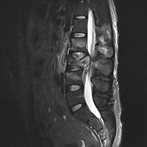
Figure 1: RMI-It shows the vertebral fracture with the occupation of the Lumbar Spine.
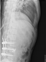
Figure 2: X-rays The vertebral fixation is shown with minimally invasive surgery.
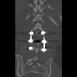
Figure 3: CT Scan: Perfect alignment of the lumbar spine.
3. Discussion
We agree with Cain, [13] in which confirmation of the diagnosis of these fractures is made with simple radiographs in A-P and lateral views, CT and nuclear magnetic resonance make it possible to specify details of this injury and include the degree of compression of the nerve structures. Thoracolumbar spinal fractures are clinically characterized by presenting a history of trauma, usually due to falls from heights, although occasionally they occur due to automobile accidents also of high intensity [13, 14]. The falls of fruit trees are observed very often, because our economy is mainly agricultural. Many authors state that patients go to the body of the guard, usually on stretchers, physical examination shows the ecchymosis and increased volume at the level of the injured vertebrae, spontaneous pain that is exacerbated by the movements and palpation, exquisite pain in the processes of said vertebrae may or may not radiate to one or both lower limbs [13]. Many authors report that when these fractures cause neurological damage by compression of the nervous structures they contain, they are added to the clinical picture. The most severe neurological defect is called paraplegia (grade A of the ASIA scale) [14, 15].
As documented in the literature reviewed, men [16] continue to contribute the most cases, which is due to the fact that they are more exposed to accidents due to the tasks they usually perform. In terms of age, the average figures are quite similar and are given to be the most active age of life [17, 18]. If urgent measures are not taken with these patients, that is to say, if the diagnosis is not made early and the vertebral fracture is unstable, it may reach a neurological injury due to a sudden movement. If there is, from the beginning, the diagnosis of the neurological lesion, the treatment must be intensive and early from the medical point of view as surgical, since the longer the nerve structures are compressed, the greater the tissue ischemia and the more irreversible the neurological damage [19, 20].
4. Conclusion
Surgeons defend the idea that in all unstable thoracolumbar spine injuries, although there are no neurological injuries, surgical treatment should be performed as soon as possible to avoid the complications of bedding and thus reduce their morbidity and mortality.
References
- Floman Y. Toracolumbar spine fractures. Philadelphia: Lippincott (1993).
- Errico BJ, Bauer RD, Waugh TR. Spinal trauma. Philadelphia: Lippincott (1991).
- Sansana ES, Mansilla MB. Fracturas del raquis toracolumbar tratadas mediante instrumentación pedicular. Rev Mapfre Med 10 (1999): 261-270.
- Benson DR, Burkus JK, Montesano PX. Unstable toracolumbar and lumbar burst fractures treated with the AO fixateaur interne. J Spinal Disord 5 (1999): 535-561.
- Baransky M, Kroczak S. Fractures of thoracic lumbar spine treatment and follow up. Ann Transplant 4 (2000): 3-4.
- Van Beek EJ. Upper thoracic spinal fractures. Injury 31 (2000): 219-223.
- Defino HL, Rodríguez-Fuente AE. Treatment of fractures of the thoracolumbar spine by combined anteroposterior fixation using the harms methods. Ear spine 7 (1998): 187-194.
- Ruan DK, Shen GB, Chui HX. Shen instrumentation for management of unstable thoracolumbarspinal fractures. Spine 23 (1999): 1324-1332.
- Watanabe T, Vaccaro AR, Welch WC. High inidence of occult nurogenic bladder dysfunction in neurologically intact patient with thoracolumbar spinal injuries. J Urol 159 (1999): 965-968.
- Rinsky L. Instrumentation of the lumbar spine. An overview. Clin Orthop 203 (2000): 209-218.
- Herdonm WA, Sullivan JA, Gross RH. Segmental spinal instrumentation with sublaminal wires. J Bone Joint Surg 69 (2000): 851-859.
- Bradford DS, Cook J. Result of spinal arthrodesis with pediele serew fixation. J Bone Joint Surg 73 (2001): 1179-1184.
- Cain JE, Dejong JT, Dinenberg AS. Pathomechanical analysis of thoracolumbar burst fractures reduction. Spine 18 (1999): 192-201.
- Garfin S, Nowery CJ. Confirmation of the posterolateral technique descompress and the thoracolumbar spine burst fractures. Spine 10 (2000): 218-226.
- Reid DC, Henderson LS, Miller JD. Etiology and clinical course of spinal fractures. J Trauma 27 (1999): 980-986.
- Sasso RC, Reuben JD. Posterior fixation of thoracolumbar spine fractures using pedicule serew. Spine 16 (2000): 134-142.
- Lindsey RW, Dick W. The fixation interne in the reduction and stabilization of thoracolumbar fractures. Clin Orthop 17 (2000): 161-166.
- Vacarro AR, Blan OG. Adult spine trauma. AM Acad Orth Surg 4 (2002): 593-608.
- Sandén B, Olerud C, Larson S. Hydroxyapatite Coating Improves Fixation of Pedicle Screws. J Bone Joint Surg 84 (2002): 387-391.
- Krepler P, Windhager R, Breschneider W, et al. Total vertebrectomy for primary malignant tumours of the spine. J Bone Joint Surg 84 (2002): 712-715.