Marianne J Middelveen1, Melissa C Fesler2, Raphael B Stricker2
1Atkins Veterinary Services, Calgary, AB, Canada.
2Union Square Medical Associates, San Francisco, CA, USA.
*Corresponding author: Raphael B Stricker, Union Square Medical Associates, San Francisco, CA, USA.
Received: 19 July 2026; Accepted: 24 July 2026; Published: 31 July 2026
Morgellons disease is a skin condition marked by lesions containing unusual fibers or filaments that emerge from or are embedded within the skin. Morgellons disease is generally regarded as a delusional disorder with fibers attributed to textile contamination introduced by compulsive scratching. However, a growing number of researchers propose that these fibers are produced by human cells in response to infection. This review traces the evolution of Morgellons fiber research, from early skepticism – due to misidentification and superficial collection of fibers – to evidence supporting their biological origin. We also present previously unpublished data and observations demonstrating the cellular origin of these fibers. Rigorous chemical, molecular, microscopic, and spectroscopic analyses confirm that Morgellons fibers are primarily composed of keratin and collagen, produced by keratinocytes and fibroblasts, and sometimes manifest as small human hairs. Unique properties include autofluorescence under ultraviolet light and abnormal eumelanin and pheomelanin pigmentation. Recent studies have detected pathogenic bacterial antigens and amyloid components within the fibers, suggesting a complex interplay between infection and filament formation. Accumulating evidence underscores the need for rigorous methodology and open-minded inquiry to examine the human cellular nature of Morgellons fibers and to address unresolved questions about pigmentation, autofluorescence and microbial involvement.
Morgellons disease; Fibers; Lyme disease; Borrelia burgdorferi; Spirochetes; Dermopathy; Borrelial dermatitis
Morgellons disease articles; Fibers articles; Lyme disease articles; Borrelia burgdorferi articles; Spirochetes articles; Dermopathy articles; Borrelial dermatitis articles.
Introduction
Morgellons disease is a dermatologic condition associated with fibers or filaments embedded in or protruding from the skin. Multiple peer reviewed studies demonstrate that these fibers originate from the deeper epidermal layers and are generally composed of normal human proteins such as keratin or collagen [1,2]. Morgellons disease has sparked controversy due to the bizarre nature and poorly understood etiology of these filaments.
Historically medical practitioners have considered Morgellons fibers or filaments to be textile in origin as they may exhibit brilliant coloration that is uncharacteristic of human skin [1,2]. This vivid appearance often contributes to the misperception that the filaments are derived from textiles rather than being cutaneous in origin. Determining the composition of the characteristic fibers seen in Morgellons disease patients has proved challenging for researchers and clinicians due to lack of standard diagnostic protocols and guidelines. The fibers may be microscopic - some as small in diameter as a red blood cell - and may be embedded deeply within the skin, making isolation and detection difficult. Differentiating Morgellons fibers from textile fibers through simple visualization techniques remains challenging due to the similarities in superficial appearance and microscopic nature of some fibers. To ensure cutaneous origin, only fibers that are embedded in or protruding from the skin or dermis should be collected for analysis.
This paper will review the complex history of Morgellons disease fibers and provide evidence for their cellular origin. The information presented will provide a framework for researchers and clinicians to translate the evidence into best practice for patients affected by this disease.
A Chronological Analysis
Morgellons Fiber Study, Charles E. Holman Foundation, 2004
About four years after Mary Leitao laid eyes on the first documented Morgellons fibers, clinical microbiologist Jenny Haverty conducted a small-scale study in 2004. Fiber samples were collected from four subjects [3]. The specimens from subject 1 and subject 4 were superficially collected and therefore largely composed of environmental contaminants. Notably, some small fibers exhibited ladder-like internal structures consistent with feline down hairs. The fibers from subject 2 were embedded within callus material, which were therefore reliably Morgellons fibers. (C. Casey, personal communication, 2025) Fibers from subject 3 were removed from lesions and were therefore likely to have been Morgellons fibers. The specimens were examined using both 400x light microscopy and fluorescence microscopy employing a 330-380 µm excitation filter and a 420 µm barrier filter (Figures 1-4).
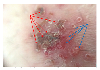
Figure 1: Blue fibers embedded inside intact calluses (red arrows). Note some calluses have separated and peeled off the skin (blue arrows). 40x magnification.
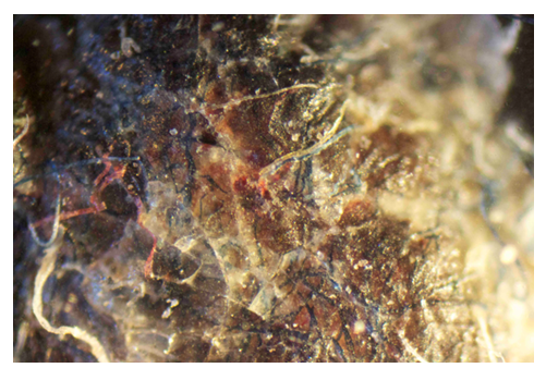
Figure 2: Blue, red and white fibers embedded within a callus. 100x magnification.
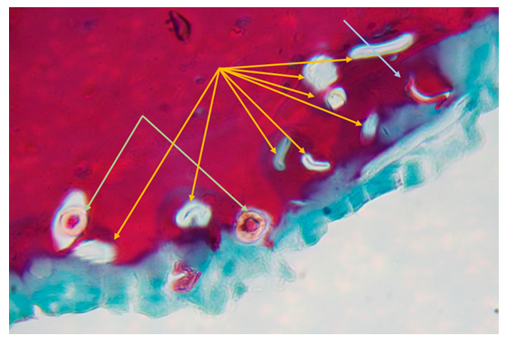
Figure 3: Morgellons callus section showing embedded sectioned fibers; both keratin positive (stained red) and collagen positive (stained green). 1000x magnification. Note morphological features exhibited by two keratin fibers showing amorphous medullas surrounded by cortices (light green arrows), and solid cortices surrounding hollow medullas exhibited by all the collagen fibers (gold arrows), and a single keratin fiber (blue arrow).
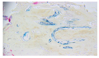
Figure 4A: Unstained Morgellons callus section with embedded blue fibers. 400x magnification.
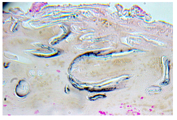
Figure 4B: Consecutive Morgellons callus section with fibers staining positively with Fontana Masson stain. 400x magnification.
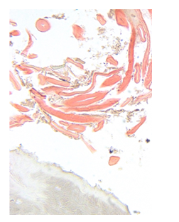
Figure 4C: Morgellons callus section showing red fibers partially embedded in a callus section with negligible Fontana Masson staining. 400x magnification.
Key findings from this study revealed that fiber diameters ranged from 7.68 µm to 33.28 µm, with clear fibers representing both the smallest and largest measurements. Blue fibers ranged from 15.36 µm to 30.72 µm, red fibers measured from 12.8 µm to 17.92 µm, and black fibers varied from 12.8 µm to 30.72 µm. For perspective, human hairs typically range from the smallest at 17 µm to 181 µm in diameter, while a human red blood cell is about 8.2 µm. The fibers were described as having tubular to ribbon-like profiles. (Figures 1-4). They did not stain positively with Calcofluor-white stain, indicating they were not composed of either cellulose or chitin. Both the white and blue fibers exhibited aqua colored autofluorescence under ultraviolet (UV) light [3]. It is noteworthy that, contrary to the findings of Haverty’s study, our observations are that red Morgellons fibers do exhibit autofluorescence when examined under UV microscopy. (Middelveen, unpublished observation, 2013) The ability to detect fluorescence depends on the specific light sources and filter sets used in fluorescent microscopes, as these instruments emit different wavelengths of UV and visible light to excite specific fluorophores. Since fluorescence requires the excitation light to match the absorption spectrum of the fluorophore, certain specimens may not fluoresce if the microscope’s light emission and filters are not appropriately matched.
To summarize the key findings:
- Some Morgellons fibers display distinctive autofluorescence.
- Chemical analysis confirmed that Morgellons fibers lack cellulose.
- Not every fiber found on the skin surface of a Morgellons patient is relevant to the disease.
- Meticulous specimen collection is critical to ensure accurate identification and analysis of Morgellons fibers.
Savely et al., 2006
In 2006, Savely et al. proposed a connection between Lyme disease and patients with Morgellons disease [4]. Systemic symptomatology was similar between the two patient groups and patients with Morgellons oftentimes had positive Western blot testing for Borrelia burgdorferi, the causative agent of Lyme disease. The authors noted that antibiotic treatment of Morgellons patients was associated with remission or resolution of their symptoms, including decreased fiber production [4].
Northern Arizona University Study, 2006
In 2006, Robert Smith, a master’s degree chemistry graduate student at Northern Arizona University, conducted a groundbreaking preliminary study on behalf of the Charles E Holman Morgellons Disease Foundation that, despite its significance, was never published. (R. Smith, email 2025) Although the research was limited to fibers collected from a single Morgellons subject, its importance cannot be overstated: it was the first to conclusively demonstrate that Morgellons fibers were of human origin and primarily composed of cytokeratin. The study also confirmed the distinctive fluorescence exhibited by blue or white Morgellons fibers and identified the presence of an unknown protein. It is noteworthy that the Morgellons fibers analysed in this study were of unknown color as the principal researcher indicated he was color blind, but he believed that they were either white or blue.
Morgellons fibers and hairs (for comparison purposes) from the same patient were first examined under white light, then subjected to UV illumination using a filter with 365 nm excitation (UV-A). The fibers exhibited fluorescence, whereas the hairs did not. For further analysis, the fibers were placed in individual scintillation vials and observed with a detector. When excited at both 305 nm and 365 nm, the fibers demonstrated fluorescence under both 460 nm and 557 nm filters. This fluorescence was transient, fading as soon as the illumination ceased. To characterize the fluorescence, a fluorometer measured the excitation and emission spectra of the Morgellons fibers, revealing a distinct fluorescence pattern - its unique spectral fingerprint. (R. Smith, email 2025)
A single fluorescent Morgellons fiber was subsequently analysed without further modification or treatment using scanning electron microscopy (SEM) at the University of Northern Arizona’s Imaging and Histology Core FacilitySEM imaging revealed that the fiber displayed cuticular scaling along the shaft and a root terminus, both morphological features of human hairs. The fiber lacked a smooth surface, indicating it was not coated with a protein monolayer. However, the Morgellons fiber exhibited fluorescence while normal human hairs were not fluorescent. (R. Smith, email 2025)
Interestingly, Morgellons fibers resisted dissolution in separate solutions of 6 M guanidine HCl, 6 M urea, and TRIzol reagent, which are potent denaturing agents used to disrupt cellular components and unfold proteins and used for protein isolation. The fibers were therefore ground using a mortar and pestle. The resulting material was suspended in a buffered solution and subjected to electrophoresis, separating the proteins based on their molecular weight. This process revealed two distinct protein bands: one at approximately 60 kDa and another at 30 kDa. Both protein bands were excised from the gel and sent to the University of Arizona Proteomics Laboratory for further characterization. The proteins were enzymatically digested with trypsin, and the resulting peptide fragments were analysed using nano-High Performance Liquid Chromatography/Mass Spectrometry (nano-HPLC/MS). Proteins were identified by comparing the mass-to-charge ratios of their fragments to known sequences in protein databases. The predominant 60 kDa protein shared sequence coverage with and was therefore identified as human serum albumin and human cytoskeletal keratin II. Given that SEM confirmed that fiber morphology was consistent with human hair, human cytoskeletal keratin II was definitely present. In contrast, the 30 kDa protein did not match any known human proteins in the databases. It is important to note that this protein exhibited the same unique fluorescent spectral fingerprint as the Morgellons fibers, strongly suggesting it was responsible for their autofluorescence. The identity of the 30 kDa protein remains unresolved. (R. Smith, email 2025) Further research is warranted, including repeating these analyses with fibers from additional Morgellons subjects and isolating and characterizing the 30 kDa protein to better understand its properties and role.
Smith also mentioned that the University of Arizona facility could not purify the protein from the buffer solution using conventional methods, as a black tar-like oil precipitated under those conditions. Smith hypothesized that other molecules might have been present in the buffered protein mixture, causing this phenomenon. (R. Smith, email 2025) The black oily precipitate is of particular interest because some Morgellons sufferers report that similar substances ooze from their lesions [2,4].
Oklahoma State University, 2006
In 2006, Dr. Randy Wymore, a pharmacology professor at Oklahoma State University (OSU), submitted fibers he identified as Morgellons fibers to the Tulsa Police Department forensic laboratory for analysis. Under the supervision of Mark Boese, the fibers were compared against the FBI’s extensive database of known carpet and clothing fibers. The forensic team determined that the submitted fibers did not match any known man-made or plant fibers in the database, leading them to hypothesize that these fibers might be a “byproduct of a biological organism”[5].
Stricker et al. 2007
In 2007, San Francisco internist Dr. Raphael Stricker and colleagues investigated the possibility that Morgellons disease might be linked to Agrobacterium spp [6], which are plant pathogens known to cause disease by transferring tumor-inducing or root-inducing DNA into plant hosts causing abnormal cell growth [7]. This hypothesis was investigated largely because identifiable pathogens had yet to be detected in Morgellons tissue, and skin biopsies from Morgellons patients showed only non-specific pathological changes or signs of inflammation. Therefore, clinicians believed the unidentified fibrous material protruding from epidermal tissue was composed of cellulose [6]. This study did not include fiber characterization, and therefore a cellulose component was not identified in Morgellons fibers.
Morgellons Disease Scientific/Medical Conference, 2011
At the 4th annual Morgellons Disease Scientific/Medical Conference in 2011, Wymore presented research that focused on the characterization of loose fibers collected from Morgellons patients [8]. He aimed to advance understanding of the disease by submitting fiber samples to commercial laboratories for compositional analysis. Most materials were identified as environmental contaminants, including synthetic textile fibers, cellulose (likely from cotton), fungal filaments, bird down, rodent hairs, and a blond human hair. Among the samples, one fiber was noteworthy for being encased in a waxy substance, which was identified as sapienic acid - a fatty acid present in human sebum, thus establishing human origin. However, further analysis of this fiber was not pursued. Wymore mentioned the cellulose composition of some fiber samples to be intriguing and worthy of further investigation.
Morgellons Disease Scientific/Medical Conference, 2012
At the 5th annual Morgellons Disease Scientific/Medical Conference in 2012, Wymore presented an update on his six-year investigation of Morgellons fibers - largely investigated by electron microscopy and spectroscopy [9]. Of the specimens analysed, 97% were conclusively identified: 31% were cellulose-based fibers (e.g., cotton or rayon), 44% were synthetic materials (such as polyester, dacron and nylon), and 22% were human or animal hair. The remaining 3% were fibers of indeterminate origin, which Wymore designated as “mystery fibers”. Subsequent analysis revealed that several of these fibers were environmental contaminants, specifically two blue-dyed fibers, a red nylon fiber, and a sponge polyester. The true “mystery fibers”, he explained, exhibited resistance to a range of chemical agents, including 6 N hydrochloric acid, 65% nitric acid, bleach, ammonia, guanidine isothiocyanate, and 2-mercaptoethanol. Despite their interesting chemical resilience, fiber origin was not established, and no evidence was provided to support a human origin, therefore an association with Morgellons disease pathology was not established. Given that most fibers collected by Wymore were identifiable environmental contaminants, there is little reason to believe that the “mystery fibers” were not also environmental in origin, as they were collected using the same methodologyWymore acknowledged that, while some fibers could not be classified as textile fibers, this limitation did not exclude the possibility that they were synthetic. Furthermore, Wymore stated that a fiber extracted from beneath unbroken skin was conclusively identified as synthetic nylon. He put forward that an unknown mechanism of synthetic material deposition into intact skin might play a role in Morgellons disease pathology. Wymore concluded his talk by saying it was not worthwhile studying fibers further and that he was moving on to the search for pathogens. Unfortunately, he failed to study any fibers identified as hairs, and in retrospect these may have been more relevant than the “mystery fibers”. Had he carefully collected only fibers of known human origin the outcome of his fiber studies might have been different.
During this presentation, Wymore clarified that the so-called “mystery fibers” - those that were resistant to heat and chemical dissolution - were the specimens submitted for forensic analysis at the Tulsa Police Department laboratory. In addition, Wymore acknowledged a significant limitation: the analytical procedures used to convince himself that the fibers were not textiles inadvertently altered them, making them unsuitable for further investigation [9]. It remains uncertain if these fibers were chemically or physically modified prior to their analysis at the Tulsa Crime Lab. Such modifications could have made the fibers difficult to compare with those catalogued in the FBI’s fiber database. Additionally, the comprehensiveness of the FBI database itself is unclear, and it is not known whether all relevant fiber types were included for comparison. Certain synthetic fibers - such as Teflon and the aramids Kevlar and Nomex - are renowned for exceptional chemical and heat resistance yet are not commonly found in household carpets or clothing, and therefore may not have been present in the database. These factors collectively cast doubt on the validity of the Tulsa Crime Lab study, as contamination, methodological limitations and database scope may have influenced the findings.
Unfortunately, the most scientifically valuable specimens for analysis, fibers and materials of confirmed human origin, were not the focus of Wymore’s investigation. Apparently, most fibers identified in his studies were textile or synthetic, and this was likely due to the institutional restrictions at OSU prohibiting the study of skin tissue. (C. Casey, personal communication 2026). As a result, study fibers were limited to those easily extracted using forceps or similar tools, increasing the likelihood of environmental contamination and preventing the analysis of fibers embedded in skin verifiably associated with Morgellons disease. (C. Casey, personal communication 2026).
Although Wymore consistently maintained that Morgellons disease was not a delusional disorder, the findings from OSU's early studies did not challenge the prevailing psychogenic perspective. The assertion that 97% of examined fibers were identified as common textiles or synthetic materials inadvertently lent support to the theory of contamination or self-implantation, reinforcing skepticism within the medical community and creating doubt about subsequent research supporting the human origin of Morgellons fibers. Of note, a recent 2025 poster presentation by Rice and Wymore has acknowledged the keratin and collagen composition of Morgellons fibers, and this study may help to dispel misconceptions and foster greater acceptance of a somatic origin of Morgellons disease among medical professionals [10].
Middelveen & Stricker, 2011
In 2011, Middelveen and Stricker published a comparative analysis revealing striking similarities between Morgellons disease and bovine digital dermatitis (BDD) [11]. Unusual filament formation was characteristic of both diseases, and both were associated with spirochetal infection. In BDD, the underlying pathology was primarily attributed to members of the genus Treponema, often accompanied by other bacterial co-infections. Likewise, in Morgellons disease, diagnoses of Lyme disease and detection of its etiologic agent, Borrelia burgdorferi suggested a spirochetal etiology, with possible involvement of additional tick-borne pathogens. Drawing on these parallels, the researchers hypothesized that Morgellons disease might share a similar pathology with BDD. Because proliferative keratinocytes in BDD lesions produced abnormal keratin fibers and Morgellons disease similarly exhibited unusual fiber formation within skin tissues where keratinocytes predominate, Middelveen and Stricker therefore proposed that keratin was a likely constituent of Morgellons fibers.
Pearson et al., 2012
In 2012, six years after first announcing their intention to investigate Morgellons disease, the Centers for Disease Control and Prevention (CDC) in Atlanta published their landmark study on Morgellons disease entitled “Clinical, Epidemiologic, Histopathologic, and Molecular Features of an Unexplained Dermopathy” [12]. The study concluded that “no common underlying medical condition or infectious source was identified” and stated that Morgellons disease was “similar to more commonly recognised conditions such as delusional infestation.” The CDC researchers examined fibers and “other materials” obtained from two sources: 4 mm punch biopsies, and specimens collected from the surface of participants’ skin. In total, 31 participants either had lesions suitable for biopsy or had material present on their skin for collection. However, the report does not clarify how many subjects underwent biopsy versus how many had only surface material collected, leaving the exact breakdown ambiguous. Deficiencies regarding fiber collection and analysis are described in detail below.
Biopsies were obtained from both normal and abnormal skin, with “abnormal” defined as areas that were either visibly altered or that exhibited unusual sensations - meaning that overt lesions were not required for classification, and even visually normal skin could be categorized as abnormal. The collected biopsies were sectioned for histological examination, focusing on birefringent materials, assessed by polarized light microscopy [12], and characteristic of many substances, including human-derived materials such as collagen, keratin, amyloid proteins, and muscle fibers, as well as non-human materials like cellulose [13,14].
Birefringent material was detected in 16 biopsied skin lesions, predominantly within the superficial scale-crust at the periphery or separate from the main tissue [12]. Fibers deeply embedded within skin were not observed, suggesting that the detected birefringent artefacts were superficial environmental contaminants rather than intrinsic to the skin itself. The study did not specify the morphological features of these materials, leaving it unclear whether they were fibers, amorphous clumps, particulates, or other forms. In only two cases did biopsies reveal birefringent material in deeper tissue - specifically, within foreign-body-type giant cells. Tissue sections containing unidentified birefringent material underwent further analysis using scanning electron microscopy with energy dispersive X-ray analysis (SEM/EDXA) to determine elemental composition, and infrared spectroscopy (IRS) to assess molecular characteristics. Most analysed materials exhibited spectral features consistent with cellulose, suggestive of cotton. However, the composition of all observed materials was not fully clarified, leaving open the possibility that some could be collagen, keratin or other human-derived substances. In addition, by restricting the analysis to birefringent fibers and materials, the study excluded structures of human origin that were not birefringent from consideration. Of the two giant cells identified, one contained material spectroscopically consistent with cotton, while the other contained silicon-based material. An important consideration is the likelihood of procedural contamination during specimen collection. As the skin was biopsied, it was likely swabbed with alcohol or another disinfectant, thereby introducing surface contamination by inadvertently transferring cotton fibers onto the skin. In other words, cellulose fibers may have been implanted by physicians during the procedure rather than by subjects via self-implantation.
Among the non-biopsy specimens, a total of 23 samples - described as either fibers or “other material” - were collected from 12 subjects [12]. The authors stated that these specimens were obtained from intact skin sites, meaning they were taken directly from the skin surface rather than from lesions or deeper skin tissues. In addition, the report does not specify how many of these samples were actual fibers versus other types of material, nor does it clarify what constituted the “other material”. Morphological descriptions of collected materials were not provided, leaving the nature of these specimens unknown. The results presented are unclear and ambiguous. The authors state, “The materials were largely composed of protein (83%), likely superficial skin or cellulose consistent with cotton fibers (43%).” This phrasing does not make grammatical or mathematical sense, and it is not evident how these percentages relate to the total number of specimens or their composition. The authors also reported that three samples contained polyamide (likely nylon), cellulose nitrate with bismuth (possibly originating from nail polish), and polyethylene. Importantly, they acknowledged that the polyethylene was consistent with contamination from the specimen container lid. This admission highlights that fact that environmental contaminants introduced during the biopsy procedure might have been some of the samples analysed, underscoring the importance of rigorous specimen handling and interpretation.
In summary, there were significant flaws in the CDC analysis of fibers and foreign material:
- The study focused on foreign material, introducing a bias against human biofibers.
- The number of specimens collected and analysed was not clearly reported, making it difficult to assess the scope and reliability of the findings.
- Only birefringent materials were analysed for composition, meaning that relevant non-birefringent materials were excluded from study.
- The superficial nature of the materials collected limited analysis to environmental contaminants, and the specimen collection protocol itself probably contributed to contamination.
- Crucially, no fibers deeply embedded within the skin were studied or observed, indicating that authentic Morgellons fibers were not present in the samples analysed.
Middelveen et al., 2012
In 2012, a later study by Middelveen et al advanced an investigation into the possible keratin composition of Morgellons fibers with a chemical and light microscopic comparison of materials collected from three affected individuals, alongside samples of normal human hair and bovine digital dermatitis (BDD) fibers [15]. The specimens varied: samples from subject 1 included white fibers embedded in scabs (calluses) from hand lesions examined; samples from subject 2 consisted exclusively of white, red, purple and blue fiber-embedded calluses from skin lesions; and specimens from subject 3 were hairs with follicular bulbs and attached filaments [15].
Key findings included the observation that fibers were either firmly attached to or deeply embedded within epithelial tissue. Notably, both white fibers (from subjects 1, 2, and 3) and blue fibers (from subject 2) exhibited fluorescence under specific lighting conditions, and some fibers from subject 1 had bulges at their ends resembling follicular bulbs. Filament formation was associated with both follicular bulbs (subject 3) and other cutaneous tissues (subjects 1 and 2). In specimens from subject 3, follicles demonstrated root-like filamentous growth from the follicular sheath and hyaline filaments extending from the sheath; numerous hair bulbs showed deformities, such as the formation of multiple hairs from a single follicle (pili multigemini), a phenomenon often linked to folliculitis [15]. Interestingly, folliculitis was observed in one participant in the CDC study [12]. Additionally we have since noted that Morgellons lesions often start as folliculitis later evolving into ulcerative filamentous lesions [2].
To assess chemical resistance, filaments deeply embedded within calluses (subjects 1 and 2) were exposed to solutions of 12% sodium hypochlorite (NaOCl), 10% sodium hydroxide (NaOH), and 10% potassium hydroxide (KOH), with observations made at intervals of 1, 10, 60, and 120 minutes. For comparison, BDD fibers and human hair were tested under the same conditions. In 12% NaOCl, Morgellons fibers showed complete dissolution within 60 minutes, whereas BDD fibers and human hair exhibited only partial dissolution even after 120 minutes. In 10% NaOH, Morgellons fibers, human hair, and BDD fibers all showed partial dissolution, but BDD fibers were more resistant, with no dissolution at one minute. In 10% KOH, all specimens demonstrated only partial dissolution after 120 minutes, with Morgellons fibers showing partial dissolution at 10 minutes and BDD fibers only after 120 minutes [15].
Additionally, callus material (subjects 1 and 2) was sectioned for histological analysis and stained with anti-pankeratin (AE1/AE3) and anti-cytokeratin (5/6) immunostains. The results revealed that filamentous strands were strongly positive for pankeratin, supporting the presence of keratin-based structures in these fibers. Additionally, the authors hypothesized that, due to the vivid hues, the blue and red coloration observed in many Morgellons fibers might result from structural coloration rather than from pigmentation,[15] which later proved not to be the case.
Two of the authors, Middelveen and Stricker, previously submitted callus samples containing embedded, vividly blue fibers to Dr. Matthew Shawkey and colleagues at the University of Akron. The findings, referenced in the above 2012 published paper revealed through scanning electron microscopy (SEM) and transmission electron microscopy (TEM) that these blue fibers exhibited cuticular scaling and morphological features characteristic of human hair.[15] This provided compelling evidence that at least some blue Morgellons fibers were in fact small human hairs, corroborating the earlier observations by Robert Smith confirming human origin and keratin composition of some Morgellons fibers - and reminiscent of the historical reference to “harsh hairs” in children described by Sir Thomas Browne in 1690 [16].
While the 2012 study by Middelveen et al. presented compelling evidence that Morgellons filaments originated from human epithelial cells and were composed of keratin and produced by keratinocytes, there were limitations in that early research: samples from subject 1 may have contained environmental contamination, as some material collected was loose and potentially exposed to external sources; and the immunohistochemical staining did not reveal the distinct morphological shapes of sectioned embedded fibers seen in cross, oblique, and longitudinal sections revealed in later studies. [Middelveen, unpublished observation, 2016] It is also worth noting that the publication omitted mentioning an important observation made at that time: Morgellons fibers are not fire resistant and combust in a manner comparable to human hair. [Middelveen, unpublished observation, 2016] Collectively, the observations contradict previous claims by Wymore that Morgellons fibers do not degrade in any solvents or detergents, and that they do not burn at 1,600 degrees Fahrenheit [17]. Another unpublished and omitted observation is that contrary to the findings reported by the Haverty study in 2004, red Morgellons fibers do exhibit autofluorescence when examined under some UV microscopes. The intensity and visibility of fluorescent materials depend on interaction between activators and specific wavelengths. Therefore, red Morgellons fibers may glow brightly under some UV wavelengths but not others, making autofluorescence dependent on a microscope’s light source and filter settings.
Middelveen et al., 2013
In 2013, Middelveen et al reported significant advancements in understanding the composition and development of Morgellons fibers [18]. This study employed light microscopy, histological analysis, and electron microscopy showing that cutaneous Morgellons filaments consist of both keratin and collagen, arising from the proliferation and activation of keratinocytes and fibroblasts. This study also reported the visual detection of spirochetes in Morgellons cutaneous tissue. To minimize the risk of environmental contamination, the study focused exclusively on analysis of fibers firmly attached to or deeply embedded within thickened calluses that had separated from the deeper layers of skin. Material unattached to skin was deliberately excluded.
Specimens were examined to assess their gross morphological characteristics [18]. Dermatological tissue from all subjects exhibited hyperplasia and parakeratotic hyperkeratosis, with pronounced keratin staining in the thickened stratum corneum. For subject 1, calluses collected from hand lesions were densely covered in white or hyaline filaments, which were attached to and projected from one side of the cutaneous tissue. Calluses from subjects 2 and 3, originated from corporeal lesions, displaying a distinct convex external surface (facing outward) and a concave internal surface (oriented down toward the dermis). On the convex surface, colored (blue or red) and white filaments measuring 10 - 40 μm in diameter protruded, extending outward. Some fibers were embedded within and distributed throughout the callus. The concave underside of some of these calluses revealed protruding keratin projections, possibly representing follicular casts. These projections varied in shape, with some exhibiting sharp tips and others appearing blunt or ballooned. Occasionally, clear, ingrown hairs or hair-like structures approximately 60 μm in diameter were observed protruding from the tips of these keratin projections. In contrast, subject 4 presented only small lesions without substantial callus formation, yielding a single fragment of dermatological tissue about 1 mm in diameter embedded with filaments.
Microscopic examination revealed that filaments viewed in longitudinal, oblique, or cross section presented as elongated, elliptical, bean-shaped, curved or rounded inclusions in or projecting from the skin, with the distinctive morphological features of a cortex and medulla. Immunohistochemical analysis using CK AE1/AE3 pan-keratin staining confirmed the presence of keratin within these filaments; however, the staining was often patchy and irregular, suggesting that Morgellons fibers are composed not only of keratin but also of another human filamentous protein, which we hypothesized to be collagen. To further explore that possibility, Gömöri trichrome staining was employed, which distinguishes keratin (red) from collagen (green). Most filaments stained positively for collagen, with keratin staining occurring less often and typically in irregular or patchy distributions. Collagen-rich fibers were readily identified as inclusions embedded within keratin-rich tissue, often adjacent to collagen-rich, green-stained dermis. Keratin-positive sectioned filaments rarely appeared in isolation, underscoring the mixed nature of Morgellons filamentous inclusions. Additionally, some sections exhibited blue-stained nuclei (via hematoxylin staining), particularly at the base of filament attachment, and those cells were continuous with the surrounding epidermal tissue, thus establishing human origin [18]. Although keratin fibers such as wool are used in textiles, collagen textile fibers are rare, are blended with cellulose, and have been only recently introduced [19]. The previous studies [2,3] demonstrated that Morgellons fibers do not contain cellulose, making it straightforward to distinguish textile collagen fibers from those found in Morgellons disease. It is therefore impossible that collagen Morgellons fibers are textiles.
Histological sectioning and transmission electron microscopy both demonstrated that most Morgellons disease filaments possess a distinct medulla encased by a cortex, characteristic of hair-like structures [18]. In histological sections, although most fibers demonstrated a hollow medulla, some keratin-rich fibers exhibited an amorphous medulla consistent with the SEM observations made by Shawkey and colleagues. [Middelveen, unpublished observation, 2016] These findings align with previous research, which identified hair-like morphology, a close association with hair follicles, and the presence of follicular-like bulbs in these filaments. The observation that some fibers not only resemble hair morphologically but are also composed of keratin supports earlier conclusions by Robert Smith and Shawkey and colleagues, confirming that at least some Morgellons fibers are, in fact, small human hairs.
Previously, structural coloration was proposed as the explanation for the vivid red and blue hues observed in Morgellons fibers. However, given that bluish discoloration of the skin has been documented in patients with pinta—a disease caused by the spirochete Treponema carateum [20]- the possibility of melanin involvement was also considered. To investigate this, the researchers employed Fontana-Masson (FM) staining on fiber-embedded callus sections, which stains argentaffin and melanin granules a blackish color. In patient 1 whose calluses had hyaline filaments, melanin staining was negative as expected. Patient 2 presented calluses with blue, red and white filaments, and blue filaments were positive for melanin, while red filaments were negative. Patient 3 had calluses containing white and blue filaments; once again, blue filaments were positive for melanin, whereas white filaments were negative [18]. Human skin contains two primary types of melanin: eumelanin, which imparts brown or black pigmentation, and pheomelanin, responsible for red or yellow hues. FM staining is primarily used to detect eumelanin. While FM can also stain pheomelanin, it does so less intensely, producing a weak yellowish rather than black color. As a result, FM pheomelanin staining can be easily missed [21]. It follows that red Morgellons fibers could be colored by pheomelanin pigmentation not detected in this study, warranting further investigation.
Shawkey and colleagues, University of Akron, 2014
In 2014, Shawkey provided an update concerning research of Morgellons fibers conducted by his laboratory at the University of Akron, (Shawkey, email 2014). Some of these findings were later included in the 2016 publication by Middelveen and Stricker [1], but here we present the full account, including previously unpublished data and insights. Four skin samples containing deeply embedded red, blue, or purple fibers were submitted for analysis by two of the authors, Middelveen and Stricker. Shawkey and his colleagues focused their study on blue fibers. Because these fibers were deeply embedded within the skin, Shawkey and colleagues concluded that they were cutaneous in origin and could not possibly be textile fibers. Transmission electron microscopy (TEM) and scanning electron microscopy (SEM) revealed that the morphology of blue fibers was consistent with human hair, exhibiting cuticular scaling, a cortex containing melanosomes, and an amorphous medulla—features typical of human hairs. SEM images revealed the presence of cuticles on the cortex confirming their identity as hairs, and fiber diameters ranging from 12 µm - 14 µm. Oblong cross sections seen in TEM images measured approximately 12 µm - 15 µm. TEM examination of fiber cross sections revealed the presence of disorganised melanosomes, a trait indicative of selective absorbance and characteristic of pigmentation rather than structural coloration [22]. The melanin deposited inside the melanosomes stained darkly, consistent with human hair [22]. Notably, the fibers did not display nanostructuring associated with structural coloration, indicating that blue coloration was solely due to pigmentation. While most melanosomes contained melanin deposits, some lacked these deposits perhaps due to ejection during TEM preparation. TEM cross sections showed cuticular scales in series of three to four, confirming the fibers’ identity as hairs. It is important to note the extremely small diameter of these hairs at 10 - 12 µm, lying somewhere between the width of human red blood cells (8.2 µm) and human monocytes (15 - 30 µm). Human hair diameter typically ranges from the smallest at 17 µm to the largest at 181 µm, with an average of about 75 µm [23].
As Morgellons blue filaments were so small, they could only be studied for reflectance by microspectroscopy, which revealed broad reflectance from 450 nm - 750 nm, with most light reflected in the spectra for violet and blue light from 380 nm - 470 nm. The broad reflectance band seen for the Morgellons fibers is characteristic of pigmentation rather than structural coloration [22,24]. In addition, the fibers demonstrated a dip in reflectance, a characteristic of selective absorbance of certain wavelengths of light by pigments, and consistent with patterns from pigmented tissues. The fibers were also studied by Raman spectroscopy; a technique used to identify and quantify substances by observing the interaction between light and chemical bonds. As shown by Raman spectroscopy, eumelanin and pheomelanin each normally have 3 major intensity peaks: eumelanin at 500 cm-1, 1380 cm-1 and 1580 cm-1, and pheomelanin at 500 cm-1, 1490 cm-1 and 2000 cm-1 [25]. Blue Morgellons fibers had only two major intensity peaks, one corresponding with eumelanin at 1580 cm-1 and one corresponding with pheomelanin at 1490 cm-1. Interestingly, the fibers also showed two additional intensity peaks, one at 1363 cm-1 representing a C-H bend (CH3 - a methyl group) and one at 1573 cm-1 representing a C-O stretch. The University of Akron’s research established the unmistakable presence of both pheomelanin and eumelanin within blue Morgellons fibers, however the absence of 2 spectral eumelanin peaks and two pheomelanin peaks suggests that their molecular structures are altered. The precise mechanism underlying their abnormal expression remains elusive, (Shawkey, email 2014).
Middelveen & Stricker, 2016
A 2016 study provided additional important insights into the composition of Morgellons fibers [1]. When sectioned embedded filaments were stained with Congo red, they exhibited apple-green birefringence under polarized light - a possible indicator of an amyloid component. In contrast, calcofluor-white staining produced negative results, confirming that these filaments are not composed of cellulose, as is typical of plant-based textile fibers like cotton or linen, nor do they contain chitin, which is found in fungal cells and insect exoskeletons. These findings directly contradict the conclusions of the 2012 CDC study, which had reported that Morgellons fibers were predominantly cotton cellulose textiles [1]. Birefringence can be exhibited by various biological substances beyond amyloid plaques. Collagen, muscle fibers, nerve bundles, and cellulose are all capable of demonstrating birefringence under polarized light microscopy [13,14]. In the context of Morgellons disease, research has shown that the fibers embedded within the skin possess a significant collagen component. Therefore, the presence of collagen may contribute to the observed birefringence in Morgellons fibers, as could an amyloid component [1]. In an attempt to elucidate coloration of Morgellons red fibers, histologically sectioned red fibers were also stained with Prussian red, ruling out red coloration by iron compounds. [Middelveen, unpublished observation, 2016]
Middelveen, 2018
A review article in 2018 [2] and a presentation by Middelveen [26] highlighted a significant challenge in the diagnosis and analysis of Morgellons disease: the frequent contamination of patient-supplied samples with extraneous artefacts. These contaminants - including pollen particles, non-infesting arthropods, feathers, and textile fibers - complicate the identification of authentic dermatological findings. In addition, many patients are confused by adhering contaminants, reporting the emergence of various materials from their skin, such as fuzzballs, hexagonal crystals, and glitter. Histological examination using Gömöri trichrome staining revealed that “fuzzballs” found in patient samples were predominantly composed of textile fibers that failed to stain for keratin or collagen. A minority of fibers in the fuzzballs did stain positively for keratin, indicating a biological origin. Analysis of hexagonal crystals by spectroscopy at Mount Allison University determined that these were man-made contaminants, consisting of cellulose or plastic cores with metallic coatings, and the “glitter” artefacts contained salts likely of human origin. Given the propensity for sticky lesions to attract and retain environmental debris, the authors emphasise the importance of rigorous specimen collection. Only fibers that are deeply embedded within skin tissue or firmly attached and clearly projecting from skin should be selected for the study and characterization of Morgellons fibers. In their experience, thickened calluses that separate and peel from the skin contain deeply embedded fibers, making them ideal candidates for reliable fiber analysis [2].
Middelveen et al., 2019
A 2019 study provided compelling evidence that Morgellons fibers contain antigens of at least two pathogenic bacteria - Borrelia burgdorferi and Helicobacter pylori - as demonstrated by positive immunohistochemical (IHC) staining of fibers with antibodies highly specific for those organisms [27]. In addition to these infectious agents, IHC analysis also revealed the presence of phospho-tau (p-tau) within Morgellons fibers. Given the well-established relationship between p-tau and amyloid-beta in amyloid pathology, this finding further supports the presence of an amyloid component in Morgellons fibers, as was previously suggested by the apple-green birefringence seen under polarized light following Congo red staining.
Middelveen et al., 2020
A 2020 study provided confirmatory immunohistochemical evidence confirming the presence of Borrelia burgdorferi proteins within Morgellons fibers [28]. Filaments exhibited strong positive staining for Borrelia burgdorferi antigens and, notably, these Borrelia-positive fibers demonstrated clear basal origin. Furthermore, cells at the base of fiber attachment stained intracellularly positive for Borrelia burgdorferi, reinforcing the association between Morgellons fibers and Borrelia infection.[28] Although the presence of bacterial antigens other than those of Borrelia spp. and H. pylori have not been detected in Morgellons fibers, other pathogens have been detected in Morgellons skin samples using PCR technology, including Helicobacter pylori, Treponema denticola, Bartonella henselae [2] and Rickettsia [29]. It is possible that some Morgellons fibers could contain antigens from these organisms as well.
Discussion
Early studies of Morgellons fibers were hampered by contamination from environmental fibers and methodological flaws, leading to skepticism in the medical community. The 2012 study by the CDC primarily analysed superficial environmental contaminants rather than authentic Morgellons fibers deeply embedded within the skin. Given the CDC’s strong reputation for scientific rigour, this publication has reinforced skepticism and made critics less willing to objectively consider emerging evidence and new research on Morgellons disease.
Morgellons fibers are often misidentified as textile contaminants due to their vivid colors, but careful collection and analysis demonstrate that they are cutaneous in origin. Research with improved specimen collection and analysis provides compelling evidence for the human origin and biological nature of Morgellons fibers. Reliable identification requires collecting fibers embedded in or protruding from the skin, rather than collecting loose, superficial specimens or those collected by patients. The embedded nature of the fibers that originate from epidermal cells excludes the hypothesis of self-implantation.
Scientific studies using microscopy, histology, and chemical analysis show that Morgellons fibers are primarily composed of keratin and collagen produced by keratinocytes and fibroblasts. Electron microscopy and immunohistochemical staining confirm that Morgellons fibers are of human cellular origin mostly arising from deeper layers of the epidermis and hair follicles, although some collagen fibers may arise in the dermis. Morgellons fibers are often continuous with surrounding skin cells, clearly showing their cellular origin. Some fibers have been confirmed to be small human hairs, with morphology consistent with human hairs.
Many Morgellons fibers exhibit strong autofluorescence under UV light, a property not seen in normal human hair. As naturally occurring blue pigments are virtually unknown in mammalian tissues, the presence of vividly colored blue filaments in Morgellons disease is scientifically intriguing. The blue coloration of fibers is due to melanin pigmentation, not synthetic dyes or structural coloration; however, the eumelanin and pheomelanin found in Morgellons fibers are altered and differ from those of normal human pigmentation. While the red coloration of Morgellons fibers is not understood, it may involve altered pigmentation by pheomelanin. The alteration of these pigments could be caused by cellular mechanisms such as infection or the resultant inflammatory processes. It is also possible that an unidentified pigment may contribute to the strange coloration. Immunohistochemical studies show that bacterial proteins (Borrelia spp. and Helicobacter spp.) are present within Morgellons filaments, and some bacteria are known to produce vivid blue and red pigments [30, 31, 32]. It is plausible that bacterial proteins present in fibers contribute to their unusual coloration, and this hypothesis deserves further consideration. Another intriguing possibility is that molecular material acquired during spirochete infection may induce animals and humans with a selective genetic background to alter gene regulation causing overproduction of protein fibers [33,34]. This hypothesis awaits further molecular study [35].
Conclusions
This analysis highlights the biological origin of Morgellons fibers, their unique chemical and physical properties, and the critical importance of meticulous methodology in their study. Our evolving understanding also brings to light ongoing controversies and underscores the necessity for further research - especially regarding unresolved questions about pigmentation, autofluorescence, involvement of microorganisms and the etiology of these mysterious fibers. Morgellons fibers possess distinctive characteristics: a cutaneous origin, pronounced fluorescence, and unusual coloration. These features challenge prevailing assumptions and demand thoughtful analysis, not skepticism or ridicule. Rather than dismissing Morgellons fibers as mere artefacts of self-implantation or symptoms of delusional behaviour, the enigmatic properties of the fibers should serve as a catalyst for scientific curiosity. The growing body of rigorous evidence demonstrating that Morgellons fibers are embedded within the skin calls for enthusiastic, objective, and open-minded investigation.
Acknowledgements
The authors thank Drs. Stewart Adams, Robert Allan, Gordon Atkins, Cheryl Bandoski, Robert Bransfield, Jennie Burke, Joseph Burrascano, George Chaconas, Douglas Demetrick, Dorte Dopfer, Katherine Filush, Agustin Franco, Christine Green, Christopher Hardy, Steven Harris, Doug Kahn, Erica Lehman, Ken Liegner, Peter Mayne, Steve McClain, Jody McMurray, Elizabeth Rasmussen, Gheorghe Rotaru, Eva Sapi, Ginger Savely, John Scott, Jyotsna Shah, Matthew Shawkey, Janet Sperling, Yean Wang and Edward Winger for helpful discussion. We also thank Michael Cook, Lorraine Johnson, Phyllis Mervine, Carl Tuttle and Karen Vanderhoof-Forschner for their input over the years. We are grateful to Cindy Casey and the Charles E. Holman Morgellons Disease Foundation for funding support. This article is dedicated to the memory of Pat Smith and Alan MacDonald.
Author Contributions
Marianne J. Middelveen, Raphael B. Stricker and Melissa C. Fesler meet criteria for authorship as recommended by the International Committee of Medical Journal Editors (ICMJE). All authors made substantial contributions to the conception, design and revisions of the current article and were involved in the analysis and interpretation of data. All authors have approved the final version.
Funding Source
Author Publication Fees were kindly provided by the Charles E. Holman Morgellons Disease Foundation, Austin TX. There was no other funding source for the article.
Conflicts of Interest
The authors have no conflicts of interest to declare.
References
- Middelveen MJ, Stricker RB. Morgellons disease: a filamentous borrelial dermatitis. Int J Gen Med 9 (2016): 349-354.
- Middelveen MJ, Fesler MC, Stricker RB. History of Morgellons disease: from delusion to definition. Clin Cosmet Investig Dermatol 11 (2018): 71-90.
- Haverty J. Charles E Holman Foundation. Morgellons Fiber Study Summary (2004). https://thecehf.org/morgellons-fiber-study-summary/
- Savely VR, Leitao MM, Stricker RB. The mystery of Morgellons disease: infection or delusion?. Am J Clin Dermatol 7 (2006): 1-5.
- Associated Press. Bizarre Morgellons condition: Is it real? NBC News (2006). https://www.nbcnews.com/health/health-news/bizarre-morgellons-condition-it-real-flna1C9476993
- Stricker RB, Savely VR, Zaltsman A, Citovsky V. Contribution of Agrobacterium to Morgellons disease. J Invest Med 55 (2007): S123.
- Lee CW, Efetova M, Englemann JC, et al. Agrobacterium tumefaciens promotes tumor induction by modulating pathogen defense in Arabidopsis thaliana. Plant Cell 21(2009): 2948-2962.
- Wymore RW. Morgellons Research Update. [Conference presentation]. 4th Annual Morgellons Disease Scientific Medical Conference (2011).
- Wymore RW. Morgellons Research Update. [Conference presentation]. 5th Annual Morgellons Disease Scientific Medical Conference (2012).
- Rice SG, Wymore RS. Investigation of the microbial and molecular correlates of Morgellons disease. [Poster presentation]. International Lyme and Associated Disease Society Conference (2025). https://scholars.okstate.edu/en/publications/molecular-analysis-of-the-systemic-dermatoses-of-morgellons-disea/
- Middelveen MJ, Stricker RB. Filament formation associated with spirochetal infection: a comparative approach to Morgellons disease. Clin Cosmet Investig Dermatol 4 (2011): 167-77.
- Pearson ML, Selby JV, Katz KA, et al. Clinical, epidemiologic, histopathologic and molecular features of an unexplained dermopathy. PLoS One 7 (2012): e29908.
- Wolman M, Kasten FH. Polarized light microscopy in the study of the molecular structure of collagen and reticulin. Histochemistry 85 (1986): 41-49.
- Sano Y. Optical anisotropy of bovine serum albumin. J Colloid Interface Sci 124 (1988): 403-407.
- Middelveen MJ, Rasmussen EH, Kahn DG, et al. Morgellons Disease: A chemical and light microscopic study. J Clin Exp Dermatol Res 3 (2012): 1.
- Kellett CE. Sir Thomas Browne and the disease called 'the morgellons'. Ann Med Hist 7 (1935): 467-479.
- McKenna M. Bizarre Morgellons remains a mystery. NBC News (2008). https://www.nbcnews.com/health/health-news/bizarre-morgellons-remains-mystery-flna1C9458899.
- Middelveen MJ, Mayne PJ, Kahn DG, et al. Characterization and evolution of dermal filaments from patients with Morgellons disease. Clin Cosmet Investig Dermatol 6 (2013): 1-21.
- Thai Son SP. From nature to fashion: from collagen to filagen fabric. (2026) https://thaisonsp.com/sustainable-fabrics/from-nature-to-fashion-from-collagen-to-filagen-fabric/
- Giacani L, Lukehart SA. The endemic treponematoses. Clin Microbiol Rev 27 (2014): 89-115.
- West KL, Proia AD, Puri PJ. Fontana-Masson stain in fungal infections. J Am Acad Dermatol 77 (2017): 1119-1125.
- Osorio D, Ham AD. Spectral reflectance and directional properties of structural coloration in bird plumage. J Exp Biol 205 (2022): 2017-2027.
- Bloxam B, Robertson M. How many microns is a human hair? (2026) https://hairrestorationtour.com/how-many-microns-is-a-human-hair/
- D’Alba L, Kieffer L, Shawkey MD. Relative contributions of pigments and biophotonic nanostructures to natural color production: a case study in budgerigar (Melopsittacus undulatus) feathers. J Exp Biol 215 (2012): 1272-1277.
- Galván I, Jorge A, Solano F, et al. Vibrational characterization of pheomelanin and trichochrome F by Raman spectroscopy. Spectrochim Acta A Mol Biomol Spectrosc 110 (2013): 55-59.
- Middelveen MJ. Morgellons Disease: What’s Relevant, What Isn’t? [Conference presentation]. 10th Annual Medical-Scientific Conference on Morgellons Disease (2017). https://www.prweb.com/releases/10th_annual_morgellons_medical_scientific_conference_is_announced_by_the_charles_e_holman_morgellons_disease_foundation/prweb14206310.htm
- Middelveen MJ, Filush KR, Bandoski C, et al. Mixed Borrelia burgdorferi and Helicobacter pylori biofilms in Morgellons disease dermatological specimens. Healthcare (Basel) 7 (2019): 70.
- Middelveen MJ, Martinez RM, Fesler MC, et al. Classification and staging of Morgellons disease: Lessons from syphilis. Clin Cosmet Investig Dermatol 13 (2020): 145-164.
- Lewis J. Evidence of bacterial co-infections in Morgellons and Lyme patients. [Conference presentation]. 11th Annual Medical-Scientific Conference on Morgellons Disease (2018). https://www.lymedisease.org/event/morgellons-conference-2018/
- Green JA, Rappoport DA, Williams RP. Studies on pigmentation of Serratia marcescens. II. Characterization of the blue and the combined red pigments of prodigiosin. J Bacteriol 72 (1956): 483-487.
- Fujikawa H, Akimoto R. New blue pigment produced by Pantoea agglomerans and its production characteristics at various temperatures. Appl Environ Microbiol 77 (2011): 172-178.
- Santos MCD, Bicas JL. Natural blue pigments and bikaverin. Microbiol Res 244 (2021): 126653.
- Mayne PJ. Clinical determinants of Lyme borreliosis, babesiosis, bartonellosis, anaplasmosis, and ehrlichiosis in an Australian cohort. Int J Gen Med 8 (2014): 15-26.
- Fesler MC, Middelveen MJ, Stricker RB. Clinical evaluation of Morgellons disease in a cohort of North American patients. Dermatol Reports 10 (2018): 7660.
- Lambert AN, Kindschuh W. Metagenomics reveals a phylogenetically informed microbial signature associated with Morgellons disease. bioRxiv (2026).04.15.718803; https://doi.org/10.64898/2026.04.15.718803.